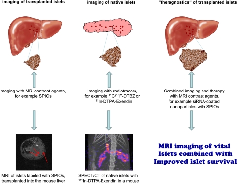
FIG. 1.
This figure shows the main approaches to imaging of β-cells at this point in time. SPIOs have been used for labeling of islets prior to transplantation since 2004 (left). Other MRI contrast agents are currently under investigation and potentially also allow for quantification by MRI spectroscopy. For the determination of the pancreatic β-cell mass, PET and SPECT imaging with highly specific radiotracers seem to be the techniques of choice today (middle). MRI imaging of native islets using the calcium analog Mg++ as contrast agent has successfully been performed for imaging of the functional pancreatic β-cells. The approach using siRNA-coated nanoparticles, providing contrast for diagnostics (MRI) and at the same time exerting therapeutic effects that increase the islet survival, is taking β-cell imaging to a new level (right). (A high-quality digital representation of this figure is available in the online issue.)

