Abstract
Lyme disease is a multisystemic disorder caused by an epizootic organism of the spirochete group, called Borrelia burgdorferi, which is transmitted to humans by ticks of the genus Ixodes. Lyme neuroborreliosis may occur during the early dissemination phase, most often as a painful meningo-radiculitis and very rarely as a radiculo-myelitis, whereas encephalomyelitis is observed in the late phase. We report the case of a patient with an early subacute poliomyelitis-like syndrome closely matching the selective involvement of the anterior horns and roots of the cervical spinal cord seen on magnetic resonance imaging. This condition improved with appropriate antibiotics.
BACKGROUND
Lyme disease is a multisystemic disorder caused by an epizootic organism of the spirochete group, called Borrelia burgdorferi (Bb), which is transmitted to humans by ticks of the genus Ixodes.1 Three sequential clinical stages have been described: (i) early localised; (ii) early disseminated; and (iii) late persistent disease. Lyme neuroborreliosis may occur during the early dissemination phase, most often as a painful meningo-radiculitis and very rarely as a radiculo-myelitis, whereas encephalomyelitis is observed in the late phase.2 We report the case of a patient with an early subacute poliomyelitis-like syndrome closely matching the selective involvement of the anterior horns and roots of the cervical spinal cord seen on magnetic resonance (MR) imaging.
CASE PRESENTATION
A previously healthy 21-year-old man presented in September 2006 with painless weakness in both arms that had increased over a period of 2 weeks. He had a short history of neck pain and stiff neck 2 months previously, which had spontaneously resolved. An indepth retrospective anamnesis failed to reveal any evidence of tick bites or erythema chronicum migrans, but the patient had undertaken open air activities in a woody countryside at the end of June.
Physical examination showed severe weakness (2/5) in the proximal muscles of both upper limbs. Strength was only mildly reduced in the hands (4.5/5) and was normal in the legs. No sensory deficit was present. Diffuse fasciculations were observed in both shoulders. Deep tendon reflexes were not elicited in the right arm. Of note, Lhermitte’s sign was absent. There were no meningeal signs or fever.
INVESTIGATIONS
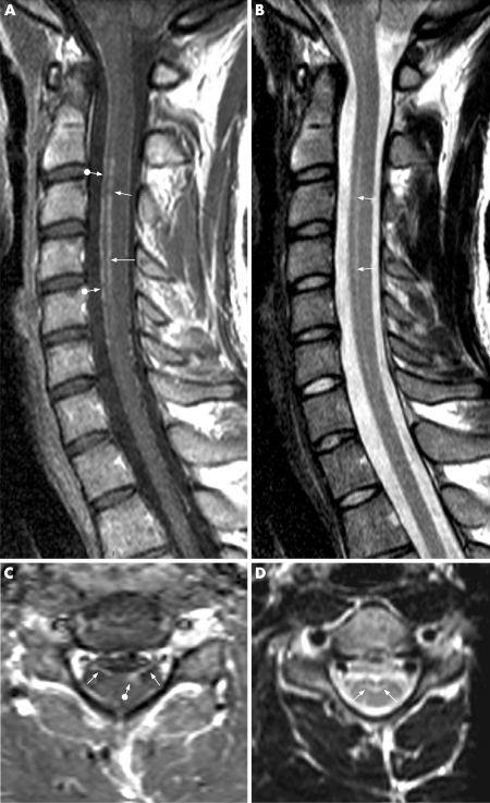
Cervical spine MR examination showed abnormally increased signal intensity within the anterior horns of the cord “grey butterfly” from C2 to C5 on the T2 weighted images (fig 1B, D), together with increased thickness and strong contrast enhancement of the anterior radicelles of the same dermatoma on post-contrast T1 weighted images (fig 1A, C). Additional abnormal post-contrast enhancement was also observed within the pia mater (fig 1A) and the anterior horns (fig 1C). Brain MR examination was unremarkable.
Figure 1. Magnetic resonance (MR) workup of the cervical spine at admission.
(A) Post-contrast mid-sagittal T1 weighted image showing abnormal enhancement of both the anterior pia mater (ball-arrowheads) and anterior horns of central grey matter from C2 to C5 (arrows). (B) Mid-sagittal T2 weighted image (similar slice location as in (A)) showing abnormal linear hypersignal intensity in the anterior horns of central grey matter in the same C2–C5 area (arrows). (C) Post-contrast transverse T1 weighted image at the C5 level showing thickened, abnormally enhancing, anterior roots due to inflammatory blood root barrier disruption (arrows), together with enhancement within the left anterior horns (ball-arrowhead). (D) T2 weighted transverse image at a comparable level showing abnormal hypersignal intensity within the anterior horns (arrows).
Neurophysiological studies demonstrated elective involvement of motor nerves at the radicular or motoneuronal level. Needle electromyography showed signs of acute denervation with spontaneous fibrillations in both deltoid and biceps brachii, with a right-sided predominance. Abnormalities were also present in the first dorsal interosseous muscles and in the extensor digitorum communis, but to a lesser degree. Sensory and motor conduction velocities of the ulnar and median nerves were normal, as well as the latency and the amplitude of the F waves.
Standard haematological tests and biochemical assays were normal. Serology was negative for HIV, hepatitis B and C, and syphilis (TPHA and VDRL tests). However, serological tests for Lyme disease (Euroimmun Anti-Borrelia plusV1sE ELISA IgG and Euroimmun Anti-Borrelia ELISA IgM, Lübeck, Germany) were strongly positive in serum for both IgM (IgM ratio of 5.74, normal value <1.0) and IgG (133 relative unit (RU)/ml, normal value <20) antibodies. The IgG and IgM positive samples were confirmed by a commercial western blot assay (Euroline-WB; Euroimmun AG) showing specific patterns (OspC for IgM antibodies, V1sE, p83, p39, p31, 30, OspC and p17 for IgG antibodies).
CSF analysis showed 79 cells/mm3 with 79% lymphocytes, 12% reactive lymphoblasts and 9% monocytes. The total protein content was elevated (98 mg/dl; normal range 20–55). Considerable intrathecal synthesis of IgM was present (intrathecal fraction at 76%) with a lower synthesis of IgG (intrathecal fraction at 17%). However, several CSF restricted oligoclonal IgG were present. Bb PCR was negative. However, native CSF contained 58 RU/ml of anti-Bb IgG antibodies confirmed by Euroline-WB assay, whereas serum diluted at the same IgG concentration was negative. Such results demonstrated intrathecally restricted production of these antibodies, which is a key feature of Lyme neuroborreliosis.3
TREATMENT
Intravenous ceftriaxone (2 g daily) was given for 2 weeks and the weakness slowly improved thereafter. The clinical examination normalised 6 weeks after the start of treatment. A follow-up MR examination performed 6 months later demonstrated complete normalisation of the spinal status.
OUTCOME AND FOLLOW-UP
Intravenous ceftriaxone (2 g daily) was given for 2 weeks and the weakness slowly improved thereafter. The clinical examination normalised 6 weeks after the start of treatment. A follow-up MR examination performed 6 months later demonstrated complete normalisation of the spinal status.
DISCUSSION
The diagnosis of Lyme neuroborreliosis was therefore biologically supported by the positive blood IgM and IgG serology for Bb, together with the strong intrathecal synthesis of IgM and IgG antibodies seen on analysis of CSF. In addition, the patient’s status dramatically improved with ceftriaxone antimicrobial treatment. The clinical presentation was, however, atypical, because of the absence of pain with nocturnal exacerbation, the purely motor involvement with absent tendon reflexes in the upper right limb and the diffuse fasciculations in the shoulders. These atypical signs and symptoms nicely corresponded to the cervical spinal cord imaging showing selective inflammatory involvement of anterior grey matter and spinal roots.
Recently, eight documented cases of early subacute Bb myelitis have been reported.2 In six of these cases, painful radicular symptoms appeared before spinal cord signs. Most patients exhibited CSF mononuclear pleocytosis and cervical spinal cord lesions on MRI. All but one patient had a favourable outcome with appropriate intravenous antibiotherapy. Absence of encephalitic involvement and cranial nerve palsy, frequent combination with meningoradiculitis and good response to antibiotherapy are key features allowing discrimination between early and late variants of Bb myelitis.
Leptomeningeal enhancement and nerve root enhancement of the cauda equina on post-contrast T1 weighted MR images have been reported in spinal Lyme neuroborreliosis.4 Enhancement of the cervical nerve roots has also been reported in this condition.5 Concomitant enhancement of the leptomeninges, anterior horns and anterior radicelles of the cervical spine has never been described to date. The close correlation between clinical and MR features was a prominent feature of this clinically and radiologically atypical case of Lyme neuroborreliosis.
LEARNING POINTS
Lyme neuroborreliosis can cause a painful radiculopathy with a transverse myelititis.
In the case described here, there was no pain and purely motor involvement which corresponded to the spinal cord segments involved on MRI.
Lyme neuroborreliosis associated myelitis responds to antibiotics.
Acknowledgments
This article has been adapted with permission from Charles V, Duprez TP, Kabamba B, Ivanoiu A, Sindic CJM. Poliomyelitis-like syndrome with matching magnetic resonance features in a case of Lyme neuroborreliosis. J Neurol Neurosurg Psychiatry 2007;78:1160–1.
Footnotes
Competing interests: None.
REFERENCES
- 1.Hengge R, Tannapfel A, Tyring SK, et al. Lyme borreliosis. Lancet Infect Dis 2003; 3: 489–500 [DOI] [PubMed] [Google Scholar]
- 2.Meurs L, Labeye D, Declercq I, et al. Acute transverse myelitis as a main manifestation of early stage II neuroborreliosis in two patients. Eur Neurol 2004; 52: 185–8 [DOI] [PubMed] [Google Scholar]
- 3.Sindic CJM, Depré A, Bigaignon G, et al. Lymphocytic meningoradiculitis and encephalomyelitis due to Borrelia burgdorferi: a clinical and serological study of 18 cases. J Neurol Neurosurg Psychiatry 1987; 50: 1565–71 [DOI] [PMC free article] [PubMed] [Google Scholar]
- 4.Demaerel P, Wilms G, Van Lierde S, et al. Lyme disease in childhood presenting as primary leptomeningeal enhancement without parenchymal findings on MR. Am J Neuroradiol 1994; 15: 302–4 [PMC free article] [PubMed] [Google Scholar]
- 5.Hattingen A, Weidaauer S, Kieslich M, et al. MR imaging in neuroborreliosis of the cervical spinal cord. Eur Radiol 2004; 14: 2072–5 [DOI] [PubMed] [Google Scholar]


