Abstract
A gastrosplenic fistula is a rare complication of gastric and splenic lymphomas which can occur spontaneously or secondary to chemotherapy. We report a case of a spontaneous gastrosplenic fistula secondary to a diffuse splenic large B cell lymphoma in a previously well 43-year-old patient. CT imaging demonstrated the fistula, which was subsequently managed with chemotherapy. The clinical management of this rare condition is discussed with a review of the literature.
Background
This case is based upon a rare complication of non-Hodgkin's lymphoma.
Different disciplines within the medical field, including gastroenterologists, radiologists and oncologists worked effectively together to manage the patient, thus underlining the importance of a multi-disciplinary approach to malignancies. Furthermore, this case emphasises the difficulty in treating gastrosplenic fistulas with a review of the literature highlighting the adoption of different approaches in their management.
Case presentation
A 43-year-old female was referred by her primary care physician to the local hospital with a 2-month history of upper abdominal pain and weight loss of 8 kg in conjunction with early satiety. Her abdominal pain was initially localised to the left upper quadrant, but after 1 month this progressed to include the epigastrum. There were no complaints of fevers or vomiting, and she had no significant past medical history.
Physical examination revealed a tender left upper quadrant mass, palpable 5 cm below the costal margin. There was no peripheral lymphadenopathy, nor any evidence of respiratory or cardiovascular disease.
The patient's admission was later complicated with an episode of haematemesis and malaena with subsequent haemodynamic compromise that required a blood transfusion. The patient was appropriately stabilised with blood products and volume support.
Investigations
Laboratory studies identified a normocytic anaemia (Hb 9.2 d/dl) with a normal platelet count (312 × 109/l), white cell count (7.2 × 10⁹/l), liver function and renal function tests, but a raised C-reactive protein (107 mg/l).
Gastroscopy revealed an ulcer measuring 2 × 2 cm at the gastric fundus with irregular edges, in addition to gastric varices with the presence of blood within the stomach. In view of these findings, the patient was subsequently admitted for further investigation.
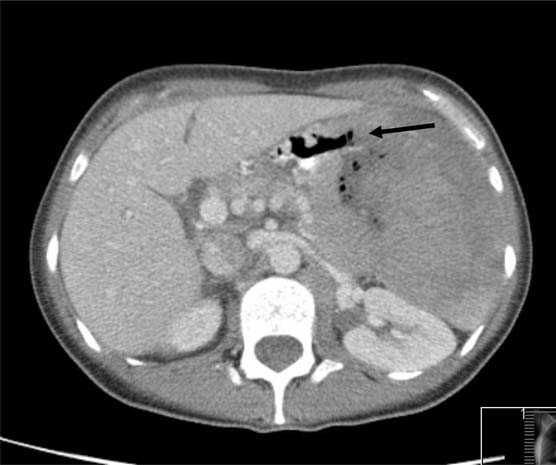
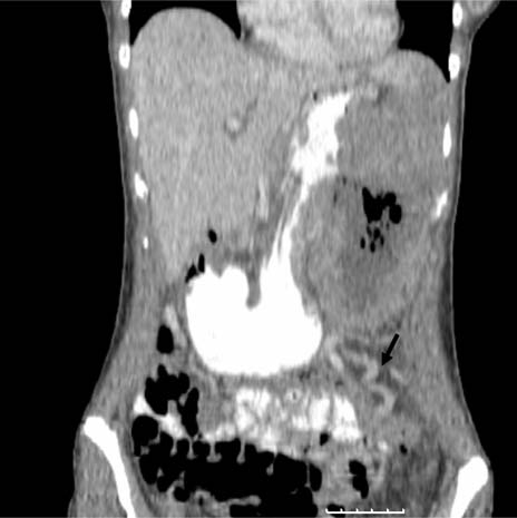
CT of the chest, abdomen and pelvis demonstrated a 18.9 cm (superior–inferior) × 10 cm (anterior–posterior) × 8.6 cm (transverse) mass centred on the spleen which directly invaded neighbouring structures including the anterior peritoneal wall, pancreatic tail, left adrenal gland, greater curve of stomach and the left gastric vessels. The superior aspect of the lesion also extended up to, but did not breach, the left hemidiaphragm. Splenic vein invasion was demonstrated causing secondary venous collaterals surrounding the mass (figure 1) indicative of the slow growing nature of the mass. The tumoural lesion was heterogeneous in appearance with both solid and cystic components including central liquefied necrosis and locules of air. A direct communication, that is, a gastrosplenic fistula, between the mass and the gastric lumen was outlined with air and oral contrast (figure 2). Importantly there was no significant evidence of local or distant significant lymphadenopathy. CT imaging therefore demonstrated a large aggressive tumoural mass invading adjacent structures centred on the spleen.
Figure 1.

Coronal reformat image with intravenous and oral contrast. Secondary collaterals (black arrow) are visible.
Figure 2.

Multislice axial image of the abdomen with intravenous and oral contrast demonstrates the gastrosplenic fistula outlined by air and contrast (black arrow).
In view of the extensive and unusual nature of disease, a tertiary opinion and referral was made. A further gastroscopy was performed with deep biopsies taken from the greater curve for histopathological analysis. Examination of the biopsies was consistent with diffuse large B cell lymphoma with no evidence of Helicobacter pylori-like organisms or intestinal metaplasia. The cells were CD20 positive, PACS5 positive, BCL6 positive, MUM1 positive and weakly CD10 positive. The proliferation index with MIB1 was 100%.
Further staging with positron emission tomography scan using [18F] fluoro-D-glucose tracer was performed identifying small volume but metabolically active lymph nodes above and below the diaphragm.
Differential diagnosis
Differential diagnosis of the CT findings included a primary splenic lymphoma given the age of the patient, splenic angiosarcoma or a malignant stromal lesion.
Treatment
Following multi-disciplinary discussion, the patient went on to receive an urgent first cycle of chemotherapy within 1 month of presentation. She was eligible for the R-CODOX-M/IVAC regime (an alternating combination of rituximab, cyclophosphamide, vincristine, doxorubicin and methotrexate with etoposide, ifosfamide and cytaribine) in view of her high international prognostic index accompanying the diffuse large B cell lymphoma.
Outcome and follow-up
The patient tolerated this regime well and is currently in complete remission according to the National Cancer Research Institute criteria at the time of this report after completing two courses of chemotherapy.
Discussion
A fistula between the stomach and spleen is a very rare clinical manifestation. Aetiologies include gastric adenocarcinomas,1 Crohn's disease,2 benign gastric ulcers3 and lymphomas.3–18 Gastric and splenic lymphomas can also fistulate with other organs, including the bronchus19 and colon.20
Extensive splenic necrosis and gastric wall infiltration is required for gastrosplenic fistula formation.9 It can occur after chemotherapy as a result of rapid regression of the tumour that has infiltrated the gastric mucosa thus resulting in a fistula.4 6 12 13 Alternatively, as in our case, it can occur spontaneously. The aggressive nature of diffuse large B cell lymphomas and the tendency for adjacent organ invasion contributes to this spontaneous fistula formation.9 11 These features were seen in our case where there was extensive infiltration of adjacent organs, including the pancreas and diaphragm.
To our knowledge, there have been only six reported cases of spontaneous gastrosplenic fistula associated with a primary splenic lymphoma.5 7–9 16 We report the seventh case of a spontaneous gastrosplenic fistula from a primary splenic non-Hodgkin's lymphoma.
The clinical presentation of diffuse large cell splenic lymphoma usually consists of left upper quadrant abdominal pain accompanied by features of a systemic illness and splenomegaly on examination.5 These characteristics were also present in our patient. Furthermore, our patient's admission was complicated with haematemesis, which has been the clinical presentation of other gastrosplenic fistulas.6 8
Multislice CT provides the best imaging modality to identify gastrosplenic fistulas10 14 due to excellent spatial resolution and accurate staging of lesions. The clinical presentation of our patient coupled with the absence of any significant past medical history or evidence of endocarditis helped to exclude other differential diagnoses such as necrotising splenic abscess. In previously described cases the spleen was shown to be heterogenous with parenchymal masses containing air, similar to our case.8 9 The fistulous tract has been outlined with oral contrast7 in some cases. One other case of gastrosplenic fistula in the literature was identified by a retrograde catheter cystography for a suspected splenic abscess that inadvertently revealed itself as a fistula.13
Radical surgical resection is the most common treatment option, which would typically demand a splenectomy and gastrectomy. However, there are reports of necessary distal pancreatectomies performed in some cases.14 Although open procedures are more commonly described, there has been a successful laparoscopic case.17 An advantage of surgical resection is that it aids in establishing a pathological diagnosis in uncertain cases.11 In contrast, there are also cases of resolution of the fistula with chemotherapy.3 18 Patients are at risk of haematemesis due to the risk of invasion into adjacent vasculature, specifically the gastric and splenic vessels. Treatment can also be supportive and in life threatening blood loss interventional radiology in the form of splenic artery embolisation8 has been described. Surgery in our patient would have been a huge undertaking with associated high mortality in view of the size of the lesion and multiple adjacent structures involved. The patient was not clinically septic and chemotherapy was therefore the treatment option offered. The presence of fistulas, risk of haemorrhage and infection highlights the challenges faced with managing tumoural fistulas.
Learning points.
-
▶
A gastrosplenic fistula is a rare complication of lymphoma.
-
▶
It can occur spontaneously or after chemotherapy.
-
▶
Multislice CT with intravenous and oral contrast demonstrated the site of pathology, complications such as varices, the fistula tract and any invasion of adjacent organs.
-
▶
Treatment modalities include surgical resection, chemotherapy or a combination of both.
Footnotes
Competing interests None.
Patient consent Obtained.
References
- 1.Krause R, Larsen CR, Scholz FJ. Gastrosplenic fistula: complication of adenocarcinoma of stomach. Comput Med Imaging Graph 1990;14:273–6 [DOI] [PubMed] [Google Scholar]
- 2.Cary ER, Tremaine WJ, Banks PM, et al. Isolated Crohn's disease of the stomach. Mayo Clin Proc 1989;64:776–9 [DOI] [PubMed] [Google Scholar]
- 3.Carolin KA, Prakash SH, Silva YJ. Gastrosplenic fistulas: a case report and review of the literature. Am Surg 1997;63:1007–10 [PubMed] [Google Scholar]
- 4.Bubenik O, Lopez MJ, Greco AO, et al. Gastrosplenic fistula following successful chemotherapy for disseminated histiocytic lymphoma. Cancer 1983;52:994–6 [DOI] [PubMed] [Google Scholar]
- 5.Harris NL, Aisenberg AC, Meyer JE, et al. Diffuse large cell (histiocytic) lymphoma of the spleen. Clinical and pathologic characteristics of ten cases. Cancer 1984;54:2460–7 [DOI] [PubMed] [Google Scholar]
- 6.Hiltunen KM, Airo I, Mattila J, et al. Massively bleeding gastrosplenic fistula following cytostatic chemotherapy of a malignant lymphoma. J Clin Gastroenterol 1991;13:478–81 [DOI] [PubMed] [Google Scholar]
- 7.Blanchi A, Bour B, Alami O. Spontaneous gastrosplenic fistula revealing high-grade centroblastic lymphoma: endoscopic findings. Gastrointest Endosc 1995;42:587–9 [DOI] [PubMed] [Google Scholar]
- 8.Bird MA, Amjadi D, Behrns KE. Primary splenic lymphoma complicated by hematemesis and gastric erosion. South Med J 2002;95:941–2 [PubMed] [Google Scholar]
- 9.Choi JE, Chung HJ, Lee HG. Spontaneous gastrosplenic fistula: a rare complication of splenic diffuse large cell lymphoma. Abdom Imaging 2002;27:728–30 [DOI] [PubMed] [Google Scholar]
- 10.Puppula S, Williams R, Harvey J, Crane MD. Spontaneous gastrosplenic fistula in primary gastric lymphoma: case report and review of literature. Clin Radiol Extra 2005;60:20–2 [Google Scholar]
- 11.Kerem M, Sakrak O, Yilmaz TU, et al. Spontaneous gastrosplenic fistula in primary gastric lymphoma: Surgical management. Asian J Surg 2006;29:287–90 [DOI] [PubMed] [Google Scholar]
- 12.Palmowski M, Zechmann C, Satzl S, et al. Large gastrosplenic fistula after effective treatment of abdominal diffuse large-B-cell lymphoma. Ann Hematol 2008;87:337–8 [DOI] [PubMed] [Google Scholar]
- 13.Aribas BK, Baskan E, Altinyollar H, et al. Gastrosplenic fistula due to splenic large cell lymphoma diagnosed by percutaneous drainage before surgical treatment. Turk J Gastroenterol 2008;19:69–70 [PubMed] [Google Scholar]
- 14.Seib CD, Rocha FG, Hwang DG, et al. Gastrosplenic fistula from Hodgkin's lymphoma. J Clin Oncol 2009;27:e15–17 [DOI] [PubMed] [Google Scholar]
- 15.García Marín A, Bernardos García L, Vaquero Rodríguez A, et al. [Spontaneous gastrosplenic fistula secondary to primary gastric lymphoma]. Rev Esp Enferm Dig 2009;101:76–8 [DOI] [PubMed] [Google Scholar]
- 16.Maillo C, Bau J. [Gastrosplenic and thoracosplenic fistula due to primary untreated splenic lymphoma]. Rev Esp Enferm Dig 2009;101:222–3 [DOI] [PubMed] [Google Scholar]
- 17.Al-Ashgar HI, Khan MQ, Ghamdi AM, et al. Gastrosplenic fistula in Hodgkin's lymphoma treated successfully by laparoscopic surgery and chemotherapy. Saudi Med J 2007;28:1898–900 [PubMed] [Google Scholar]
- 18.Moghazy KM. Gastrosplenic fistula following chemotherapy for lymphoma. Gulf J Oncolog 2008;3:64–7 [PubMed] [Google Scholar]
- 19.Cameron EW, Colby JM, Swanson RS. Gastrobronchial fistula in untreated lymphoma. J Thorac Imaging 1996;11:150–2 [DOI] [PubMed] [Google Scholar]
- 20.Naschitz JE, Yeshurun D, Horovitz IL, et al. Spontaneous colosplenic fistula complicating immunoblastic lymphoma. Dis Colon Rectum 1986;29:521–3 [DOI] [PubMed] [Google Scholar]


