Abstract
We present a case of a chondral lesion after anterior cruciate ligament (ACL) reconstruction caused by femoral cross-pin breakage and intra-articular migration of the fragment. A 20-year-old man initially underwent ACL reconstruction using a hamstring autograft. The RigidFix bioabsorbable cross-pin (DePuy Mitek) was used for the femoral fixation. The patient returned to a pre-injury level of activity (professional soccer player) 6 months postoperatively. However, 20 months postoperatively, the patient presented with effusion and lateral joint-line pain after practice, without signs of instability in clinical examination. Conservative treatment failed and at re-arthroscopy a chondral lesion of the lateral femoral and tibial condyle was found, which had been caused by the broken femoral cross-pin. The fragment was removed and the symptoms resolved. Orthopaedic surgeons should be aware of this complication when using a bioabsorbable cross-pin for femoral fixation in ACL reconstruction.
BACKGROUND
In our opinion this case reveals a new cause of chondral injury. The fact that the cause is related to anterior cruciate ligament (ACL) reconstruction using a bioabsorbable femoral cross-pin is very interesting due to the popularity of the technique.
CASE PRESENTATION
A 20-year-old professional soccer player initially sustained an injury to his right knee that resulted in an ACL tear. The patient underwent ACL reconstruction in our department using a hamstring autograft. The RigidFix cross-pin system (DePuy Mitek) was used for femoral fixation and the BiointraFix system (DePuy Mitek) for tibial fixation. The patient returned to a pre-injury level of activity 6 months postoperatively, after having followed the usual accelerated postoperative rehabilitation regimen successfully.
Twenty months postoperatively, the patient presented with effusion and lateral joint-line pain after practise. He showed no signs of instability and he was treated with activity modification, non-steroidal anti-inflammatory drugs and physical therapy. The symptoms resolved but soon after his return to athletic activities he had recurrent effusion and lateral joint-line pain.
INVESTIGATIONS
Magnetic resonance imaging studies did not demonstrate any noticeable pathology and the patient underwent a diagnostic arthroscopy.
TREATMENT
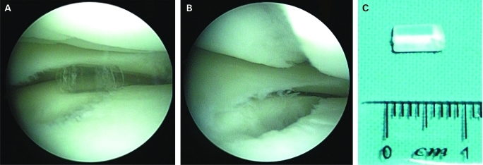
A broken pin (fig 1A) was found as a loose body in the lateral compartment causing chondral erosion in both the lateral femoral (grade I) and tibial (grade II) condyle (fig 1B). A thorough examination failed to reveal any other intra-articular pathology and the broken pin was removed. The graft was found intact and well incorporated in both femoral and tibial tunnels.
Figure 1.
(A) Arthroscopic view of the broken cross-pin. (B) Arthroscopic view of the lateral femoral and tibial condyle chondral injury. (C) Measurement of the fragment.
OUTCOME AND FOLLOW-UP
The patient’s symptoms resolved and he, eventually, returned to the pre-injury level of activity. The patient is still asymptomatic 2 years after re-arthroscopy.
DISCUSSION
More than 150 ACL reconstructions per year are performed in our institution. We have used this femoral fixation technique in 750 ACL reconstructions over the last 7 years and this is the first case of femoral cross-pin breakage that we are aware of.
Two more cases of femoral RigidFix bioabsorbale cross-pin breakages have been reported in the literature.1,2 The type and level of activity is not clearly specified in both cases. In the first case,1 it is reported that the patient was participating in competitive sports and in the second case,2 the patient sustained an ACL tear while performing a martial arts manoeuvre. In the case reported by Cosssey and Paterson1 a bone-patella tendon-bone graft was used for the ACL reconstruction, while in the case reported by Han et al2 an allogenic hamstring tendon graft was used for the ACL reconstruction.Therefore, this is the first case reported in the literature with femoral cross-pin breakage using a hamstring tendon autograft.
Cossey and Paterson1 made no comment regarding the cause of the femoral cross-pin breakage. They found two fragments of the cross-pin, one of which was the tip of the cross-pin, and they had to scope the patient twice in order to remove the fragments. In the case reported by Han et al2 the tip of the cross-pin was also found as a loose body in the knee joint cavity. The authors suggested two reasons that could have contributed to cross-pin breakage. First, the vertically oriented tibia tunnel might have resulted in a vertically oriented femoral tunnel (transtibial technique) and posterior wall blowout. Secondly, the direction of cross-pins was quite posterior. Both reasons could have led the tip of the cross-pin outside the femoral bone in the joint space. This hypothesis is in accordance with the fact that the fragment they found was the tip of the femoral cross-pin.
We reviewed the medical file and video of the ACL reconstruction, as well as the postoperative magnetic resonance imaging films, to detect technical errors that could have contributed to cross-pin breakage. It seems that, the above-mentioned scenario is unlikely to have happened in our case, so we have to look for another explanation. Femoral tunnel orientation (we use the anteromedial portal) and cross-pin direction were properly drilled and placed. Moreover, the fragment we found (fig 1C) came from the middle part of the cross-pin and it was similar in length to the diameter of the femoral tunnel (7 mm approximately).
We assume that the intra-tunnel part of the cross-pin was weakened more quickly than the rest of the cross-pin (hydrolysis) due to communication with the intra-articular fluid and it eventually broke and migrated to the lateral compartment of the knee. Graft fixation into the femoral tunnel, as close to the articular cavity as possible, has the advantage of minimising the effect of windscreen (tunnel enlargement due to graft motion distal to the fixation point). On the other hand, the possibility of weakening and breakage of the absorbable implants due to communication with the intra-articular fluid should be further evaluated.
LEARNING POINTS
Orthopaedic surgeons should be aware of this rare complication when using bioabsorbable cross-pins for femoral fixation in ACL reconstruction.
Accurate surgical technique is of great importance in order to avoid cross-pin breakage.
Intra-tunnel weakening and breakage of the femoral cross-pin is a possibility that should be kept in mind.
Footnotes
Competing interests: none.
Patient consent: Patient/guardian consent was obtained for publication.
REFERENCES
- 1.Cossey AJ, Paterson RS. Loose intra-articular body following anterior cruciate ligament reconstruction. Arthroscopy 2005; 21: 348–50 [DOI] [PubMed] [Google Scholar]
- 2.Han I, Kim YH, Yoo JH, et al. Broken bioabsorbable femoral cross-pin after anterior cruciate ligament reconstruction with hamstring tendon graft. Am J Sports Med 2005; 33: 1742–5 [DOI] [PubMed] [Google Scholar]


