Abstract
Acute lymphoblastic leukaemia and acute myeloblastic leukaemia are the most common malignancies diagnosed in children. Facial palsy is an acute peripheral palsy involving the facial nerve and is an unusual presentation of childhood acute leukaemia. We present three cases (a 9-year-old boy, a 14-year-old boy and a 10-year-old boy) of acute leukaemia with initial presentation of facial palsy. It is important for physicians to recognise the neurological manifestations of childhood leukaemia and extensive work-up should be carried out to exclude secondary causes of facial palsy.
Background
Acute lymphoblastic leukaemia (ALL) and acute myeloblastic leukaemia (AML) are the most common malignancies diagnosed in children and arise within bone marrow precursors of lymphoid and myeloid lineages. ALL accounts for one fourth of all childhood cancer and approximately 75% of all cases of childhood leukaemia, with an annual incidence of about 30 cases per million people and a peak incidence in children aged 2–5 years. AML comprises approximately 15–20% of childhood leukaemia.1,2
Facial palsy is an acute, peripheral, lower motor neuron facial nerve paralysis with a usually favourable prognosis. Its causes are unknown, although it appears to be a polyneuritis with possible infectious, inflammatory, autoimmune and metabolic aetiologies.3 In addition, facial palsy is an unusual presentation of leukaemia and other lymphoid and myeloid malignancies where facial neuritis has secondary involvement.3–7
We present three cases of childhood acute leukaemia where facial palsy was the first manifestation of disease.
Case presentation
Case 1
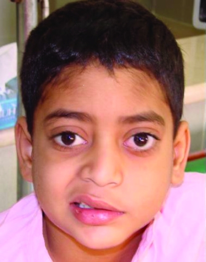
A 9-year-old boy was admitted with a chief complaint of decreased vision and pain in the left eye of 2 months’ duration (figure 1).
Figure 1.
Left facial palsy as initial presentation in a patient with acute lymphoblastic leukaemia.
Corticosteroid treatment was administered for 15 days without improvement. The patient developed malaise, fatigue, anorexia, nausea and vomiting. He had also developed bone pain 3 weeks before admission and fever 2 weeks before admission. Magnetic resonance imaging (MRI) revealed T2 hypersignal changes at the left side of the facial nerve suggesting acute inflammation.
On admission complete blood count (CBC) showed white blood cells (WBC) 261.9×10³/μl, haemoglobin (Hb) 9.2 g/dl, platelets 150×103/μl with blast cells 92%, lymphocytes 6%, neutrophils 1% and myelocytes 1%.
Bone marrow aspiration and immunophenotyping showed T cell ALL. Cerebrospinal fluid (CSF) analysis revealed pleocytosis with total cells 142/mm3 and 8% blast cells, sugar 68 mg/dl and protein 93 mg/dl, suggesting central nervous system (CNS) involvement.
The patient underwent a initial session of chemotherapy (vincristine, daunorubicin, prednisolone and l-asparginase) but developed tumour lysis syndrome, with serum calcium 5.3 mg/dl, serum phosphorus 9 mg/dl, blood urea nitrogen 68 mg/dl, serum creatinine 6 mg/dl, blood sodium 138 mEq/l and blood potassium 4.5 mEq/l, necessitating haemodialysis. After finishing treatment, the patient achieved complete remission and the facial palsy improved.
Case 2
A 14-year-old boy presented with isolated acute onset of right peripheral facial palsy without any other symptoms and signs. The patient was treated with prednisolone, 1 mg/kg/day for 2 weeks. After 2 weeks the patient showed relative improvement in facial paralysis but complained of bone pain, backache, fever and mild hepatosplenomegaly. No abnormalities were seen in the general and neurological exams, except for slight right peripheral facial palsy. Brain MRI with and without contrast revealed normal results. CBC revealed WBC 6.2×10³/μl, Hb 11.5 g/dl and platelets 238×103/μl, and sedimentation rate was 23 mm/h. Bone marrow aspiration showed 90% blast cells indicating acute lymphoblastic leukaemia. Flow cytometry revealed T cell ALL.
CSF analysis revealed total cells 150/mm3 with lymphoblasts 30%, lymphocytes 60% and segmented cells 10%, protein 75 mg/dl and sugar 65 mg/dl.
The patient underwent chemotherapy, ultimately recovered and was discharged after 6 weeks of hospitalisation. During a 7-month follow-up, the patient was found to be in remission and was receiving maintenance therapy.
Case 3
A 10-year-old boy was admitted with isolated acute onset of left peripheral facial palsy without any other positive physical findings. The patient was treated as a case of facial palsy with prednisolone, 1 mg/kg/day for 4 weeks without any significant improvement. He discontinued his medication himself. Two weeks later he developed spontaneous nose bleeding and was referred to the paediatric emergency ward. He had a history of headache for the previous few weeks. At the time of admission he was pale, irritable and febrile, with left facial palsy, multiple purpuro-ecchymotic lesions over his body and mild gum hypertrophy. Brain computed tomography (CT scan) with contrast was normal. MRI, performed with and without gadolinium, was normal. CBC showed WBC 11.2×10³/μl, Hb 6.8 g/dl, platelets 28×103/μl with blast cells 42%, neutrophils 34%, lymphocytes 14%, monocytes 5%, pro-myelocytes 3% and meta-myelocytes 2%. Bone marrow aspiration showed cellular marrow with 80% blast cells suggesting acute myelo-monocytic leukaemia (AML-M4). Bone marrow cytogenetic study showed 46XY. CSF analysis revealed pleocytosis with total cells 145/mm3 and 10% blast cells, protein 110 mg/dl and sugar 57 mg/dl which suggested CNS involvement. The patient underwent chemotherapy and cranial radiotherapy but few months later he experienced bone marrow relapse. He received anti-CD30 antibody (Myelotarg) without significant response and finally died due to disseminated fungal infection.
Discussion
Facial palsy is an idiopathic acute peripheral palsy involving the facial nerve which supplies all the muscles used for facial expression. Facial palsy has been described in patients of all ages with an incidence of 2.7 per 100 000 in children under 10 years of age.3
There have been some reports of an association between facial palsy and acute leukaemia in the literature.3–7 Ozçakar et al reported a 20-year-old man with ALL who presented with bilateral facial nerve involvement4 and Schattner et al described a 36-year-old man with T-ALL, with bilateral 7th nerve palsy who had normal MRI results.5 Krishnamurthy et al presented two 11-month-old infants and a 6-year-old boy with acute leukaemia whose initial manifestation was lower motor neuron facial palsy.7 The first diagnosis was facial palsy and MRI revealed no evidence of facial nerve or meningeal enhancement in the infants but did reveal left facial nerve enhancement in the older boy. Krishnamurthy et al concluded that the negative results of MRI in infants may reflect the fact that thin slicing of the facial canal was not performed. CSF analysis was normal in the three patients without leukaemic cells. Facial palsy was improved in one infant and an older child after 6-month follow-up, but one infant had no improvement in facial palsy 2 months after chemotherapy and radiotherapy despite complete remission.7
In this report, we also describe three cases of facial palsy as the first manifestation of childhood leukaemia. Facial palsy was improved in one patient after successful chemotherapy and in another patient there was relative improvement after prednisolone therapy and completely cure after chemotherapy. The third patient died before any improvement.
Surrounding meningeal involvement of the nerve and direct leukaemic infiltration of the tympanic cavity and temporal bone can cause injury to the facial nerve in patients with leukaemia.
Although brain MRI is well known to prove cranial nerve involvement in facial palsy, it is shown that brain MRI and CSF cytology does not provide definite results in all patients. The routine use of steroid treatment in patients suspected of having idiopathic facial palsy may provide partial remission but may lead to delay in the actual diagnosis of acute leukaemia. All such patients must therefore be followed up and examined for any new signs or symptoms of leukaemia.
In conclusion, acute leukaemia should be considered early in the differential diagnosis of facial palsy and intensive CNS therapy based on chemotherapy and radiotherapy is recommended in patients with leukaemia and facial palsy even when the results of MRI and CSF cytology are negative.
Learning points
It is important to recognise neurological manifestation of childhood leukaemia because a delay in diagnosis can have an adverse effect on survival.
Extensive work-up should be carried out to exclude secondary causes for facial palsy in childhood.
Normal MRI and CSF analysis does not rule out nerve involvement and intensive central nervous system therapy must be considered in children with leukaemia and facial palsy involvement.
Footnotes
Competing interests: None.
Patient consent: Patient/guardian consent was obtained for publication.
REFERENCES
- 1.Pui CH, Relling MV, Downing JR. Acute lymphoblastic leukemia. N Engl J Med 2004; 350: 1535–48 [DOI] [PubMed] [Google Scholar]
- 2.Löwenberg B, Downing JR, Burnett A. Acute myeloid leukemia. N Engl J Med 1999; 341: 1051–62 [DOI] [PubMed] [Google Scholar]
- 3.Bilavsky E, Scheuerman O, Marcus N, et al. Facial paralysis as a presenting symptom of leukemia. Pediatr Neurol 2006; 34: 502–4 [DOI] [PubMed] [Google Scholar]
- 4.Ozçakar L, Akinci A, Ozgöçmen S, et al. Bell’s palsy as an early manifestation of acute lymphoblastic leukemia. Ann Hematol 2003; 82: 124–6 [DOI] [PubMed] [Google Scholar]
- 5.Schattner A, Kozack N, Sandler A, et al. Facial diplegia as the presentation manifestation of acute lymphoblastic leukemia. Mt Sinai J Med 2001; 68: 406–9 [PubMed] [Google Scholar]
- 6.Sawada H, Matsui M, Udaka F, et al. Adult T-cell leukemia initially manifesting as facial diplegia. Am J Hematol 1989; 32: 61–5 [DOI] [PubMed] [Google Scholar]
- 7.Krishnamurthy S, Weinstock AL, Smith SH, et al. Facial palsy, an unusual presenting feature of childhood leukemia. Pediatr Neurol 2002; 27: 68–70 [DOI] [PubMed] [Google Scholar]


