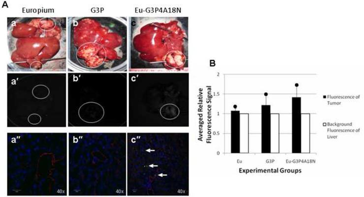
Figure 4. In vivo cannulation with Eu3+, G3P and Eu-G3P4A18N with luminescence imaging and analysis.
(A) Gross photographs, luminescence and confocal microscopic images at 40× magnification of the livers containing tumors (circles) that were injected in the spleen 15– 20 days prior to infusion and excised at 0 h time point. Photos (a), (a′) and (a″) were from infusion of Eu3+ only at 0 h. Photos (b), (b′) and (b″) were from infusion of G3P (non-functionalized dendrimer without Eu3+) and at 0 h. Photos (c), (c′) and (c″) were from liver infused with Eu-G3P4A18N at 0 h. Arrows show the luminescence of Eu-G3P4A18N in the confocal microscopic images in photo (c″). (B) Average tumor luminescence was corrected for background autofluorescence in the resulting graph. Two rats per each experimental group were used.

