
Figure 2.
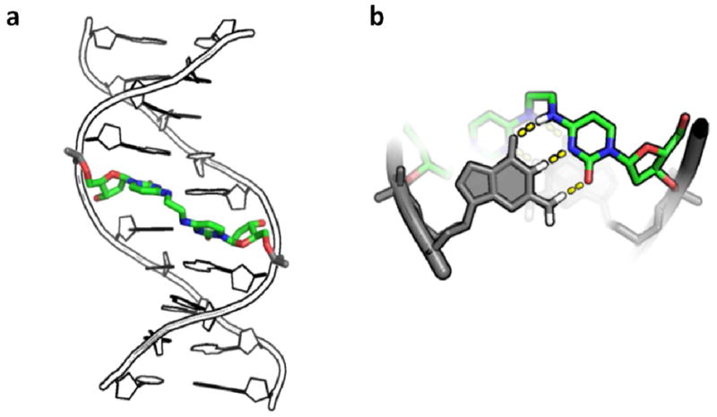
Structure of DNA duplex containing the N4C-ethyl-N4C crosslink. (a) Crystallographic model of duplex DNA (pdb accession name 2OKS) containing a central staggered 5′–CG-3′ N4C-ethyl-N4C interstrand crosslink in the major groove. (b) Structural detail of crosslinked cytosines looking down the DNA helical axis. Dashed lines denote the Watson-Crick hydrogen bonding groups of cytosine that are free to pair with an opposing guanine base shown in grey. For reference, the DNA major and minor grooves are located at the top and bottom of the structure, respectively.