Abstract
PB2 627K is a determinant of influenza host range and contributes to the pathogenicity of human-, avian-, and mouse-adapted influenza viruses in the mouse model. Here we used mouse and pig models to analyze the contribution of a swine-origin and avian-origin PB2 carrying either 627K or 627E in the background of the classical swine H1N1 (A/Swine/Iowa/15/30; 1930) virus. The results showed PB2 627K is crucial for virulence in the mouse model, independent of whether PB2 is derived from an avian or swine influenza virus (SIV). In the pig model, PB2 627E decreases pathogenicity of the classical 1930 SIV when it contains the swine-origin PB2, but not when it possesses the avian-origin PB2. Our study suggests the pathogenicity of SIVs with different PB2 genes and mutation of codon 627 in mice does not correlate with the pathogenicity of the same SIVs in the natural host, the pig.
Keywords: swine influenza virus, PB2, pathogenicity
Introduction
Swine influenza caused by influenza A virus is one of the most important infectious diseases in pigs. To date, there are 16 hemagglutinin (HA) and 9 neuraminidase (NA) subtypes of influenza A virus, all of which have been isolated from aquatic birds (Alexander, 2000; Fouchier et al., 2005; Webster et al., 1992). Influenza A virus can also infect a large variety of mammalian species including humans, horses, dogs, cats, and sea mammals. Because host range is restricted, only certain subtypes of influenza A virus have been established and maintained in mammalian species; for example, only three subtypes (i.e., H1N1, H3N2, and H1N2) of influenza A viruses are consistently isolated from pigs worldwide. Pigs have been suggested to play an important role in transmission between birds and humans by acting as the “mixing vessel” for influenza viral reassortment (Ma, Kahn, and Richt, 2009; Scholtissek, 1994) because of the species’ susceptibility to infection with both human and avian influenza viruses. This susceptibility may be due to the fact that porcine tracheal cells have viral receptors for mammalian and avian viruses (Ito et al., 1998).
With the recent introduction of genes from human and avian influenza viruses into swine influenza viruses (SIVs), the major viruses now circulating through North American swine herds are triple-reassortant viruses (i.e., H3N2, H1N1, and H1N2). These triple reassortant SIVs have similar, conserved triple reassortant internal genes, including the avian PA and PB2 polymerase genes, the human PB1 polymerase gene, and the swine nucleoprotein (NP), matrix (M), and nonstructural protein (NS) genes. The novel avian/human polymerase complex encoded by these genes can accept multiple HA and NA types and may endow a selective advantage to SIVs. The recently characterized novel swine H2N3 and swine human-like H1 viruses are examples of viruses that demonstrate the flexibility of the avian/human polymerase complex to accept various HAs and NAs (Ma et al., 2007; Vincent et al., 2009). The 2009 pandemic H1N1 virus is a reassortant virus that contains NA and M genes from Eurasian SIVs and the other 6 genes from North American triple reassortant SIVs (Smith et al., 2009). The common feature of the pandemic H1N1 and North American triple reassortant viruses is that these viruses have a novel polymerase complex (i.e., avian PA and PB2 and human PB1).
The polymerase complex is responsible for transcription and replication of the viral genome and is a major determinant of host range and pathogenicity (Naffakh et al., 2008; Neumann and Kawaoka, 2006). A single residue at position 627 in the PB2 protein is largely responsible for determining host range and pathogenicity of influenza viruses (Hatta et al., 2001; Subbarao, London, and Murphy, 1993). PB2 that is derived from human influenza viruses usually possesses a lysine (K) at position 627 whereas that derived from avian influenza viruses predominantly possesses a glutamic acid (E) at this position (Chen et al., 2006). In North American SIVs, almost all pre-1998 isolates are classical H1N1 viruses containing PB2 627K whereas most post-1998 isolates are triple reassortant H1N1 or H3N2 viruses containing PB2 627E (Bussey et al., 2010). The pandemic H1N1 virus contains E at position 627 of its PB2 protein (Garten et al., 2009). It is unclear how the avian PB2 with 627E in SIVs affects viral replication, pathogenicity, and interspecies transmission. In this study, a reassortant virus (i.e., 1930-Tx/98PB2-627E) was generated by reverse genetics in the genetic background of the classical H1N1 A/Swine/Iowa/15/30 (1930) virus, which carries the PB2 gene from an H3N2 triple reassortant A/Swine/Texas/4199-2/98 (Tx/98) virus. The PB2 gene of the Tx/98 virus is avian in origin (Zhou et al., 1999). In addition, reassortant viruses (1930-Tx/98PB2-627K, 1930-PB2-627E) with a single amino acid (aa) substitution at position 627 in the avian-origin or swine-origin PB2 were generated. The growth properties and pathogenicity profiles of reassortant viruses carrying avian-origin or swine-origin PB2 with 627 E or K were evaluated in vitro and in vivo.
Results
Growth characteristics of the wild-type and reassortant viruses
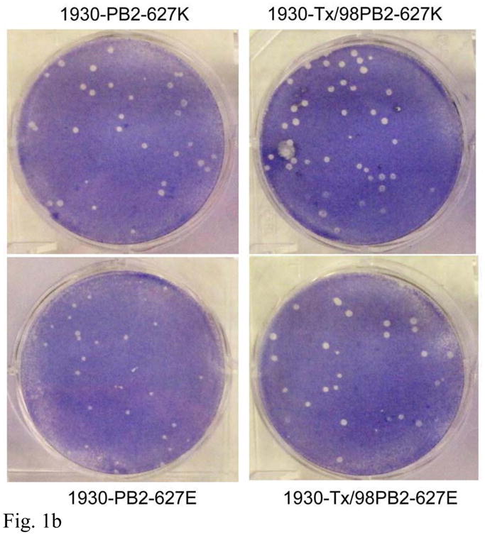
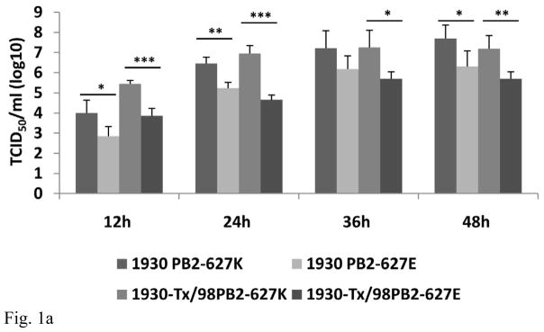
The avian-origin Tx/98 PB2 shows 96.8% homology at the nucleotide level and 95.5% homology at the aa level with the swine-origin 1930 virus PB2. There are 34 aa difference between the avian-origin Tx/98 and swine-origin 1930 PB2 proteins (Table 1). The reassortant 1930-Tx/98PB2-627E and 1930-Tx/98PB2-627K viruses formed similar size plaques as the wild-type 1930-PB2-627K virus did; however, the 1930-PB2-627E virus with the single substitution at position 627 formed smaller plaques than the other three viruses did (Figure 1a). In MDCK cells, the virus carrying the avian-origin PB2 gene with 627K (1930-Tx/98PB2-627K) grew to significantly higher titers (p < 0.05) than did the respective virus with PB2 627E (Figure 1b). Similarly, the virus containing the swine-origin PB2 gene with 627K (1930-PB2-627K) grew to significantly higher titer (p < 0.05) than did the respective virus with the swine-origin PB2 627E except for the time point 36h post inoculation (Figure 1b).
Table 1.
Comparison of the avian-origin Tx/98 and swine-origin 1930 PB2 at the amino acid level
| 22* | 64 | 65 | 66 | 82 | 114 | 122 | 136 | 147 | 184 | 199 | 225 | 243 | 265 | 271 | 312 | 382 | |
| Tx/98 PB2 | K | M | D | M | N | V | V | R | T | T | A | G | M | N | A | E | I |
| 1930 PB2 | R | I | E | T | S | I | A | G | I | M | S | S | L | S | T | K | V |
| 421 | 467 | 468 | 475 | 478 | 480 | 539 | 588 | 590 | 591 | 627 | 637 | 645 | 660 | 661 | 684 | 702 | |
| Tx/98 PB2 | V | M | I | L | I | V | I | T | S | R | E | T | L | K | A | A | K |
| 1930 PB2 | I | L | T | M | V | I | V | A | G | Q | K | A | M | R | T | T | R |
Position of the amino acid
Figure 1.

Characterization of reassortant and PB2 single-substitution viruses in vitro. (a) Plaque size in MDCK cells 2 days after infection; (b) Virus growth in MDCK cells at different time points. The results are representative of four independent experiments and values represent the log10 geometric mean TCID50/mL ± SEM (*P < 0.05; **P < 0.01; ***P < 0.001).
Mouse pathogenicity of reassortant SIVs with PB2 mutations
Mice inoculated with 1930-Tx/98PB2-627K or the wild-type 1930-PB2-627K virus developed clinical disease (i.e., weight loss, dyspnea, rough fur, and lethargy); this disease resulted in death of most of the inoculated mice (Table 2). Mice inoculated with the 1930-Tx/98PB2-627E virus containing the avian-origin PB2 with 627E developed similar clinical illnesses but without mortality. In contrast, mice inoculated with the 1930-PB2-627E virus did not show any clinical signs and appeared similar to the mock-inoculated mice. Mice in either the 1930-Tx/98PB2-627E group or the 627K groups who survived the infections did not fully recover from them by the end of the experiment. Compared to mock-inoculated controls, four inoculated groups had significantly greater (p < 0.05) microscopic lung lesions (Table 2). The 1930-PB2-627K 1930-Tx/98PB2-627K and 1930-Tx/98PB2-627E caused significantly greater microscopic lung lesions (p < 0.05) than did the 1930-PB2-627E (Table 2). No significant difference in microscopic lung lesion was evident between the 1930-Tx/98PB2-627K-inoculated mice and the 1930-Tx/98PB2-627E-inoculated mice (Table 2). Although viral RNA was detected in the lung homogenate from each infected mouse, virus was only isolated from the lungs of mice inoculated with the wild-type 1930-PB2-627K (mean virus titer = 103.97 TCID50) and from the lungs of 9 mice inoculated with 1930-Tx/98PB2-627K (mean virus titer = 102.83 TCID50). The viral RNA measured in the lungs of mice infected with PB2-627K (i.e., 1930-PB2-627K and 1930-Tx/PB2-627K) groups was 106 fold higher than that in the lungs of mice infected with PB2-627E (i.e., 1930-PB2-627E and 1930-Tx/98PB2-627E) groups (data not shown). Sequence analysis showed that position 627 (E or K) of the PB2 protein of respective viruses isolated from infected mouse lungs represents the same amino acid as the challenge viruses, indicating this specific amino acid is stable for at least one in vivo passage.
Table 2.
Mortality and lesions in mice inoculated with PB2 single-substituted or parental viruses
| Virus | Mortalitya | Average no. of days p.i. until deathb | Histopathologic Score (0–3)c |
|---|---|---|---|
| 1930-PB2-627K | 8/10d | 7.80 ± 1.09d | 2.75 ± 0.34d |
| 1930-PB2-627E | 0/10d | 14.00 ± 0.00d | 1.10 ± 0.21d, f, g |
| 1930-Tx/98PB2-627E | 0/10e | 14.00 ± 0.00e | 2.05 ± 0.24f |
| 1930-Tx/98PB2-627K | 9/10e | 7.30 ± 0.86e | 2.30 ± 0.26g |
| Control | 0/10 | 14.00 ± 0.00 | 0.00 ± 0.00 |
Mortality = number of mice that died after challenge/total number of mice in group
Values are the mean ± SEM
Significant difference between 2 groups (P < 0.05).
Pig pathogenicity of reassortant SIVs with PB2 mutations
No acute respiratory signs were observed in either pig challenge experiment. Necropsy revealed macroscopic lung lesions (i.e., plum-colored, consolidated areas) in all virus-inoculated pigs but none in the mock-inoculated control pigs (Table 3). On day 3 p.i., there was no statistically significant difference in the extent of macroscopic lung lesions among any of the virus-inoculated groups; however, viruses with swine-origin PB2 induced more extensive macroscopic lesions on average than did the viruses with avian-origin PB2 gene. On day 5 p.i., the 1930-PB2-627K group had significantly greater (P < 0.05) lung lesions than did the 1930-PB2-627E and 1930-Tx/98PB2-627K groups, but there was no significant difference between the lesions of the 1930 virus with avian-origin PB2 (i.e., avian-origin PB2 627K and 627E) groups (Table 3). On day 3 p.i., viruses carrying either the avian-origin PB2 627K (1930-Tx/98PB2-627K) or the swine-origin PB2 627K (1930-PB2-627K) replicated more efficiently (i.e. to higher titer) in pigs’ lungs than did the virus containing the swine-origin PB2 627E (P < 0.01); however, there were no significant differences found on day 5 p.i. (Table 3). No significant difference on virus titers in pigs’ lungs was found between 1930-Tx/98PB2-627K and 1930-Tx/98PB2-627E viruses on days 3 and 5 p.i. (Table 3). Lungs from virus-inoculated pigs that were euthanized on day 3 p.i. or day 5 p.i. exhibited mild to moderate interstitial pneumonia and acute to subacute necrotizing bronchiolitis with slight lymphocytic cuffing of bronchioles and vessels. Only one (20%, 1/5) nasal swab collected from a pig inoculated with the 1930-Tx/98PB2-627K virus on day 5 p.i. was positive for virus isolation and no virus was detected in other nasal swab samples collected on days 3 and 5 p.i. from any virus challenge groups in either experiment (data not shown). Sequence analysis showed that position 627 (E or K) of the PB2 protein of viruses isolated from nasal swab and BALF samples collected form infected pigs represents the same amino acid as the challenge viruses.
Table 3.
Virus titers in BALF and macroscopic and microscopic lung lesions in influenza virus-inoculated pigs
| Virus | Virus titer in BALF (TCID50/mL)a | Lung lesion score (%)b | Histopathologic score (0–3)b | |||
|---|---|---|---|---|---|---|
| Day 3 p.i. | Day 5 p.i. | Day 3 p.i. | Day 5 p.i. | Day 3 p.i. | Day 5 p.i. | |
| Avian-origin PB2 | ||||||
| Mock | 0.00 ± 0.00 | 0.00 ± 0.00 | 0.00 ± 0.00 | 0.00 ± 0.00 | 0.40 ± 0.40 | 0.40 ± 0.24 |
| 1930-Tx/98PB2-627E | 3.76 ± 0.51 | 3.92 ± 0.48 | 15.97 ± 4.46 | 14.01 ± 3.10 | 2.90 ± 0.40 | 2.60 ± 0.51 |
| 1930-Tx/98PB2-627K | 5.17 ± 0.55e | 3.65 ± 0.17 | 16.40 ± 3.37 | 9.97 ± 2.17f | 2.60 ± 0.40 | 2.50 ± 0.45 |
| Swine-origin PB2 | ||||||
| Mock | 0.00 ± 0.00 | 0.00 ± 0.00 | 0.00 ± 0.00 | 0.00 ± 0.00 | 0.70 ± 0.12 | 0.60 ± 0.19 |
| 1930-PB2-627E | 2.96 ± 0.14c, e | 3.99 ± 0.30 | 23.31 ± 4.13 | 10.26 ± 2.29d | 2.40 ± 0.19 | 2.30 ± 0.30 |
| 1930-PB2-627K | 4.25 ± 0.41c | 4.39 ± 0.10 | 24.49 ± 4.08 | 22.29 ± 5.46d, f | 3.00 ± 0.16 | 2.90 ± 0.10 |
Values represent the log10 geometric mean BALF TCID50/mL ± SEM
Values represent the mean ± SEM
There is a significant difference between the 2 groups (P < 0.05)
Discussion
Avian PB2 with 627E were introduced into North American SIVs more than 10 years ago, and this avian signature (i.e., 627E) remains stable rather than changing to the mammalian signature (i.e., 627K). In this study, we found that the PB2 protein with aa 627K is a key determinant for efficient viral replication in MDCK cells and for virulence in mice. These findings confirm previous results showing that PB2 627K is essential for virulence of the 1997 H5N1 and other highly pathogenic H5N1 avian influenza viruses in mice (Hatta et al., 2001; Hatta et al., 2007; Liu et al., 2005).
The virus containing the avian-origin PB2 with 627E was able to infect mice and pigs, indicating that other important aa mutations can compensate for the absence of 627K in the PB2 protein as reported previously (Li et al., 2009). Our sequence comparison of PB2s from the Tx/98 and 1930 viruses with those from mouse-adapted viruses revealed that both Tx/98 and 1930 PB2s contain the same aa (i.e., 333T, 482K, 701D, and 714S), which are completely different from the conserved PB2 aa signatures (i.e., 333I, 482A, 701N, and 714R) normally required in mouse-adapted viruses (Naffakh et al., 2008), suggesting that other mutations can also confer adaptation of the avian-origin PB2 to mice and other mammalian species. Normally, the avian-origin PB2 contains E at aa position 627 and alanine (A) at aa position 271 whereas the swine-origin PB2 contains K and threonine (T) at those respective positions (Bussey et al., 2010; Naffakh et al., 2008). The avian PB2 271A residue plays a key role in enhanced polymerase activity and viral growth of influenza A viruses in mammalian host cells and likely contributes to virus spread and cross-species transmission of pandemic H1N1 viruses (Bussey et al., 2010). Therefore, PB2 271A might compensate for the presence of 627E in the 1930-Tx/98PB2-627E virus and in currently circulating SIVs. The avian-origin PB2 in the currently circulating SIVs and in the pandemic H1N1 viruses contains a conserved serine (S) at position 590 and a conserved arginine (R) at position 591, called the SR polymorphism, which increase polymerase activity and viral replication in human and porcine cells if present with PB2 627E (Mehle and Doudna, 2009). This SR polymorphism is considered to be an adaptive strategy of the avian influenza virus polymerase for replication in humans (Mehle and Doudna, 2009). Because the Tx/98 avian-origin PB2 contains this SR polymorphism, it could contribute to both mammalian adaptation and disease-causing capabilities of the 1930-Tx/98PB2-627E virus.
In pig challenge experiments, the 1930 viruses with PB2 627K (i.e., 1930-Tx/98PB2-627K and 1930-PB2-627K) were able to replicate in pig lungs and induce severe macroscopic and histopathologic lung lesions whether in the avian-origin or swine PB2 background. Additionally, when the classical 1930 virus contains an avian-origin PB2, either 627K or 627E allow for pathogenicity in the pig model. This result is different from our discovery in the mouse model and in a previous study that 627K in the H5N1 avian PB2 polymerase is crucial for replication in pigs (Manzoor et al., 2009). Our comparisons of the histopathologic lung lesions and virus titers caused by each virus revealed that the 627E residue of PB2 decreases the pathogenicity of the classical 1930 virus in pigs when it contains the swine-origin PB2 but not when it contains the avian-origin PB2.
Thus, the importance of the aa at position 627 in PB2 appears to differ among different virus strains and in different animal species. These results correlate with previous observations in which substituting the E at position 627 in PB2 with K does not change virus replication and virulence of the 2009 pandemic H1N1 virus in mice (Herfst et al., 2010; Zhu et al., 2010) or the pathogenicity and transmission of the virus in ferrets (Herfst et al., 2010). Furthermore, although the majority of the lethal H5N1 viruses for ferrets and mice possess PB2 627K, some H5N1 viruses with PB2 627E are lethal in ferrets (Katz et al., 2000; Maines et al., 2005; Salomon et al., 2006). Whether PB2 is derived from swine or avian influenza viruses, 627K of PB2 is crucial for virulence of the classical H1N1 SIV in the mouse model. In the pig model, PB2 627E decreases pathogenicity of the classical 1930 SIV when it contains the swine-origin PB2, but not when it possesses the avian-origin PB2. Our study suggests that the pathogenicity of SIVs with different PB2 genes and mutation of codon 627 in mice does not correlate with the pathogenicity of the same SIVs in the natural host, the pig.
Materials and methods
Cells and eggs
Human embryonic kidney (HEK)-293T cells were maintained in Opti–modified Eagle’s medium (MEM) supplemented with 10% fetal calf serum (FCS) (HyClone; Logan UT) and 1% antibiotics (Invitrogen; Carlsbad, CA), and Madin-Darby canine kidney (MDCK) cells were maintained in Dulbecco’s MEM (DMEM) with 5% FCS, L-glutamine (Invitrogen), MEM vitamins (Invitrogen), and 1% antibiotics (Invitrogen). Cells were inoculated with the respective viruses in MEM infecting medium containing 0.3% bovine albumin (Sigma; St. Louis, MO), 1μg/mL TPCK-treated trypsin (Sigma), L-glutamine, MEM vitamins, and 1% antibiotics (Invitrogen). Ten-day-old embryonated chicken eggs were inoculated with virus via the allantoic cavity for viral amplification.
Construction of mutated PB2-containing plasmids
To change the aa at position 627 of avian-origin PB2 (Tx/98) from E to K, the GeneTailor Site-Directed Mutagenesis system (Invitrogen) was used according to the manufacturer’s recommendations. Briefly, 100 ng pHW-Sw-PB2 containing the PB2 gene from the Tx/98 virus (Solorzano et al., 2005) was methylated by incubating it with DNA methylase at 37°C for 1 hour, and then it was amplified in a mutagenesis reaction with a pair of overlapping primers (i.e., Tx/98-PB2 627K-Fw and Tx/98-PB2 627K-Rw) (primer sequences are available upon request). The mutagenesis mixture (2 μL) was used to transform competent E. coli cells, and plasmids were prepared using the Qiagen Plasmid Mini kit (Qiagen, Valencia, CA). The mutation of the resulting plasmid (i.e., pHW-Sw-PB2-627K) was confirmed by sequencing.
To change the aa at position 627 of the swine PB2 (i.e., 1930) from K to E, 2 PCR reactions for PB2 were performed using the universal forward PB2 primer (Hoffmann et al., 2001) and the reverse PB2 mutation primer, which contains the mutated nucleotide; pDZ-PB2 containing the PB2 gene from the 1930 virus was used as a template (or the forward PB2 mutation primer containing the mutated nucleotide and the universal reverse PB2 primer were used) (primer sequences are available upon request). The full-length PB2 was amplified by using PB2 universal primers and 2 partial PCR products as templates. The full-length PB2 was partially digested by Bsa I (NEB, Ipswich, MA) and then cloned into the BsmB I (NEB) sites of a pHW2000 vector. The mutation of the resulting plasmid (i.e. pHW-PB2-627E) was confirmed by sequencing.
Generation of wild-type 1930, reassortant and PB2 mutated viruses
An eight plasmid (i.e., pDZ-HA, -NA, -PB1, -PB2, -PA, -NP, -M, and -NS) reverse-genetics system was used to generate the wild-type 1930 virus as previously described (Ma et al., 2010). To rescue the 1930 (i.e., 1930-PB2-627K) virus’ codon 627 of PB2 from a K to an E, pHW-PB2-627E was used to replace pDZ-PB2. The reassortant virus (i.e., 1930-Tx/98PB2-627E) carrying the avian-origin PB2 gene from the Tx/98 virus in the genetic background of the 1930 virus was rescued by using the pHW-Sw-PB2 replacing pDZ-PB2. To generate the 1930-Tx/98PB2 (1930-Tx/98PB2-627E) virus with an E to K substitution at codon 627 of PB2, pHW-Sw-PB2-627K was used to replace pDZ-PB2. The sequence of each rescued reassortant viruses was confirmed, and each challenge virus was diluted in MEM to a final concentration of 105 50% tissue culture infective dose (TCID50).
Analysis of viral replication
To analyze viral replication efficiency, a monolayer of MDCK cells in 6-well plates was inoculated with each virus at a multiplicity of infection (MOI) of 0.01. Supernatant from infected cells was collected at different time points. Virus titer was determined by inoculating confluent monolayers of MDCK cells in 96-well plates as previously described (Richt et al., 2003), and the TCID50 per milliliter was calculated for each sample by the method of Reed and Muench (Reed and Muench, 1938).
Experiments in mice
To compare the relative virulence of the different reassortant viruses in mice, experiments were conducted using 6- to 7-week-old Balb/c mice at the National Animal Disease Center (NADC), Ames, Iowa. Mouse experiments were conducted in compliance with the NADC’s Institutional Animal Care and Use Committee. Fifty mice were randomly allocated to 5 treatment groups. Mice were anesthetized with isoflurane USP (Phoenix Pharmaceutical, Inc.; St. Joseph, MO) and intranasally inoculated with a 50 μl volume containing either virus-free MEM (i.e., negative control) or 105 TCID50 of a designated virus as depicted in Table 2. Weights were recorded daily, and general health status was observed twice daily. After the onset of disease, the general health status was observed thrice daily. Severely affected mice were euthanized immediately, and all other mice were euthanized on day 14 post-inoculation (p.i.). At necropsy, the right lung was stored at −80°C for later analysis, and the left lung was fixed in 10% formalin for histopathologic analysis. Virus detection was performed in a 10% tissue homogenate in PBS that had been homogenized twice for 1 min in a Mini BeadBeater-8 (Biospec Products; Bartlesville, OK). The homogenate was centrifuged at 640 × g for 5 minutes, and the supernatant was transferred to 1.5 mL reaction tubes for virus and RNA isolation. Real-time RT-PCR was used to detect viral RNA as described previously (Richt et al., 2004).
Experiments in pigs
Four-week-old porcine reproductive and respiratory syndrome virus– and SIV–negative pigs were used in this study, and all pig experiments were approved by the Institutional Animal Care and Use Committee at Kansas State University. The pathogenicity of viruses containing avian-origin or swine PB2 with 627E or 627K was investigated via two separate pig challenge experiments; the inoculation protocol has been described previously (Richt et al., 2003). In the first experiment, 10 pigs were inoculated with 1930-Tx/98PB2-627E and 10 with 1930-Tx/98PB2-627K. In the second experiment, 10 pigs were inoculated with 1930-PB2-627K and 10 with 1930-PB2-627E. In each experiment, 10 age-matched control pigs were mock-inoculated with serum-free MEM.
Five inoculated pigs and 5 control pigs were necropsied on days 3 and 5 p.i. in each experiment. Nasal swabs were taken on days 0, 3, and 5 p.i., placed in 2 mL MEM and stored at − 80°C. Blood was collected from all pigs on days 0 and at the time of necropsy. Bronchioalveolar fluid (BALF) was obtained by lavage of each lung with 50 mL MEM. Viral load in BALF was determined in a 96-well plate as previously described (Richt et al., 2003). To determine virus shedding, nasal swabs were tested for infectious virus by inoculating10-day-old embryonated chicken eggs.
Lung examinations of experimental pigs
At necropsy, lungs were removed in toto. A single experienced veterinarian recorded the percentage of gross lesions on each lobe, and a mean value for the seven lobes was calculated for of each animal (Richt et al., 2003). For histopathologic examination, tissue samples from the trachea, the right cardiac lung lobe, and other affected lobes were fixed in 10% buffered formalin, processed, and stained with hematoxylin and eosin. Lung sections were examined by a veterinary pathologist in a blinded fashion and given a score of 0 to 3 to reflect the severity of bronchial epithelial injury as described previously (Ma et al., 2007; Richt et al., 2003).
Statistical analysis
Macroscopic and microscopic pneumonia scores, mortality, and average number of days p.i. until death were analyzed by using analysis of variance (ANOVA) in GraphPad Prism version 5.0 (GraphPad software Inc. La Jolla, CA); a P-value of 0.05 or less was considered significant. Those response variables shown to have a significant effect by treatment group were subjected to comparisons for all pairs by using the Tukey–Kramer test. Pair-wise mean comparisons between inoculated and control groups were made using the Student’s t-test.
Acknowledgments
We thank Cherise Guess for her editorial assistance and Adolfo García-Sastre for critical discussions of the manuscript; Deb Clouser, Audree Gottlob, Darlene Sheffer, Tom Wegman, and Jacob Elder from Kansas State University for assisting the pig studies and Kevin Hassall, Hannah Polashek, Trudy Tatum, and David Panthen from NADC for assisting the mouse studies and providing technical support. We also thank the DNA sequence unit and histopathology core unit at NADC and the histopathology section at the Department of Diagnostic Medicine/Pathobiology at Kansas State University for their assistance. This project was partially funded by the National Institute Allergy and Infectious Disease, National Institutes of Health, Department of Health and Human Services, under contract number HHSN266200700005C.
Footnotes
Publisher's Disclaimer: This is a PDF file of an unedited manuscript that has been accepted for publication. As a service to our customers we are providing this early version of the manuscript. The manuscript will undergo copyediting, typesetting, and review of the resulting proof before it is published in its final citable form. Please note that during the production process errors may be discovered which could affect the content, and all legal disclaimers that apply to the journal pertain.
References
- Alexander DJ. A review of avian influenza in different bird species. Vet Microbiol. 2000;74(1–2):3–13. doi: 10.1016/s0378-1135(00)00160-7. [DOI] [PubMed] [Google Scholar]
- Bussey KA, Bousse TL, Desmet EA, Kim B, Takimoto T. PB2 residue 271 plays a key role in enhanced polymerase activity of influenza A viruses in mammalian host cells. J Virol. 2010;84 (9):4395–406. doi: 10.1128/JVI.02642-09. [DOI] [PMC free article] [PubMed] [Google Scholar]
- Chen GW, Chang SC, Mok CK, Lo YL, Kung YN, Huang JH, Shih YH, Wang JY, Chiang C, Chen CJ, Shih SR. Genomic signatures of human versus avian influenza A viruses. Emerg Infect Dis. 2006;12 (9):1353–60. doi: 10.3201/eid1209.060276. [DOI] [PMC free article] [PubMed] [Google Scholar]
- Fouchier RA, Munster V, Wallensten A, Bestebroer TM, Herfst S, Smith D, Rimmelzwaan GF, Olsen B, Osterhaus AD. Characterization of a novel influenza A virus hemagglutinin subtype (H16) obtained from black-headed gulls. J Virol. 2005;79 (5):2814–22. doi: 10.1128/JVI.79.5.2814-2822.2005. [DOI] [PMC free article] [PubMed] [Google Scholar]
- Garten RJ, Davis CT, Russell CA, Shu B, Lindstrom S, Balish A, Sessions WM, Xu X, Skepner E, Deyde V, Okomo-Adhiambo M, Gubareva L, Barnes J, Smith CB, Emery SL, Hillman MJ, Rivailler P, Smagala J, de Graaf M, Burke DF, Fouchier RA, Pappas C, Alpuche-Aranda CM, Lopez-Gatell H, Olivera H, Lopez I, Myers CA, Faix D, Blair PJ, Yu C, Keene KM, Dotson PD, Jr, Boxrud D, Sambol AR, Abid SH, St George K, Bannerman T, Moore AL, Stringer DJ, Blevins P, Demmler-Harrison GJ, Ginsberg M, Kriner P, Waterman S, Smole S, Guevara HF, Belongia EA, Clark PA, Beatrice ST, Donis R, Katz J, Finelli L, Bridges CB, Shaw M, Jernigan DB, Uyeki TM, Smith DJ, Klimov AI, Cox NJ. Antigenic and genetic characteristics of swine-origin 2009 A(H1N1) influenza viruses circulating in humans. Science. 2009;325(5937):197–201. doi: 10.1126/science.1176225. [DOI] [PMC free article] [PubMed] [Google Scholar]
- Hatta M, Gao P, Halfmann P, Kawaoka Y. Molecular basis for high virulence of Hong Kong H5N1 influenza A viruses. Science. 2001;293 (5536):1840–2. doi: 10.1126/science.1062882. [DOI] [PubMed] [Google Scholar]
- Hatta M, Hatta Y, Kim JH, Watanabe S, Shinya K, Nguyen T, Lien PS, Le QM, Kawaoka Y. Growth of H5N1 influenza A viruses in the upper respiratory tracts of mice. PLoS Pathog. 2007;3 (10):1374–9. doi: 10.1371/journal.ppat.0030133. [DOI] [PMC free article] [PubMed] [Google Scholar]
- Herfst S, Chutinimitkul S, Ye J, de Wit E, Munster VJ, Schrauwen EJ, Bestebroer TM, Jonges M, Meijer A, Koopmans M, Rimmelzwaan GF, Osterhaus AD, Perez DR, Fouchier RA. Introduction of virulence markers in PB2 of pandemic swine-origin influenza virus does not result in enhanced virulence or transmission. J Virol. 2010;84 (8):3752–8. doi: 10.1128/JVI.02634-09. [DOI] [PMC free article] [PubMed] [Google Scholar]
- Hoffmann E, Stech J, Guan Y, Webster RG, Perez DR. Universal primer set for the full-length amplification of all influenza A viruses. Arch Virol. 2001;146 (12):2275–89. doi: 10.1007/s007050170002. [DOI] [PubMed] [Google Scholar]
- Ito T, Couceiro JN, Kelm S, Baum LG, Krauss S, Castrucci MR, Donatelli I, Kida H, Paulson JC, Webster RG, Kawaoka Y. Molecular basis for the generation in pigs of influenza A viruses with pandemic potential. J Virol. 1998;72 (9):7367–73. doi: 10.1128/jvi.72.9.7367-7373.1998. [DOI] [PMC free article] [PubMed] [Google Scholar]
- Katz JM, Lu X, Tumpey TM, Smith CB, Shaw MW, Subbarao K. Molecular correlates of influenza A H5N1 virus pathogenesis in mice. J Virol. 2000;74 (22):10807–10. doi: 10.1128/jvi.74.22.10807-10810.2000. [DOI] [PMC free article] [PubMed] [Google Scholar]
- Li J, Ishaq M, Prudence M, Xi X, Hu T, Liu Q, Guo D. Single mutation at the amino acid position 627 of PB2 that leads to increased virulence of an H5N1 avian influenza virus during adaptation in mice can be compensated by multiple mutations at other sites of PB2. Virus Res. 2009;144 (1–2):123–9. doi: 10.1016/j.virusres.2009.04.008. [DOI] [PubMed] [Google Scholar]
- Liu J, Xiao H, Lei F, Zhu Q, Qin K, Zhang XW, Zhang XL, Zhao D, Wang G, Feng Y, Ma J, Liu W, Wang J, Gao GF. Highly pathogenic H5N1 influenza virus infection in migratory birds. Science. 2005;309 (5738):1206. doi: 10.1126/science.1115273. [DOI] [PubMed] [Google Scholar]
- Ma W, Kahn R, Richt J. The pig as a mixing vessel for influenza viruses: Human and veterinary implications. J Mol Genet Med. 2009;3(1):158–166. [PMC free article] [PubMed] [Google Scholar]
- Ma W, Lager KM, Lekcharoensuk P, Ulery ES, Janke BH, Solorzano A, Webby RJ, Garcia-Sastre A, Richt JA. Viral reassortment and transmission after coinfection of pigs with classical H1N1 and triple reassortant H3N2 swine influenza viruses. Journal of General Virology. 2010;91:2314–2321. doi: 10.1099/vir.0.021402-0. [DOI] [PMC free article] [PubMed] [Google Scholar]
- Ma W, Vincent AL, Gramer MR, Brockwell CB, Lager KM, Janke BH, Gauger PC, Patnayak DP, Webby RJ, Richt JA. Identification of H2N3 influenza A viruses from swine in the United States. Proc Natl Acad Sci U S A. 2007;104(52):20949–54. doi: 10.1073/pnas.0710286104. [DOI] [PMC free article] [PubMed] [Google Scholar]
- Maines TR, Lu XH, Erb SM, Edwards L, Guarner J, Greer PW, Nguyen DC, Szretter KJ, Chen LM, Thawatsupha P, Chittaganpitch M, Waicharoen S, Nguyen DT, Nguyen T, Nguyen HH, Kim JH, Hoang LT, Kang C, Phuong LS, Lim W, Zaki S, Donis RO, Cox NJ, Katz JM, Tumpey TM. Avian influenza (H5N1) viruses isolated from humans in Asia in 2004 exhibit increased virulence in mammals. J Virol. 2005;79 (18):11788–800. doi: 10.1128/JVI.79.18.11788-11800.2005. [DOI] [PMC free article] [PubMed] [Google Scholar]
- Manzoor R, Sakoda Y, Nomura N, Tsuda Y, Ozaki H, Okamatsu M, Kida H. PB2 protein of a highly pathogenic avian influenza virus strain A/chicken/Yamaguchi/7/2004 (H5N1) determines its replication potential in pigs. J Virol. 2009;83(4):1572–8. doi: 10.1128/JVI.01879-08. [DOI] [PMC free article] [PubMed] [Google Scholar]
- Mehle A, Doudna JA. Adaptive strategies of the influenza virus polymerase for replication in humans. Proc Natl Acad Sci U S A. 2009;106(50):21312–6. doi: 10.1073/pnas.0911915106. [DOI] [PMC free article] [PubMed] [Google Scholar]
- Naffakh N, Tomoiu A, Rameix-Welti MA, van der Werf S. Host restriction of avian influenza viruses at the level of the ribonucleoproteins. Annu Rev Microbiol. 2008;62:403–24. doi: 10.1146/annurev.micro.62.081307.162746. [DOI] [PubMed] [Google Scholar]
- Neumann G, Kawaoka Y. Host range restriction and pathogenicity in the context of influenza pandemic. Emerg Infect Dis. 2006;12 (6):881–6. doi: 10.3201/eid1206.051336. [DOI] [PMC free article] [PubMed] [Google Scholar]
- Reed L, Muench H. A simple method of estimating fifty percent endpoints. Am J Hyg. 1938;27:493–7. [Google Scholar]
- Richt JA, Lager KM, Clouser DF, Spackman E, Suarez DL, Yoon KJ. Real-time reverse transcription-polymerase chain reaction assays for the detection and differentiation of North American swine influenza viruses. J Vet Diagn Invest. 2004;16 (5):367–73. doi: 10.1177/104063870401600501. [DOI] [PubMed] [Google Scholar]
- Richt JA, Lager KM, Janke BH, Woods RD, Webster RG, Webby RJ. Pathogenic and antigenic properties of phylogenetically distinct reassortant H3N2 swine influenza viruses cocirculating in the United States. J Clin Microbiol. 2003;41 (7):3198–205. doi: 10.1128/JCM.41.7.3198-3205.2003. [DOI] [PMC free article] [PubMed] [Google Scholar]
- Salomon R, Franks J, Govorkova EA, Ilyushina NA, Yen HL, Hulse-Post DJ, Humberd J, Trichet M, Rehg JE, Webby RJ, Webster RG, Hoffmann E. The polymerase complex genes contribute to the high virulence of the human H5N1 influenza virus isolate A/Vietnam/1203/04. J Exp Med. 2006;203(3):689–97. doi: 10.1084/jem.20051938. [DOI] [PMC free article] [PubMed] [Google Scholar]
- Scholtissek C. Source for influenza pandemics. Eur J Epidemiol. 1994;10 (4):455–8. doi: 10.1007/BF01719674. [DOI] [PubMed] [Google Scholar]
- Smith GJ, Vijaykrishna D, Bahl J, Lycett SJ, Worobey M, Pybus OG, Ma SK, Cheung CL, Raghwani J, Bhatt S, Peiris JS, Guan Y, Rambaut A. Origins and evolutionary genomics of the 2009 swine-origin H1N1 influenza A epidemic. Nature. 2009;459 (7250):1122–5. doi: 10.1038/nature08182. [DOI] [PubMed] [Google Scholar]
- Solorzano A, Webby RJ, Lager KM, Janke BH, Garcia-Sastre A, Richt JA. Mutations in the NS1 protein of swine influenza virus impair anti-interferon activity and confer attenuation in pigs. J Virol. 2005;79 (12):7535–43. doi: 10.1128/JVI.79.12.7535-7543.2005. [DOI] [PMC free article] [PubMed] [Google Scholar]
- Subbarao EK, London W, Murphy BR. A single amino acid in the PB2 gene of influenza A virus is a determinant of host range. J Virol. 1993;67 (4):1761–4. doi: 10.1128/jvi.67.4.1761-1764.1993. [DOI] [PMC free article] [PubMed] [Google Scholar]
- Vincent AL, Ma W, Lager KM, Gramer MR, Richt JA, Janke BH. Characterization of a newly emerged genetic cluster of H1N1 and H1N2 swine influenza virus in the United States. Virus Genes. 2009;39:176–185. doi: 10.1007/s11262-009-0386-6. [DOI] [PubMed] [Google Scholar]
- Webster RG, Bean WJ, Gorman OT, Chambers TM, Kawaoka Y. Evolution and ecology of influenza A viruses. Microbiol Rev. 1992;56 (1):152–79. doi: 10.1128/mr.56.1.152-179.1992. [DOI] [PMC free article] [PubMed] [Google Scholar]
- Zhou NN, Senne DA, Landgraf JS, Swenson SL, Erickson G, Rossow K, Liu L, Yoon K, Krauss S, Webster RG. Genetic reassortment of avian, swine, and human influenza A viruses in American pigs. J Virol. 1999;73 (10):8851–6. doi: 10.1128/jvi.73.10.8851-8856.1999. [DOI] [PMC free article] [PubMed] [Google Scholar]
- Zhu H, Wang J, Wang P, Song W, Zheng Z, Chen R, Guo K, Zhang T, Peiris SM, Chen H, Guan Y. Substitution of lysine at 627 position in PB2 protein does not change virulence of the 2009 pandemic H1N1 virus in mice. Virology. 2010;401:1–5. doi: 10.1016/j.virol.2010.02.024. [DOI] [PMC free article] [PubMed] [Google Scholar]


