Figure 7.
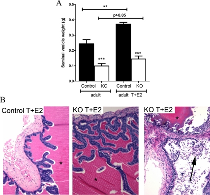
Gross morphology and histology of adult PTM-ARKO mice exposed to exogenous T and E2. A, Quantification of SV weight in adult PTM-ARKO mice exposed to T and E2 for 13 wk. Note the increase in SV weight in control but not KO mice after exposure to T+E2. B, Hematoxylin and eosin staining of adult PTM-ARKO and control SVs exposed to T+E2, with dense esinophilic seminal secretion in the lumen of both the PTM-ARKO and control SV (*). Note the densely packed cell nuclei in both the stroma and epithelia in control and PTM-ARKO SVs exposed to T+E2; this is more pronounced in KOs than controls, with desquamation of epithelial cells obvious in some KO SVs (arrow). Values are means ± sem (n = 3 mice). ***, P < 0.001, compared with control littermates; **, P < 0.01, compared with untreated control mice.

