Abstract
The binding specificities of a panel of avian influenza virus subtype H5 hemagglutinin (HA) proteins bearing mutations at key residues in the receptor binding site were investigated. The results demonstrate that two simultaneous mutations in the receptor binding site resulted in H5 HA binding in a pattern similar to that shown by human viruses. Coexpression of the ion channel protein, M2, from most avian and human strains tested protected H5 HA conformation during trafficking, indicating that no genetic barrier to the reassortment of the H5 surface antigen gene with internal genes of human viruses existed at this level.
Influenza A viruses with at least 15 antigenically distinct hemagglutinin (HA) proteins and nine different neuraminidase subtypes can be isolated from avian species (29). Some of the molecular changes which occur when an avian influenza virus adapts to a new host (e.g., humans), such as changes in receptor recognition and mutations in polymerase protein PB2, have already been characterized (11, 15, 24, 29), but the genetic restrictions which define the species barrier are still not fully understood. The consequence of the adaptation of a new antigenic subtype for replication and transmission in humans is pandemic influenza, which carries a heavy morbidity and mortality toll.
A major determinant of host range is the affinity of the viral HA protein for the host cell sialic acid (SA) receptor. In the natural avian host, SA is joined to the sugar chain through an α2—3 linkage, and viruses isolated from birds possess HAs with high affinity for this type of sugar. On the other hand, in the human respiratory tract, terminal SA is linked through an α2—6 bond. Viruses circulating in humans have acquired mutations in their HAs which result in the loss of affinity for α2—3 SA and the concomitant increase in α2—6 binding (4, 21, 28). This has been particularly well characterized for the H3 subtype, which crossed from ducks into the human population in 1967 and 1968 and caused the 1968 (Hong Kong) influenza pandemic. HAs from viruses isolated early in the pandemic differed from their avian progenitors by a change from glutamine to leucine at residue 226 and from glycine to serine at residue 228 in the HA receptor binding site (RBS) (15). This may represent the minimum change necessary for the H3 subtype to establish itself in the new human host.
In 1997, a highly virulent H5N1 virus spread to live poultry markets in Hong Kong. Eighteen people became infected, and six deaths resulted (3). The viruses recovered from these individuals were identical in all eight RNA segments to those isolated from the chickens at the same time, indicating for the first time that a virus seemingly unadapted for mammalian replication could replicate in humans (1, 25). However, these avian viruses did not transmit between humans and this may be why a pandemic did not ensue.
The crystal structure of an H5 HA from A/Duck/Singapore/3/97 virus, which is closely related to the HAs of the viruses isolated in Hong Kong in 1997, such as the primary human isolate A/HK/156/97, has been determined (9). The width of the receptor binding pocket is less than that for the previously studied human H3 HA protein. It is hypothesized that changes at residues 226 and 228 may allow H5 HA to better interact with the human form of the SA receptor. We have used cloned H5 HA proteins to test whether such mutations indeed result in human-receptor binding characteristics.
The full-length cDNA encoding the H5 HA protein from the human index isolate of the outbreak, A/HK/156/97, was amplified by reverse transcription and PCR from viral RNA and cloned into the expression plasmid pcDNA3 (2). A series of mutations in the H5 HA cDNA which altered the nucleotides encoding the RBS, specifically at residues 226, 227, and 228, were engineered. At position 226, the mutations changed glutamine to either leucine or valine (the Q226L and Q226V mutants), and at residue 228, glycine was changed to serine (the G228S mutant). Changes at residues 226 and 228 were also generated in combination to create LSS and VSS mutants. We also generated mutation S227I. This change was present in a subset of the H5 viruses isolated from humans in Hong Kong (11).
A hemadsorption assay was used to measure red blood cell (RBC) binding to exogenously expressed H5 HA. Following transfection of Vero cells with 1 μg of appropriate plasmids and 3 μl of Lipofectamine and infection with a recombinant fowlpox virus (FPV)-expressing T7 RNA polymerase, cells were treated with 5.5 mU of bacterial neuraminidase/ml for 1 h (6). This treatment was necessary because the sugar modifications on the HA protein itself are sialylated and, in the absence of viral neuraminidase, the SA will block access to the RBS (19). It was also necessary to coexpress the HK156 M2 protein with HK156 HA in order to facilitate cell surface transport of the functional protein. A 0.2-μg amount of the appropriate M2 expression plasmid was cotransfected with each of the HA mutants.
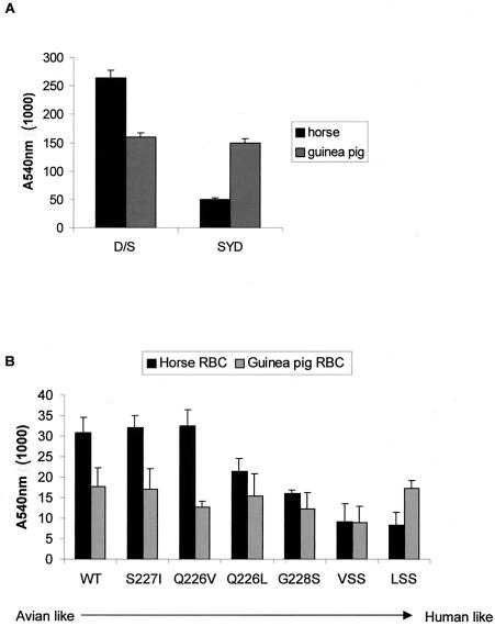
We measured the binding of HA to different linkages of SA by analyzing differential levels of binding to erythrocytes of different species which vary in the types of SA they display. Horse RBCs exclusively display α2—3 terminally linked SA, whereas guinea pig RBCs predominantly display SA of the α2—6 linkage and express approximately 1/10 of their SA in the α2—3 linkage (13, 16; R. Harvey and W. S. Barclay, unpublished data). Accordingly, cells infected with apathogenic H5 avian virus, A/Duck/Singapore/3/97, bound horse RBCs efficiently and also hemadsorbed smaller amounts of guinea pig RBCs, possibly via the α2—3 SA which they display. On the other hand, cells infected with the human H3 subtype strain A/Sydney/5/97 bound only small amounts of horse RBCs but bound larger amounts of guinea pig RBCs due to specific binding of α2—6 SA (Fig. 1A). Each of the H5 HA mutants was assessed for its ability to hemadsorb horse or guinea pig RBCs (Fig. 1B). By using fluorescence-activated cell scanning analysis of cell surface expression, we confirmed that the mutations did not significantly alter the ability of the HA proteins to be transported to the plasma membrane (data not shown). As expected, HK156 wild-type HA showed a typical avian-virus-like pattern of efficient binding to α2—3-linked SA. The S227I or Q226V changes did not affect receptor binding. The Q226L and G228S changes each decreased the binding to horse RBCs without any change in the binding of guinea pig RBCs. The LSS and VSS double mutants showed a considerable loss in their ability to hemadsorb horse erythrocytes, and in the case of the LSS mutations, this was not at the expense of overall SA affinity since binding to guinea pig RBCs was high. In fact, the LSS mutant behaved very much like the human-adapted H3 HA protein of A/Sydney/5/97 virus that was used as a control in this assay (Fig. 1A).
FIG. 1.
Hemadsorption of horse or guinea pig erythrocytes by avian or human influenza A virus HA. (A) Vero cells were infected with either A/Duck/Singapore/97 (D/S) or A/Sydney/5/97 (SYD) at a multiplicity of infection of >1. Twenty-four hours postinfection, a 0.5% suspension of horse or guinea pig erythrocytes was added for 1 h at room temperature. Following washing with phosphate-buffered saline, Vero cells and bound erythrocytes were lysed. The absorbance of the clarified lysate was measured at 540 nm. (B) Vero cells were infected with FPV directing the expression of T7 polymerase and were then transfected with plasmids directing the expression of mutant H5 HA (1 μg) and HK156 M2 (0.2 μg) proteins. Forty-eight hours posttransfection, cells were treated with 5.5 mU of bacterial neuraminidase (Vibrio cholerae sialidase; Roche) per ml for 1 h. Then a 0.5% suspension of horse or guinea pig erythrocytes was added, and after 1 h of incubation at room temperature, cells were washed and lysed for absorbance measurement. Cells infected with FPV T7 but untransfected were processed in the same way, and the mean absorbance from these wells was subtracted as background. All transfections were performed in triplicate, and mean absorbance values with standard errors are shown. WT, wild type.
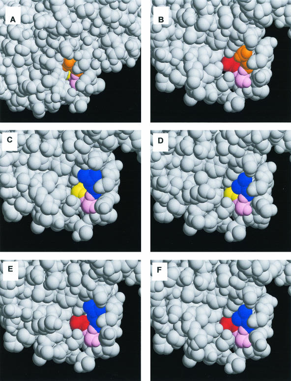
For Fig. 2, we used MODELLER (22) to model the mutations onto the structure of the A/Duck/Singapore/3/97 H5 HA protein. A comparison of panels B to F with the wild-type structure in panel A shows the increase in the width of the RBS pocket, which is predicted to allow greater access of the more bulky α2—6 SA linkage and concomitantly decrease the stabilizing interactions of HA with the α2—3 SA-conjugated receptor. The latter effect may be important for the resistance of virus to naturally occurring inhibitors in human sera and respiratory secretions which display α2—3-linked SA. Ha et al. (10) have recently described the structure of an avian H3 HA that may be a progenitor to human H3 strains. A comparison with the human H3 structure shows that there are structurally significant differences in the human HA RBS that cause the pocket to open. Moreover, the Q226L and G228S substitutions are also predicted to cause some rearrangement of other residues in the RBS. Whether similar rearrangements occur in the H5 HA to accompany the 226 and 228 mutations we have engineered in this study could only be addressed by a crystallization of mutant H5 proteins.
FIG. 2.
Modeling of point mutations into the receptor binding site of H5 HA. (A) Wild type; (B) G228S mutant; (C) Q226L mutant; (D) Q226V mutant; (E) LSS double mutant; (F) VSS double mutant. Native residues 226Q, 227S, and 228G are shown in orange, pink, and yellow, respectively. Mutations to a hydrogen bonding residue (serine) are shown in red, and mutations to a hydrophobic residue (leucine-valine) are shown in blue.
Matrosovich et al. (15) identified human isolates from early in the Asian influenza pandemic of 1957 that still carried the avian-virus-like residues at positions 226 and 228. Other strains from that year contained just one of the two changes associated with human viruses, the Q→L mutation at position 226, and already showed altered receptor binding characteristics. By 1958, only 1 year into the pandemic, simultaneous humanizing mutations at residues 226 and 228 were fixed in the H2 human virus lineage. This example serves to illustrate the proposition that receptor binding changes are not a prerequisite for early zoonotic events but are strongly selected for during the circulation of new subtypes of viruses in humans. Applying this proposition to the H5 situation, it can be hypothesized that if the H5 viruses which transmitted to humans in 1997 had continued to circulate, changes at HA residues 226 and 228 might have been selected. The data presented in this study illustrate that the H5 protein can tolerate these changes and that they do alter the characteristics of the protein towards those of an HA with higher affinity for human receptors. It is likely that the changes occur in a stepwise fashion, as reported for the above-described H2 example, and we show here that both single changes Q226L and G228S affect α2—3 SA binding. It is possible that the mutations we have introduced into H5 HA would not be tolerated in the context of infectious virus. Previously, it has been shown that changes in the RBS can affect the pH of fusion of HA (5). To fully establish the viability and characteristics of viruses carrying these mutations, it would be necessary to introduce them into recombinant viruses by using reverse-genetics techniques (7, 17). However, such viruses would require isolation and handling under high containment conditions, which limits easy experimental confirmation.
It has recently been suggested that certain internal gene products from human viruses, such as the matrix protein M1 and/or the ion channel protein M2, encoded on RNA segment 7, and certain avian HA subtypes are incompatible (23). Reassortment with HA genes like the HK/97 H5 HA is even more restricted than for other HAs because of a string of basic amino acids at the HA1/HA2 cleavage site which render the HA susceptible to cleavage by ubiquitous intracellular proteases, such as furin. Although this susceptibility to cleavage increases the tissue tropism of the virus, it also places the newly synthesized HA at risk for premature fusogenic conformational change. This possibility is avoided by viral expression of an ion channel protein, M2, which allows protons to flow out from the late Golgi apparatus, thus maintaining this compartment at a higher pH than usual (18, 27). It was previously shown that the potency of the M2 ion channel varies among virus strains and that some reassortants are not viable due to an incompatibility between the gene products of segment 4, encoding HA, and segment 7, encoding M2 (8). In addition, functional transport of the multibasic H7 Rostock HA protein required four times as much of the human A/Udorn/72 M2 ion channel as of the homologous Rostock avian M2 protein (27). We therefore asked whether human M2 proteins from strains circulating in 1997 and later could facilitate transport of functional multibasic H5 HA to the mammalian cell surface.
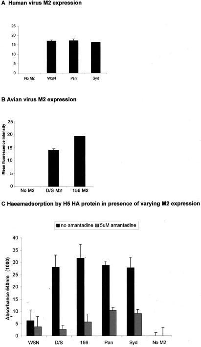
We cloned the M2 open reading frames from avian and human viral sources into pcDNA3 and cotransfected equal concentrations (0.2 μg) of plasmid DNA with 1 μg of wild-type HK156 H5 HA plasmid. We used M2-specific antisera to check that the levels of expression of each M2 protein were similar (Fig. 3A and B). Although it is not possible to make a complete comparison because different antibodies were needed to analyze the expression of human or avian M2 proteins due to differences in their sequences, the results showed that all M2 proteins were expressed well and that within the two sets analyzed, expression levels were equivalent. Significantly, the two human M2 genes cloned from H3N2 viruses circulating in 1997 and 1999, A/Sydney/5/97 and A/Panama/99, were as effective in this assay as the homologous M2 ion channel derived from the human index isolate HK156 or the avian virus M2 gene cloned from A/Duck/Singapore/3/97 virus (Fig. 3C). To confirm that the ion channel activity was indeed responsible, we repeated each assay in the presence of amantadine, a drug that inhibits the ion channel activity of M2. Following transfection of the two HA and M2 plasmids, 5 μM amantadine was added and the cells were incubated for a further 48 h before the hemadsorption assay was performed. The facilitative effect of M2 on HA transport was inhibited (Fig. 3C). The ability of human M2 protein to support H5 HA transport is significant because it indicates that there was no genetic barrier operating at this level to prevent the generation of an avian-human reassortant virus containing segment 4 RNA, encoding a novel HA subtype, mixed with a human segment 7 RNA.
FIG. 3.
(A and B) Human and avian virus M2 protein expression. Forty-eight hours following transfection of 0.2 μg of plasmid DNA, cells were incubated with primary antibodies against M2. A mouse monoclonal antibody (14C2) was used to detect the human M2 proteins (A), and a rabbit polyclonal antiserum raised against a swine influenza virus M2, kindly provided by P. Heinen, was used to detect the avian M2 proteins (B). Fluorescein isothiocyanate-conjugated secondary antibody was used, and the mean fluorescence intensity of each sample was measured by fluorescence-activated cell sorter analysis. (C) Representation of the ability of different M2 proteins to support hemadsorption by H5 HA. Vero cells were transfected with 1 μg of plasmid directing the expression of HK156 HA and 0.2 μg of plasmid expressing different avian or human viral M2 proteins and incubated either with (grey bars) or without (black bars) 5 μM amantadine. Forty-eight hours posttransfection, cells were treated with 5.5 mU of bacterial neuraminidase (V. cholerae sialidase) per ml, and a 0.5% suspension of horse erythrocytes was added. After 1 h of incubation at room temperature, cells were washed and lysed for absorbance measurement at 540 nm. Cells infected with FPV T7 but untransfected were processed in the same way, and the mean absorbance from these wells was subtracted as background. All transfections were performed in triplicate, and mean absorbance values with standard errors are shown. D/S, A/Duck/Singapore/3/97; 156, A/HK/156/97; Pan, A/Panama/99; Syd, A/Sydney/5/97.
Interestingly, we found that WSN M2 was much less active in supporting the functional transport of H5 HA in the hemadsorption assay even though it was expressed to the same amount as the other human virus M2 channels. This finding suggests that the ion channel activity of this protein is low. The WSN M2 protein has an S-to-N change at residue 31, which is associated with resistance to the drug amantadine. However, it should be stated that amantadine resistance does not necessarily result in the loss of ion channel activity since several strains of avian influenza virus with multibasic HA cleavage sites have acquired mutations which lead to drug resistance (reviewed in reference 12). We did not determine the levels of activity of different M2 proteins. However, bearing in mind that M2 is highly expressed in the infected cells and that we coexpressed M2 from only one-fifth the amount of plasmid DNA used for HA expression in these studies, it is not likely that our results have been affected by the overexpression of M2. It is possible that by transfecting increased amounts of the WSN M2 protein, H5 HA transport would be rescued, but how these levels of M2 and HA expression relate to virus infection is not clear. It is already known that some M2 ion channels are not active enough to support the transport of multibasic HA in a viral context (8). It might be important to consider this during the designing of recombinant viruses to be used as pandemic vaccines: several groups have recently described the recovery of viruses containing human internal proteins and avian H5 HA genes (14, 26) with the multibasic site H5 HA removed (because of obvious safety considerations). Since influenza virus has a known capacity to acquire mutations and insertions around this region (20), coupling such engineered HA genes with an incompatible M2 protein may limit the risk of reversion during vaccine propagation. The hemadsorption assay that we have presented here is a functional test for monitoring the ability of exogenous proteins to mediate changes in the pHs of intracellular compartments and assist in the transport of viral membrane proteins. As such, it may prove useful for the analysis of potential proton channel activity bestowed by different M2 proteins as well as by other viral or host cell proteins.
Acknowledgments
We thank Mike Skinner, Institute for Animal Health, Compton, United Kingdom, for the gift of FPV T7 recombinant virus; R. A. Lamb, Northwestern University, for the gift of 14C2; Paul Heinen, formerly of ID Lelystad and currently of the Health Protection Agency, for the gift of antiserum to avian M2 proteins; and James White, University of Reading, for assistance with establishment of the hemadsorption assay.
R.H. was a recipient of a PHLS studentship.
REFERENCES
- 1.Bender, C., H. Hall, J. Huang, A. Klimov, N. Cox, A. Hay, V. Gregory, K. Cameron, W. Lim, and K. Subbarao. 1999. Characterization of the surface proteins of influenza A (H5N1) viruses isolated from humans in 1997-1998. Virology 254:115-123. [DOI] [PubMed] [Google Scholar]
- 2.Boom, R., C. J. A. Sol, M. M. M. Salimans, C. L. Jansen, P. M. E. Wertheim-van Dillen, and J. van der Noordaa. 1990. Rapid and simple method for purification of nucleic acids. J. Clin. Microbiol. 28:495-503. [DOI] [PMC free article] [PubMed] [Google Scholar]
- 3.Claas, E. C., A. D. Osterhaus, R. van Beek, J. C. De Jong, G. F. Rimmelzwaan, D. A. Senne, S. Krauss, K. F. Shortridge, and R. G. Webster. 1998. Human influenza A H5N1 virus related to a highly pathogenic avian influenza virus. Lancet 351:472-477. [DOI] [PubMed] [Google Scholar]
- 4.Connor, R. J., Y. Kawaoka, R. G. Webster, and J. C. Paulson. 1994. Receptor specificity in human, avian, and equine H2 and H3 influenza virus isolates. Virology 205:17-23. [DOI] [PubMed] [Google Scholar]
- 5.Daniels, R. S., S. Jeffries, P. Yates, G. C. Schild, G. Rogers, J. C. Paulson, S. A. Wharton, A. R. Douglas, J. J. Skehel, and D. C. Wiley. 1987. The receptor binding and membrane fusion properties of influenza virus variants selected using antihaemagglutinin monoclonal antibodies. EMBO J. 6:1459-1465. [DOI] [PMC free article] [PubMed] [Google Scholar]
- 6.Feldmann, A., M. K. H. Schäfer, W. Garten, and H.-D. Klenk. 2000. Targeted infection of endothelial cells by avian influenza virus A/FPV/Rostock/34 (H7N1) in chicken embryos. J. Virol. 74:8018-8027. [DOI] [PMC free article] [PubMed] [Google Scholar]
- 7.Fodor, E., L. Devenish, O. G. Engelhardt, P. Palese, G. G. Brownlee, and A. García-Sastre. 1999. Rescue of influenza A virus from recombinant DNA. J. Virol. 73:9679-9682. [DOI] [PMC free article] [PubMed] [Google Scholar]
- 8.Grambas, S., M. S. Bennett, and A. J. Hay. 1992. Influence of amantadine resistance mutations on the pH regulatory function of the M2 protein of influenza A viruses. Virology 191:541-549. [DOI] [PubMed] [Google Scholar]
- 9.Ha, Y., D. J. Stevens, J. J. Skehel, and D. C. Wiley. 2001. X-ray structures of H5 avian and H9 swine influenza virus hemagglutinins bound to avian and human receptor analogs. Proc. Natl. Acad. Sci. USA 98:11181-11186. [DOI] [PMC free article] [PubMed] [Google Scholar]
- 10.Ha, Y., D. J. Stevens, J. J. Skehel, and D. C. Wiley. 2003. X-ray structure of the hemagglutinin of a potential H3 avian progenitor of the 1968 Hong Kong pandemic influenza virus. Virology 309:209-218. [DOI] [PubMed] [Google Scholar]
- 11.Hatta, M., P. Gao, P. Halfmann, and Y. Kawaoka. 2001. Molecular basis for high virulence of Hong Kong H5N1 influenza A viruses. Science 293:1840-1842. [DOI] [PubMed] [Google Scholar]
- 12.Hay, A. 1992. The action of adamantanamines against influenza A viruses: inhibition of the M2 ion channel protein. Semin. Virol. 3:21-30. [Google Scholar]
- 13.Ito, T., Y. Suzuki, L. Mitnaul, A. Vines, H. Kida, and Y. Kawaoka. 1997. Receptor specificity of influenza A viruses correlates with the agglutination of erythrocytes from different animal species. Virology 227:493-499. [DOI] [PubMed] [Google Scholar]
- 14.Li, S., C. Liu, A. Klimov, K. Subbarao, M. L. Perdue, D. Mo, Y. Ji, L. Woods, S. Hietala, and M. Bryant. 1999. Recombinant influenza A virus vaccines for the pathogenic human A/Hong Kong/97 (H5N1) viruses. J. Infect. Dis. 179:1132-1138. [DOI] [PubMed] [Google Scholar]
- 15.Matrosovich, M., A. Tuzikov, N. Bovin, A. Gambaryan, A. Klimov, M. R. Castrucci, I. Donatelli, and Y. Kawaoka. 2000. Early alterations of the receptor-binding properties of H1, H2, and H3 avian influenza virus hemagglutinins after their introduction into mammals. J. Virol. 74:8502-8512. [DOI] [PMC free article] [PubMed] [Google Scholar]
- 16.Medeiros, R., N. Escriou, N. Naffakh, J. C. Manuguerra, and S. van der Werf. 2001. Hemagglutinin residues of recent human A(H3N2) influenza viruses that contribute to the inability to agglutinate chicken erythrocytes. Virology 289:74-85. [DOI] [PubMed] [Google Scholar]
- 17.Neumann, G., T. Watanabe, H. Ito, S. Watanabe, H. Goto, P. Gao, M. Hughes, D. R. Perez, R. Donis, E. Hoffmann, G. Hobom, and Y. Kawaoka. 1999. Generation of influenza A viruses entirely from cloned cDNAs. Proc. Natl. Acad. Sci. USA 96:9345-9350. [DOI] [PMC free article] [PubMed] [Google Scholar]
- 18.Ohuchi, M., A. Cramer, M. Vey, R. Ohuchi, W. Garten, and H.-D. Klenk. 1994. Rescue of vector-expressed fowl plague hemagglutinin in biologically active form by acidotropic agents and coexpressed M2 protein. J. Virol. 68:920-926. [DOI] [PMC free article] [PubMed] [Google Scholar]
- 19.Ohuchi, M., A. Feldmann, R. Ohuchi, and H. D. Klenk. 1995. Neuraminidase is essential for fowl plague virus hemagglutinin to show hemagglutinating activity. Virology 212:77-83. [DOI] [PubMed] [Google Scholar]
- 20.Perdue, M. L., M. Garcia, D. Senne, and M. Fraire. 1997. Virulence-associated sequence duplication at the hemagglutinin cleavage site of avian influenza viruses. Virus Res. 49:173-186. [DOI] [PubMed] [Google Scholar]
- 21.Rogers, G. N., J. C. Paulson, R. S. Daniels, J. J. Skehel, I. A. Wilson, and D. C. Wiley. 1983. Single amino acid substitutions in influenza haemagglutinin change receptor binding specificity. Nature 304:76-78. [DOI] [PubMed] [Google Scholar]
- 22.Sali, A., and T. L. Blundell. 1993. Comparative protein modelling by satisfaction of spatial restraints. J. Mol. Biol. 234:779-815. [DOI] [PubMed] [Google Scholar]
- 23.Scholtissek, C., J. Stech, S. Krauss, and R. G. Webster. 2002. Cooperation between the hemagglutinin of avian viruses and the matrix protein of human influenza A viruses. J. Virol. 76:1781-1786. [DOI] [PMC free article] [PubMed] [Google Scholar]
- 24.Shaw, M., L. Cooper, X. Xu, W. Thompson, S. Krauss, Y. Guan, N. Zhou, A. Klimov, N. Cox, R. Webster, W. Lim, K. Shortridge, and K. Subbarao. 2002. Molecular changes associated with the transmission of avian influenza A H5N1 and H9N2 viruses to humans. J. Med. Virol. 66:107-114. [DOI] [PubMed] [Google Scholar]
- 25.Subbarao, K., A. Klimov, J. Katz, H. Regnery, W. Lim, H. Hall, M. Perdue, D. Swayne, C. Bender, J. Huang, M. Hemphill, T. Rowe, M. Shaw, X. Y. Xu, K. Fukuda, and N. Cox. 1998. Characterization of an avian influenza A (H5N1) virus isolated from a child with a fatal respiratory illness. Science 279:393-396. [DOI] [PubMed] [Google Scholar]
- 26.Subbarao, K., H. Chen, D. Swayne, L. Mingay, E. Fodor, G. Brownlee, X. Xu, X. Lu, J. Katz, N. Cox, and Y. Matsuoka. 2003. Evaluation of a genetically modified reassortant H5N1 influenza A virus vaccine candidate generated by plasmid-based reverse genetics. Virology 305:192-200. [DOI] [PubMed] [Google Scholar]
- 27.Takeuchi, K., and R. A. Lamb. 1994. Influenza virus M2 protein ion channel activity stabilizes the native form of fowl plague virus hemagglutinin during intracellular transport. J. Virol. 68:911-919. [DOI] [PMC free article] [PubMed] [Google Scholar]
- 28.Vines, A., K. Wells, M. Matrosovich, M. R. Castrucci, T. Ito, and Y. Kawaoka. 1998. The role of influenza A virus hemagglutinin residues 226 and 228 in receptor specificity and host range restriction. J. Virol. 72:7626-7631. [DOI] [PMC free article] [PubMed] [Google Scholar]
- 29.Webby, R. J., and R. G. Webster. 2001. Emergence of influenza A viruses. Philos. Trans. R. Soc. Lond. B 356:1817-1828. [DOI] [PMC free article] [PubMed] [Google Scholar]


