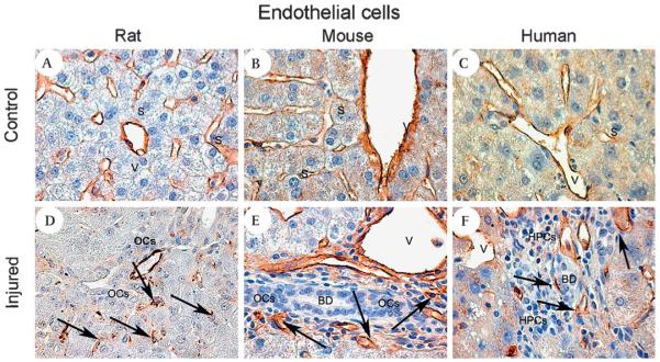
Figure 3.
Endothelial cells. Photomicrographs illustrating rodent and human liver tissue to visualise the partner cells of the hepatic stem cell niche. In control liver tissue (A–C) the endothelial cell-specific marker von Willebrand factor (vWF; brown) highlights veins and sinusoids. (A) Normal rat liver; (B) normal mouse liver; (C) control human donor liver. In the injured liver (D–F) endothelial cells (black arrows) appear in close contact with the bile ducts and the oval cells (OCs). (D) 2-Acetylaminofluorene and subsequent partial hepatectomy (2AAF/PH) rat, day 9 after the PH—vWF (brown); (E) hepatitis B surface antigen-tg/retrorsine treated mouse—vWF (brown); (F) biopsy specimen from hepatitis C virus cirrhotic liver—vWF (brown). Magnification (A–F) ×40.

