Abstract
Objective
To develop a rapid quantitative real-time polymerase chain reaction (PCR) to detect herpes simplex virus (HSV) in the genital secretions of women that may be used in labor.
Methods
Samples of genital secretions from women in labor, swabs of active genital lesions, and swabs of buffer solution were analyzed using a newly developed rapid HSV PCR assay to detect HSV glycoprotein B gene and quantitate virion copy number. A previously validated TaqMan PCR to detect HSV glycoprotein B gene was performed as the comparator gold standard. Positivity determination that optimized sensitivity and specificity was determined with receiver operating characteristic (ROC) curves.
Results
The median time to result for rapid HSV PCR was 2 hours (range 1.5–3.5 hours). A positivity determination rule that required both wells of the rapid test to detect 150 copies or greater of HSV per ml maximized specificity (96.7%) without appreciable loss of sensitivity (99.6%). Among positive samples, the correlation between the rapid test and TaqMan for the quantity of HSV isolated was excellent (R=0.96, p<.001). The rapid test had a positive predictive value of 96.7% and a negative predictive value of 99.6% in a population with HSV shedding prevalence of 10.8%, based on the prevalence of genital HSV previously found among HSV-2 seropositive women in labor.
Conclusion
Rapid HSV PCR provides results with excellent sensitivity and specificity within a timeframe that could inform clinical decision making for identifying infants at risk of neonatal HSV infection.
Neonatal HSV occurs in up to 1 per 3200 live births (1), with an estimated incidence of 1,500 cases in the United States annually (2). Neonatal HSV causes disseminated or CNS disease in approximately 50% of cases; up to 30% of these neonates die and up to 40% of survivors have neurologic damage, despite anti-viral therapy (2). Current guidelines for prevention of neonatal herpes recommend visual inspection for maternal herpetic genital lesions at the time of labor with cesarean delivery if lesions are present (3); this strategy has not resulted in an appreciable reduction in incidence (4). The main mode of transmission to the neonate is exposure to infected maternal secretions at the time of passage through the birth canal. Isolation of HSV from the maternal genital tract at the time of labor is the primary risk factor for neonatal HSV with an estimated relative risk of >300 for infection (1). As such, prevention of neonatal exposure to HSV during birth by identifying women with HSV in the genital tract during labor is a potential approach for reducing risk of infection. Interventions that could be tested among women shedding HSV during labor include cesarean delivery, anti-viral prophylaxis for mother and/or the neonate, and enhanced observation for signs of neonatal HSV infection.
Currently, there is no clinically useful method to detect HSV in the genital tract at the time of labor. Lesions consistent with genital herpes indicate an increased risk of HSV, however, both sensitivity and specificity of genital lesions for viral shedding is poor (5–7). Most women with HSV in the genital tract at the time of labor are without signs or symptoms of genital herpes (1). In the past, viral cultures of genital swabs collected in the third trimester of pregnancy were used to identify women at risk of perinatal HSV transmission. However, results from these prenatal specimens rarely correlated with culture results of swabs collected at the time of delivery, and thus were not helpful in predicting neonatal exposure (8). Therefore, this practice was abandoned in the late 1980s (3). Viral cultures to detect HSV from genital swabs collected at the time of labor are not practical because results are not available in a time frame that would allow for effective interventions. Moreover, false negative viral cultures were reported in up to 30% cases of neonatal HSV. In most reports, PCR- based assays detected HSV in these negative viral cultures (1, 5).
Real-time quantitative PCR has proved to be sensitive and reproducible in the diagnosis and management of vial diseases (9). However, due to the complexity of the procedures involved and the potential of contamination, clinical diagnostic PCR usually is performed in state-of-the-art clinical laboratories. Transfer of specimens from clinical sites to comprehensive clinical laboratories often takes hours or even days. Once received, the entire analytic procedure, including nuclear acid extraction and 45 cycles of Taqman PCR, takes about 4 hours. Although point-of-care (POC) molecular diagnostic systems are available for adaptation (10), to adopt one of these systems for specific pathogen detection will heavily involve the commercial proprietary issues, higher cost per test and less flexibility for any in house development. As such, we chose to develop an in-house, sensitive, specific, rapid, automated, self-contained PCR assay using the extraction and PCR equipment available commercially that could be utilized to detect HSV-1 or HSV-2 from samples taken in the delivery room and reported back rapidly (within 2 hours) to clinicians providing care of the laboring woman and newborn. The assay is designed to be utilized by any trained laboratory technician, so as to be feasibly implemented in at any hour of the day in a clinical laboratory serving obstetrical patients.
MATERIALS AND METHODS
Sample collection
Swab samples for analysis were derived from 3 different sources. First, a buffer only solution was used as a negative control group (N=299). For this group, vials were filled with 1 mL of buffer solution and a sterile swab was inserted. Second, swabs were collected from non-pregnant patients with herpetic genital lesions (N=211) who presented to the Virology Research Clinic and placed in a vial containing 1 mL of PCR digestion buffer. Third, mucocutaneous genital swab samples from pregnant women presenting in labor at the University of Washington Medical Center were included (N=206). These swabs were collected between October 2006 and March 2009 from women who were aged 18 or older, presented in labor, and understood English. Verbal consent was obtained for collection of genital secretions on Dacron swabs at the time of clinically indicated routine speculum examination at admission. Each swab was placed in a vial containing 1 mL of PCR digestion buffer; no identifying information was retained with the sample so that the samples were anonymous. University of Washington Institutional Review Board approved all study procedures.
Laboratory
HSV rapid PCR reagents
To make the rapid PCR procedure as simple as possible to be carried out in a less stringent laboratory setting and performed by less experienced technologists, the reagents for the samples and controls were prepared and aliquoted by experienced PCR technologists in the Molecular Diagnostic Laboratory. All the reagents were quality control tested before being sent to the Microbiology Laboratory where the rapid PCR procedure was performed.
Each aliquot contained enough reagents to perform a single run of PCR reactions for 4 clinical samples, one negative control sample, one positive control sample, no DNA control, and 5 standard curve points in duplicate. An appropriate amount of ABI Taqman Fast Universal PCR Master Mix (2x) also was pre-aliquot accordingly.
DNA Extraction
DNA was extracted from 200 µl of buffer using the EZ1 Virus Kit on Qiagen BioRobot EZ1 (Qiagen, Germantown MD) and eluted into 100 µl of AVE buffer. This machine was chosen because of its speed, small size, ability to extract 6 samples simultaneously, ease of operation, and minimization of cross contamination by use of a single reagent cartridge for each sample. Ten µl of eluted DNA was then used for PCR. To control contamination and DNA recovery, one negative control and one positive control were included in each extraction run.
Rapid PCR Assay
The ABI7500 Fast machine was used to run PCR reactions. The glycoprotein B gene primers and probe were used for this study as described previously (11, 13). These primers and probes pick up both HSV-1 and HSV-2 and have been validated in a variety of clinical and investigative studies (11, 14, 15). The gB probe was labeled with FAM at 5’-end and TAMRA at 3’-end. Each 30 µl rapid HSV PCR reaction contained 15 µl of 2x ABI Taqman Fast Univeral PCR Master Mix, 830 nM primer, 100 nM probe. The PCR conditions were 95°C for 20 seconds, followed by 45 cycles of 95°C for 3 seconds and 60°C for 30 seconds. All the PCR reactions were spiked with internal control, as described below, to monitor for false negative results. The rapid test was run in duplicate wells and we reported the average value of the two wells when both wells were positive. However, to determine the cutoff for positivity, we considered the sensitivity and specificity with requiring only one vs. both wells to be positive. The duration of the procedure was recorded for each PCR run.
Real-Time TaqMan PCR Assay
After the rapid real-time HSV PCR was performed at the Microbiology Laboratory, samples were sent to Molecular Diagnostic Laboratory where they were re-analyzed using the standard real-time TaqMan HSV PCR assay (11). Briefly, DNA was extracted from 200µl buffer using Qiagen QIAmp 96 Blood kit and eluted into 100ul. Ten µl of DNA was then used for real-time TaqMan PCR with gB primer/probe. Each 30 µl reaction contained 15 µl of QuantiTech Multiplex PCR Master Mix (2x) (Qiagen), and 830 nM primer, 100 nM probe and EXO internal control. ABI 7900 HT was used to run the assay. The PCR conditions were 52°C for 2 min, 95°C for 15 min and followed by 45 cycles of 94°C for 1 min and 60°C for 1 min. Each PCR run contained several negative controls, including two reaction mixtures without DNA and several specimens that are known to contain no HSV DNA, a positive control and a standard dilution curve DNA.
Statistical Analysis
We hypothesized that results of the rapid HSV PCR would not vary significantly from those of standard 4-hour HSV PCR. An a priori sample size calculation determined that analyzing 650 specimens could determine a specificity of at least 97% with a precision of 1.5%, assuming that 518 of these specimens did not contain HSV using standard HSV PCR as the gold standard. Positivity determination that optimized sensitivity and specificity was determined with receiver operating characteristic (ROC) curves. Under any reasonable sensitivity assumptions (at least 80%), specificity of 97% provides a positive predictive value of at least 76% at a baseline HSV standard PCR positive rate of 10.8%, a rate of positivity observed in our previous cohort of pregnant women (1, unpublished data).
We estimated the sensitivity and specificity of the rapid test to detect HSV DNA, using the standard TaqMan HSV PCR result as the gold standard, and obtained confidence intervals using the logit transformation. A TaqMan HSV PCR result of > 150 HSV copies/ml was considered positive, consistent with the cutoff currently utilized in our standard PCR assay for clinical diagnoses (12). We ascertained whether the results of the rapid test and the standard TaqMan test varied significantly for each specimen and describe the distribution of virion copy numbers of HSV DNA for specimens with detectable HSV DNA. Receiver operating characteristic curves were used to evaluate the summary measure for the two wells and the range of potential cutoffs for positivity determination. Since both the TaqMan and the Rapid PCR quantitate copies of HSV DNA per ml, we compare both the quantity obtained as well as the binary indicator of response: positive or negative. Positive predictive values (PPV) and negative predictive values (NPV) of the assay were estimated in the target population of interest (laboring women) based on demonstrated accuracy over all samples and the previously observed prevalence of HSV DNA.
RESULTS
We tested 732 swabs on 732 unique persons; 16 (2%) swabs were excluded because the negative controls in the runs were positive, and the remaining 716 were valid and used for analyses. The median time to perform the Rapid Test from initiation of test procedures to result was 2 hours (interquartile range 1.75–2.33 hours, range 1.5–3.5 hours).
Using a cutoff of 150 copies/ml, our Rapid PCR assays detected HSV DNA in 207 of 716 (28.9%, 95% CI 25.7–32.3%) swabs, compared to 212 of 716 (29.6%, 95% CI 26.4–33.1%) in the TaqMan assay. Below, we provide additional detail on sensitivity and specificity that result in establishment of the cutoff for PCR positivity as both wells positive at 150 copies per ml or greater.
Buffer only
We evaluated the specificity of the Rapid PCR using samples containing buffer only. Seven (7/299, 2.3%, 95% CI 1.1–4.8%) samples yielded HSV in at least one well. None of the 7 was positive in the TaqMan assay. However, the median amount of HSV DNA in the samples with HSV detected was low (106 copies/ml, range 47–147 copies/ml). In addition, none of the buffer only samples had HSV detected in both wells. Thus, using positivity in both wells to define a positive test minimized the potential for false positive test results related to low level contamination.
Genital lesion swabs
211 swabs were obtained from genital lesions, and 202 (95.7%, 95% CI 92.0–97.8%) tested positive by the Rapid Assay. The concordance between Rapid PCR and our standard TaqMan results varied by the cutoff for the Rapid Test. For example, at a cutoff for Rapid PCR at 150 copies/ml, both tests were positive in 201 samples, neither in 8 samples, and the results were discordant in 2 samples, one of which was positive only by Rapid PCR and the other only by TaqMan. Thus, overall, 209 samples (99.1%, 95% CI 96.3–99.8%) had concordant results.
Labor and delivery samples
We collected 206 samples from pregnant women presenting to Labor and Delivery; 5 (2.4%, 95% CI 1.0–5.7%) tested positive by the Rapid assay and 10 (4.9%, 95% CI 2.6–8.8%) tested positive by TaqMan. Using 150 copies/ml the cutoff for positive results and requiring both wells to be positive for Rapid PCR, both tests were positive in 4 samples, both tests were negative in 195 samples, and the results were discordant in 7, with only Rapid PCR being positive in 1 and only TaqMan positive in 6. The median amount of HSV DNA in the samples that were positive by TaqMan only was 431 copies/ml (range 160–1901 copies/ml). The amount of HSV DNA in the one sample that was positive solely in the rapid assay was 596 copies/ml.
Cut-off determination
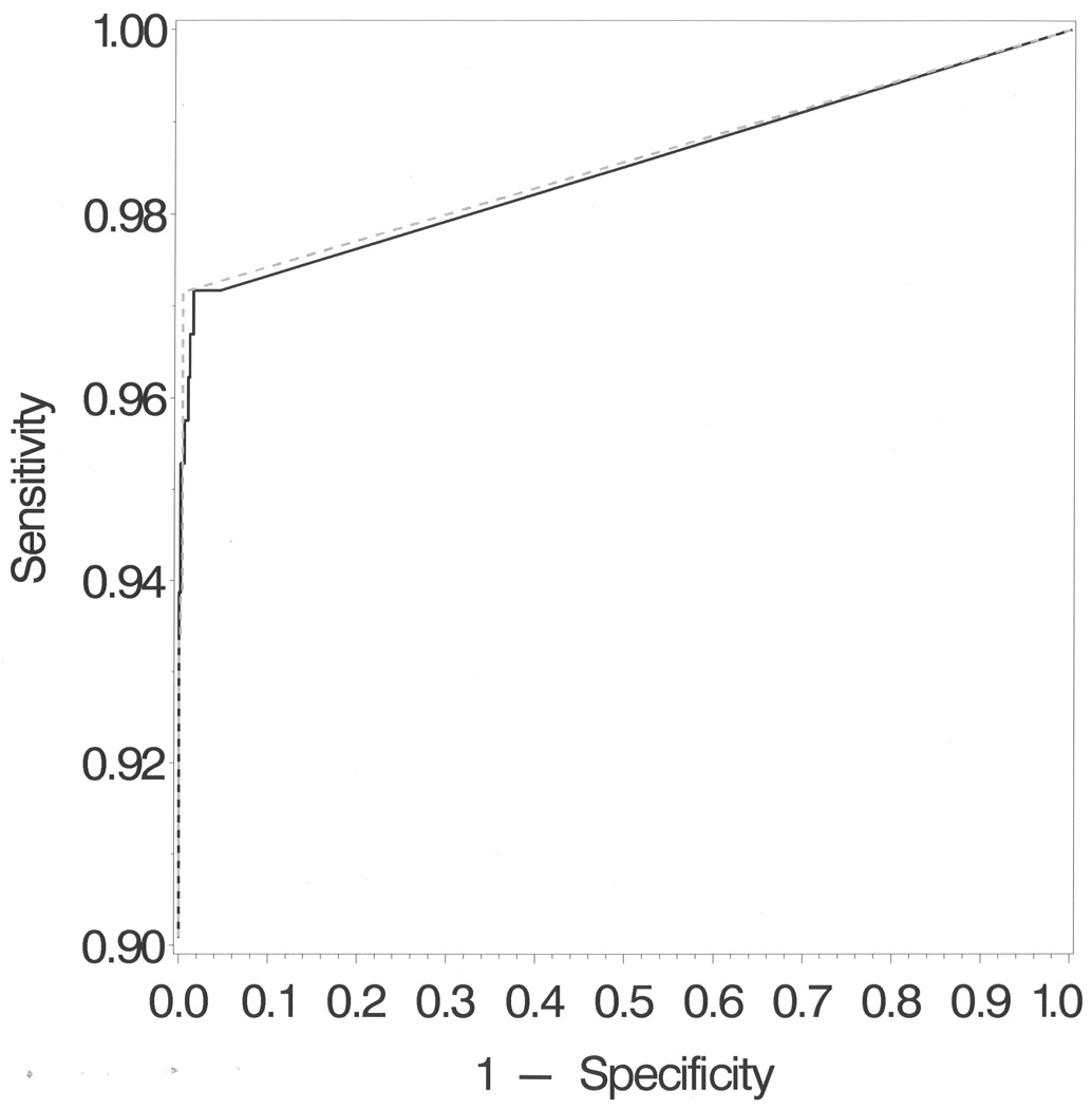
Using the above data in aggregate, we determined the sensitivity and specificity of the test using 3 different interpretation strategies applied to cut-off values from 50 to 500 copies/ml (Table I). Strategy A averaged the quantitative values of both wells of the rapid test into a single value that is compared to the cut-off value to determine test positivity. Strategy B used the quantitative data from both wells separately with both wells required to exceed the cut-off value of 150 copies/ml for the specimen to be considered positive. Strategy C used the average of both wells after excluding specimens with one positive and one negative well, with positive defined as >0 copies/ml. As shown in Figure 1 and Table I, comparisons of accuracy rates suggest that Strategy B that requires both wells to detect 150 copies or greater of HSV per ml to consider the test positive maximized specificity (96.7%, 95% CI 93.2–98.4%), without appreciable loss of sensitivity (99.6%, 95% CI 98.4–99.9%). The ROC curves shown in Figure 1 indicate that strategy B (the gray dashed line) is closer to the top left corner at all cutoffs relative to strategy A and that the standard Taqman cutoff of 150 copies is near optimal.
Table 1.
Comparison of HSV PCR Results by Rapid PCR and TaqMan Assay using 3 Different Cut-off Strategies.
| Rapid PCR Interpretation Strategy |
Cut-off (c/ml) |
Rapid PCR Result |
TaqMan Positive |
Result Negative |
Sensitivity |
Specificity |
|---|---|---|---|---|---|---|
| A | 50 | Positive | 206 | 19 | 97.2 | 96.2 |
| Negative | 6 | 485 | ||||
| B | Positive | 206 | 4 | 97.2 | 99.2 | |
| Negative | 6 | 500 | ||||
| C | Positive | 206 | 5 | 97.2 | 99.2 | |
| Negative | 6 | 499 | ||||
| A | 100 | Positive | 206 | 12 | 97.2 | 97.6 |
| Negative | 6 | 492 | ||||
| B | Positive | 206 | 4 | 97.2 | 99.2 | |
| Negative | 6 | 500 | ||||
| C | Positive | 206 | 6 | 97.2 | 99.2 | |
| Negative | 6 | 500 | ||||
| A | 150 | Positive | 206 | 8 | 97.2 | 98.4 |
| Negative | 6 | 496 | ||||
| B | Positive | 205 | 2 | 96.7 | 99.6 | |
| Negative | 7 | 502 | ||||
| C | Positive | 206 | 4 | 97.2 | 99.2 | |
| Negative | 6 | 500 | ||||
| A | 250 | Positive | 203 | 4 | 95.8 | 99.2 |
| Negative | 9 | 500 | ||||
| B | Positive | 199 | 1 | 93.9 | 99.8 | |
| Negative | 13 | 503 | ||||
| C | Positive | 203 | 3 | 95.8 | 99.4 | |
| Negative | 9 | 501 | ||||
| A | 500 | Positive | 199 | 1 | 93.9 | 99.8 |
| Negative | 13 | 503 | ||||
| B | Positive | 197 | 0 | 92.9 | 100.0 | |
| Negative | 15 | 504 | ||||
| C | Positive | 199 | 1 | 93.9 | 99.8 | |
| Negative | 13 | 503 | ||||
Strategy A averages the quantitative values of both wells of the rapid test into a single value that is compared to the cut-off value to determine test positivity.
Strategy B uses the quantitative data from both wells separately with both wells required to exceed cut-off for the specimen to be considered positive.
Strategy C compares the average of both wells after excluding specimens with one positive(defined as >0 c/ml) and one negative to the stated cut-off values.
Figure 1.
ROC curve using strategy A (average value of wells must exceed threshold, black line) and strategy B (values in both wells must exceed threshold, dashed line) with thresholds ranging from 50 to 500 copies/ml. The top left point on the dashed line indicates the optimal threshold of 140 copies/ml corresponding to a sensitivity of 97.2% and specificity of 99.6%. We chose to use 150 copies because it matches that used for Taqman and the performance is nearly identical: sensitivity of 96.7% and specificity of 99.6%.
Correlations in the quantity of HSV DNA detected in the 2 well of Rapid PCR and between Rapid PCR and Taqman
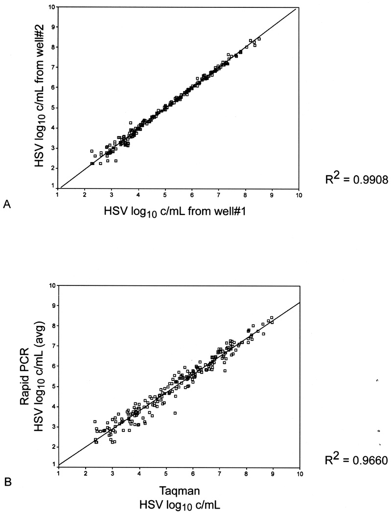
Among the Rapid PCR results, the quantitative difference between the 2 wells run for each sample varied from 0 to 0.8 log copies/ml (Figure 2A). Among positive samples (≥ 150 copies/ml) in both wells (N=207), the correlation between the number of copies of HSV DNA detected by Rapid PCR and TaqMan was excellent with a Spearman correlation coefficient of 0.99, p<.001 (Figure 2B).
Figure 2.
A: Correlation between Well #1 and Well #2 from Rapid HSV PCR (N=207 samples that had ≥150 c/ml in both wells, R= 0.99, p <.001.) B: Correlation between Quantitative Results of Rapid test and TaqMan (N= 205 samples positive at ≥150 c/ml by both Rapid PCR and TaqMan; R=0.96, P<.001)
Positive predictive and negative predictive value
Using the accuracy rates for each cutoff (Table I), we also computed the expected PPV and NPV for a prevalence of 10.8%, which was the prevalence of HSV shedding noted in our studies among HSV-2 seropositive women in labor and without genital lesions (1, unpublished data). We calculated a positive predictive value of 96.7% and negative predictive value of 99.6% using Strategy B (the cutoff of 150 copies/ml and both wells positive). Estimates of positive and negative predictive value using other cutoff values at both a 10.8% and 5% prevalence (for comparison) are shown in Table II.
Table 2.
Positive and negative predictive value of HSV Rapid PCR in a population with HSV shedding prevalence of 10.8%*
| Rapid PCR Positivity |
using 10.8% prevalence | using 5% prevalence | ||
|---|---|---|---|---|
| Cut-off† | PPV | NPV | PPV | NPV |
| 0 | 93.6% | 99.7% | 86.5% | 99.9% |
| ≥50 | 93.6% | 99.7% | 86.5% | 99.9% |
| ≥150 | 96.7% | 99.6% | 92.7% | 99.8% |
| ≥250 | 98.3% | 99.3% | 96.1% | 99.7% |
| ≥500 | 100.0% | 99.1% | 100.0% | 99.6% |
Based on the prevalence previously found among women in labor, and with a prevalence of 5%, based on the rate in our smaller subset.
PPV is positive predictive value, NPV is negative predictive value.
DISCUSSION
We developed and validated a sensitive and specific rapid HSV real-time PCR assay procedure that can be performed in 2 hours from receipt of sample to result reporting. By using the automatic extraction machine, pre-packed nuclear acid extraction reagents, and premixed and pre-aliquot PCR reagents, the procedure is self contained and can be performed in a regular laboratory by technologists with very little PCR training. Although the procedure is not performed by the clinical staff within the vicinity of the patients as other point of care (POC) devices, as defined by CAP, the procedure is a useful POC alternative since the technique is easy and fast, and the assay is sensitive and specific.
Weighing the consequences of a false positive, which may potentially expose the patient to unnecessary interventions, and a false negative, which may result in transmission of neonatal HSV, we determined that positivity in both wells maximized specificity and low copy number cut-off maximized sensitivity. The threshold amount of virus that may result in transmission is unknown; however, we have reported transmission of culture negative, PCR positive cases at between 500 – 1000 copies of DNA/swab sample in our standard TaqMan assay (1).
Ideally, a rapid test for HSV detection in labor should be combined with a serologic test for HSV-2 antibodies. Those women who lack HSV-2 antibodies but are shedding HSV at the time of labor are a very high risk (30–50%) of transmitting the infection to their neonate (1). In this situation, caesarean delivery is appropriate, and some experts would also administer acyclovir to the neonate, especially if the membranes were ruptured (13). The risk of HSV transmission to the neonate is much lower if the woman has antibodies to HSV-2 and the infant is exposed to HSV at the time of delivery. A course of intrapartum acyclovir may reduce further the already low risk of neonatal infection. Our approach would parallel the successful strategies for Group B streptococcus prevention (14) and HIV prevention (15), in which providing the infant with pre-exposure prophylaxis with an antibiotic or an antiviral prevents the pathogen from establishing productive infection. In animal models, preloading mice with acyclovir prior to HSV exposure prevents infection (16), although the relevance of these models to human HSV infection is unclear. Our rapid assay was developed as a tool to help organize scientific evaluations to define how best to manage infants exposed to HSV at delivery.
Our approach uses a type-common probe. Although type-specific PCR assays have been developed, they may be somewhat less sensitive (17), and it is not clear that such are needed for this use. Women who have HSV-2 detected in the genital tract are at high risk for infecting their infant if they are seronegative and experiencing a new HSV infection. In contrast, women who have HSV-1 detected in the genital tract are at high risk for infecting their infants regardless whether they have new HSV-1 infection or reactivation of HSV-1 (1). As such, detection of HSV in the genital tract during labor, combined with a negative serologic test for HSV-2, represents a high risk scenario regardless whether the virus detected is HSV-1 or HSV-2. In contrast, women who are HSV-2 seropositive and are shedding HSV in labor are at low, albeit not absent risk.
Studies that have evaluated the interest of women in being tested for HSV during pregnancy suggest that women are willing, and that some expect it as part of the routine care (21, 22). Even when asked specifically about a hypothetical HSV test that would be administered during labor, 85% of women expressed interest (18). While theoretical agreement to testing may not always correspond to consent for actual testing, experience at our institution where HSV antibody testing has been incorporated into routine prenatal care suggest that women rarely object to this test, which is similar to the high acceptability of HIV testing when incorporated into routine medical care for pregnant women (19). Concerns about routine serologic testing for HSV have been raised, both from the point of view of shortcomings of the commercially available serologic assays, as well as cost-benefit calculations. The use of rapid PCR for HSV in labor can obviate some of these concerns because only women who are HSV PCR positive need to be tested for HSV-2 antibody, and rapid bedside antibody tests are commercially available (25, 26).
In summary, we have developed a rapid test for HSV in genital secretions that can be resulted within 2 hours, a time frame that allows the physician to institute appropriate interventions to reduce the risk of HSV transmission to the neonate. While the technical development of this test needs to be followed by clinical studies among pregnant women, and appropriate management strategies for women who are HSV positive in labor need to be evaluated, we hope that this tool will allow clinical studies to be conducted toward defining effective strategies for reducing the burden of neonatal HSV in the United States.
Acknowledgements
Funded by NIH Grant AI-30731; also, Dr. Wald is supported by K24 AI 071113.
The authors thank Dr. Brad Cookson for laboratory support and Ellen Laschansky for diligent study coordination.
Footnotes
Publisher's Disclaimer: This is a PDF file of an unedited manuscript that has been accepted for publication. As a service to our customers we are providing this early version of the manuscript. The manuscript will undergo copyediting, typesetting, and review of the resulting proof before it is published in its final citable form. Please note that during the production process errors may be discovered which could affect the content, and all legal disclaimers that apply to the journal pertain.
Financial Disclosure: Anna Wald: consultant to AiCuris, Astellas, Immune Design, Medigene; grants received or grants pending from GlaxoSmithKline; received honoraria from Merck Vaccines. Rhoda Morrow: consultant to Biokit USA (advises their technical representatives to accurately answer client questions about HSV diagnostic tests; consultant to Roche Diagnostics (advise research and development current state of HSV diagnostic tests available, their advantages and disadvantages; received payment for the development of educational presentations, including service on speakers’ bureaus, from Abbott Laboratories and Diasorin (gave two presentations for Abbott and two for Diasorin on laboratory diagnosis of HSV; travel or accommodation expenses reimbursed by Diasorin and Abbott. Lawrence Corey: head of scientific advisory board for Immune Design; consultant to AiCuris, which is developing treatments for HSV and cytomegalovirus infections; consultant to GenPhar (gave advice on developing a recombinant adenovirus for the prevention of Ebola and Marburg viruses); listed as a coinventor on several patents describing antigens and epitopes to which T-cell responses to HSV-2 are directed (these proteins have the potential to be used in candidate vaccines against HSV); owns less than 1% of stock in Immune Design. The other authors did not report any potential conflicts of interest.
References
- 1.Brown ZA, Wald A, Morrow RA, Selke S, Zeh J, Corey L. Effect of serologic status and cesarean delivery on transmission rates of herpes simplex virus from mother to infant. JAMA. 2003;289(2):203–209. doi: 10.1001/jama.289.2.203. [DOI] [PubMed] [Google Scholar]
- 2.Kimberlin DW. Neonatal herpes simplex infection. Clin Microbiol Rev. 2004;17(1):1–13. doi: 10.1128/CMR.17.1.1-13.2004. [DOI] [PMC free article] [PubMed] [Google Scholar]
- 3.American College of Obstetricians and Gynecologists. Practice Bulletin. Management of genital herpes in pregnancy. 2007;Number 82(109):1233–1248. [Google Scholar]
- 4.Morris SR, Bauer HM, Samuel MC, Gallagher D, Bolan G. Neonatal herpes morbidity and mortality in California, 1995–2003. Sex Transm Dis. 2008;35(1):14–18. [PubMed] [Google Scholar]
- 5.Cone R, Hobson A, Brown Z, et al. Frequent detection of genital herpes simplex virus DNA by polymerase chain reaction among pregnant women. JAMA. 1994;272:792–796. [PubMed] [Google Scholar]
- 6.Waldhuber MG, Denham I, Wadey C, Leong-Shaw W, Cross GF. Detection of herpes simplex virus in genital specimens by type-specific polymerase chain reaction. Int J STD AIDS. 1999;10(2):89–92. doi: 10.1258/0956462991913691. [DOI] [PubMed] [Google Scholar]
- 7.Gardella CBZ, Wald A, Morrow RA, Selke S, Krantz E, Corey L. Poor correlation between genital lesions and detection of herpes simplex virus in women in labor. Obstet Gynecol. 2005 Aug;106(2):268–274. doi: 10.1097/01.AOG.0000171102.07831.74. [DOI] [PubMed] [Google Scholar]
- 8.Arvin A, Hensleigh P, Prober C, et al. Failure of antepartum maternal cultures to predict the infant's risk of exposure to herpes simplex virus at delivery. N Engl J Med. 1986;315:796–800. doi: 10.1056/NEJM198609253151303. [DOI] [PubMed] [Google Scholar]
- 9.Ryncarz AJ, Goddard J, Wald A, Roizman B, Corey L. Development of a high throughput quantitative assay for detecting HSV DNA in clinical samples. J Clin Microbiol. 1999;37:1941–1947. doi: 10.1128/jcm.37.6.1941-1947.1999. [DOI] [PMC free article] [PubMed] [Google Scholar]
- 10.Holland CA, Kiechle FL. Point-of-care molecular diagnostic systems--past, present and future. Curr Opin Microbiol. 2005;8(5):504–509. doi: 10.1016/j.mib.2005.08.001. [DOI] [PubMed] [Google Scholar]
- 11.Jerome KR, Huang ML, Wald A, Selke S, Corey L. Quantitative stability of DNA after extended storage of clinical specimens as determined by real-time PCR. J Clin Microbiol. 2002;40(7):2609–2611. doi: 10.1128/JCM.40.7.2609-2611.2002. [DOI] [PMC free article] [PubMed] [Google Scholar]
- 12.Magaret AS, Wald A, Huang ML, Selke S, Corey L. Optimizing PCR positivity criterion for detection of herpes simplex virus DNA on skin and mucosa. J Clin Microbiol. 2007;45(5):1618–1620. doi: 10.1128/JCM.01405-06. [DOI] [PMC free article] [PubMed] [Google Scholar]
- 13.Pediatrics AA. Herpes Simplex. In: LK P, editor. Red Book: 2009 Report of the Committee on Infectious Diseases. 28th ed. Elk Grove Village, IL: American Academy of Pediatrics; 2009. pp. 363–373. [Google Scholar]
- 14.Schuchat A. Neonatal group B streptococcal disease--screening and prevention. N Engl J Med. 2000;343(3):209–210. doi: 10.1056/NEJM200007203430310. [DOI] [PubMed] [Google Scholar]
- 15.Achievements in public health. Reduction in perinatal transmission of HIV infection--United States, 1985–2005. MMWR Morb Mortal Wkly Rep. 2006;55(21):592–597. [PubMed] [Google Scholar]
- 16.Dobson AT, Little BB, Scott LL. Prevention of herpes simplex virus infection and latency by prophylactic treatment with acyclovir in a weanling mouse model. Am J Obstet Gynecol. 1998;179(2):527–532. doi: 10.1016/s0002-9378(98)70390-4. [DOI] [PubMed] [Google Scholar]
- 17.Corey L, Huang ML, Selke S, Wald A. Differentiation of herpes simplex virus types 1 and 2 in clinical samples by a real-time taqman PCR assay. J Med Virol. 2005;76(3):350–355. doi: 10.1002/jmv.20365. [DOI] [PubMed] [Google Scholar]
- 18.Schalkwyk J, Amiri N, Lalji S, Gardella C, Wald A, Money D. Acceptance of a rapid herpes test in labour: survey of attitudes of patients and health care providers. J Obstet Gynaecol Can. 2008;30(9):776–780. doi: 10.1016/S1701-2163(16)32941-3. [DOI] [PubMed] [Google Scholar]
- 19.Chou R, Smits AK, Huffman LH, Fu R, Korthuis PT. Prenatal screening for HIV: A review of the evidence for the U.S. Preventive Services Task Force. Ann Intern Med. 2005;143(1):38–54. doi: 10.7326/0003-4819-143-1-200507050-00009. [DOI] [PubMed] [Google Scholar]


