Abstract
Taking the issue of tumor categorization a step forward and establish molecular imprints to accompany histopathological assessment is a challenging task. This is important since often patients with similar clinical and pathological tumors may respond differently to a given treatment. Protease-activated receptor-1 (PAR1), a G protein-coupled receptor (GPCR), is the first member of the mammalian PAR family consisting of four genes. PAR1 and PAR2 play a central role in breast cancer. The release of N-terminal peptides during activation and the exposure of a cryptic internal ligand in PARs, endow these receptors with the opportunity to serve as a “mirror-image” index reflecting the level of cell surface PAR1&2-in body fluids. It is possible to use the levels of PAR-released peptide in patients and accordingly determine the choice of treatment. We have both identified PAR1 C-tail as a scaffold site for the immobilization of signaling partners, and the critical minimal binding site. This binding region may be used for future therapeutic modalities in breast cancer, since abrogation of the binding inhibits PAR1 induced breast cancer. Altogether, both PAR1 and PAR2 may serve as molecular probes for breast cancer diagnosis and valuable targets for therapy.
1. Introduction
The classification of a tumor differentiation level is routinely based on histopathological criteria whereby poorly differentiated tumors generally exhibit the worst prognoses. However, the underlying molecular pathways that regulate the level of breast tumor development are as yet poorly described. Until now the pathological tissue criteria that entail tissue traits have not been defined by an appropriate set of genes. A challenging task is to take the issue of breast tumor categorization a step forward and establish molecular imprints to accompany histopathological assessment. This is important since often patients with similar clinical and pathological tumors may have a markedly different outcome in response to a given treatment. These differences are encoded by and stem from the tumor genetic profile [1]. Individual gene signature may complement or replace the traditional pathological assessment in evaluating tumor behavior and risk. This is the basis for optimizing our approach to personalized care whereby genomic finger prints may refine the prediction of the course of disease and the response to treatment [2]. Oncotype Dx is a clinically validated and widely used multigene assay (there are also other commercially available gene panels such as Mammaprint; Agendia Amsterdam, Netherland, and THEROS H/I; Biotheranostics, San Diego, CA), that quantifies the likelihood of breast cancer recurrence. This gene profile has been developed specifically for women with hormone receptor-positive (estrogen and progesterone receptor; ER, PR) and lymph node-negative disease. The gene profile consists of 21 genes that are associated with disease recurrence. Sixteen are cancer-related genes and 5 serve as reference genes. This gene panel is used to calculate the recurrence score (RS), a number that correlates with the specific likelihood of breast cancer recurrence within 10 years from the original diagnosis. Therefore, an ongoing goal is to identify important genes that play a central part in breast cancer biology and determine their relative function during the course of breast cancer progression [3]. Identification of these genes will significantly contribute to the prospect of treatment making choices.
Protease-activated receptor-1 (PAR1), a G protein-coupled receptor (GPCR), is the first and prototype member of the mammalian PAR family consisting of four genes. The activation of PAR1 involves the release of an N-terminal peptide and the exposure of an otherwise hindered ligand, resulting in an exclusive mode of activation. This mode of activation serves as a general paradigm for the entire PAR family [4–6]. While a well-known classical observation points to a close link between hyperactivation of the coagulation system and cancer malignancies, the molecular mechanism that governs procoagulant tumor progression remains poorly defined [7–10]. Thrombin is a main effector of the coagulation cascade. In addition to cleaving fibrinogen, it also activates cells through at least three PARs: PAR1, PAR3, and PAR4. In contrast, PAR2 is activated by multiple trypsin-like serine proteases including the upstream coagulant proteases VIIa—tissue factor (TF) and Xa, but not by thrombin. It is now becoming well established that human Par1, hPar 1, plays a central role in epithelial malignancies [13, 14, 16]. PAR2, the second member of the family, is also emerging with central assignments in breast cancer [11, 12]. High levels of hPar 1 expression are directly correlated with epithelia tumor progression in both clinically obtained biopsy specimens and a wide spectrum of differentially metastatic cell lines [13, 14]. PAR1 also plays a role in the physiological invasion process of placental cytotrophoblasts during implantation into the uterus deciduas [15]. Trophoblast invasion shares many features with the tumor cell invasion process. It differs, however, by the time-limited hPar 1 expression, which is confined to the trophoblast-invasive period and is shut off immediately thereafter, when there is no need to invade [13]. This strongly supports the notion that the hPar 1 gene is part of an invasive gene program. Surprisingly, the zinc-dependent matrix-metalloprotease 1 (MMP-1), a collagenase that efficiently cleaves extra cellular matrix (ECM) and basement membrane components, has been shown to specifically activate PAR1 [16]. PAR1-MMP1 axis may thus provide a direct mechanistic link between PAR1 and tumor metastasis. The mechanism that leads to hPar 1 gene overexpression in tumor is yet unclear and under current extensive investigation. Although the impaired internalization of PAR1 that results with persistent signaling and invasion was previously suggested for several breast cancer lines [17], an imbalanced expression between hPar 1 repressors and activators was proposed, suggesting transcriptional regulation [18]. We found that the mechanism of hPar 1 overexpression involves enhanced transcriptional activity, whereby enhanced RNA chain elongation takes place in the aggressive cancer cells as compared with the nonaggressive, low metastatic potential cells [19]. Indeed, we have identified the Egr-1 transcription factor as a critical DNA-binding protein eliciting hPar 1 expression in prostate cancer cells and the wt p53 tumor suppressor as an hPar 1 transcription repressor [19, 20]. The wt form of p53 thus acts as a fine-tuning regulator of hPar 1 in cancer progression.
2. Prognostic Parameters of PARs
The PARs act as delicate sensors of extra cellular protease gradient to allow the cells to respond to a proteolytically modified environment. The fact that PAR1 gene and protein overexpression are associated with the aggressiveness of a tumor, in vivo, reflect its potential role in cancer dissemination. Furthermore, it assigns PAR1 as an attractive target for anticancer therapy. On the other hand, the release of an N-terminal peptide during activation and the exposure of an otherwise cryptic internal ligand in PARs endow these receptors with the opportunity to serve as a “mirror-image” index reflecting in body fluids the level of PARs on the surface of cancer cells. Hence, PAR1 and PAR2 peptides in the blood directly imitate PAR expression serving as a faithful indicator for the extent of cancer progression. While the overexpression of both PAR1 and PAR2 takes place on the surface of cancer cells that are being constantly turned over in the body, yet there is no current information as to the half -life of the released peptides. It is envisioned that measuring the level of released peptides may underline the severity of cancer. Another aspect is that the followup levels of PAR1-released peptides may be instrumental in demonstrating the effectiveness of a given treatment. For example, determining the level of the released PAR1 and PAR2, through repeated measurements in the blood stream, may serve as a base line for a patient, and a sensitive indicator for response to a treatment. If the released PAR peptides are becoming gradually low and finally disappear, it may reassure that the tumor is indeed regressing until finally the cancer disappears. In contrast, if the level remains unchanged, it may indicate that the tumor is progressing despite of a given treatment. A critical aspect, however, that needs to be addressed is the prospect of high released PAR1&2 peptides present during inflammation [21, 22]. Therefore, the repeated followup of PAR released peptides is necessary for the purpose of demonstrating that during inflammation the high PAR-released peptide level is transient and disappears when the inflammatory response is over. In contrast, in the case of a tumor, the level of PAR-released peptides remains constantly high. The relative contribution of PAR1 versus PAR2 during the process of tumor progression is as yet unknown and is under current investigation. One approach to decisively address this issue is by immunohistological staining (of anti-PAR1 and anti-PAR2 antibodies, separately) utilizing tissue microarray biopsy specimens on a large pool of primary breast cancer biopsy specimens representing invasive carcinoma. Such analysis will determine the relative percentage of PAR-positive individuals in a given cancer patient pool. Whether PARs join the triple negative population (ER-, PR-, and Her-2/Neu, an indicator of disease aggressiveness)—or perhaps stands independently as a prognostic marker—needs to be evaluated.
3. PARs as Target for Therapy
Importantly, PAR1 cellular trafficking and signal termination appear to occur in a different mode than other GPCRs. Instead of recycling back to the cell surface after ligand stimulation, activated PAR1 is sorted to the lysosomes where it is degraded [23, 24]. While cellular trafficking of PAR1 impinges on the extent and mode of signaling, the identification of individual PAR1 signaling partners and their contribution to breast cancer progression remain to be elucidated.
We have adopted the approach of utilizing a truncated form of hPar 1 gene devoid of the entire cytoplasmic tail to demonstrate the significant role of PAR1 signaling in breast tumor progression. This was demonstrated in a xenograft mice model of mammary gland tumor development, in vivo [25]. Along this line of evidence, we have identified PAR1 C-tail as a scaffold site for the immobilization of signaling partners. In addition to identifying key partners, we have determined the hierarchy of binding and established a region in PAR1 C-tail critical for breast cancer signaling. This minimal binding domain may provide a potent platform for future therapeutic vehicles in treating breast cancer. The above-described outcome is a brief summary of the detailed experimental approach illustrated bellow.
The functional outcome of MCF7 cells overexpressing various hPar 1 constructs in vivo was assessed by orthotopic mammary fat pad tumor development. MCF7 cells overexpressing either persistent hPar1 Y397Z or wt hPar 1 constructs (e.g., MCF7/Y397Z hPar 1; MCF7/wt hPar 1) markedly enhanced tumor growth in vivo following implantation into the mammary glands, whereas MCF7 cells overexpressing truncated hPar 1, devoid of the entire cytoplasmic tail, behaved similarly to control MCF7 cells in vector-injected mice, which developed only very small tumors. The tumors obtained with MCF7/wt hPar 1 and MCF7/Y397Z hPar 1 were 5 and 5.8 times larger, respectively, than tumors produced by the MCF7/empty vector-transfected cells. Histological examination (H&E staining) showed that while both MCF7/wt hPar 1 and MCF7/Y397Z hPar 1 tumors infiltrated into the fat pad tissues of the breast, the MCF7/Y397Z hPar 1 tumors further infiltrated the abdominal muscle. In contrast, tumors produced by empty vector or truncated hPar 1-transfected cells were capsulated, with no obvious cell invasion. Tumor growth can also be attributed to blood vessel formation [26, 27]. The hPar 1-induced breast tumor vascularization was assessed by immunostaining with antilectin and anti-CD31 antibodies, showing that both MCF7/Y397Z hPar hPar 1 and MCF7/wt hPar 1 tumors were intensely stained. In contrast, only few blood vessels were found in the small tumors of empty vector or truncated hPar 1. Thus, both MCF7/wt hPar1 and MCF7/Y397Z hPar 1 cells were shown to effectively induce breast tumor growth, proliferation, and angiogenesis, while the MCF7/truncated hPar 1 and MCF7/empty vector-expressing cells had no significant effect. This experimental results highlight the significance of PAR1 signaling in PAR1-induced breast cancer progression.
4. Antibody Array for Protein-Protein Interactions Reveals Signaling Candidates
Next, in order to identify specific PAR1 signaling components, the following approach was utilized. To detect the putative mediator(s) linking PAR1 to potential signaling pathway, we examined a custom-made antibody-array membranes. When aggressive breast carcinoma MDA-MB-435 cells (with high hPar 1 levels) were incubated with the antibody-array membranes before and after PAR1 activation (15 minutes), the following results were obtained. Several activation-dependent proteins which interact with PAR1, including ICAM, c-Yes, Shc, and Etk/Bmx, were identified. Of these proteins, we chose to focus here on Etk/Bmx and Shc.
The epithelial tyrosine kinase (Etk), also known as Bmx, is a nonreceptor tyrosine kinase that is unique by virtue of being able to interact with both tyrosine kinase receptors and GPCRs [28]. This type of interaction is mainly attributed to the pleckstrin homology (PH) which is followed by the Src homology SH3 and SH2 domains and a tyrosine kinase site [29]. Etk/Bmx-PAR1 interactions were characterized by binding of lysates exhibiting various hPar1 forms to GST-PH-Etk/Bmx. While Y397Z hPar 1and wt hPar 1 showed specific association with Etk/Bmx, lysates of truncated hPar1 or JAR cells (lacking PAR1) exhibited no binding. A tight association between the PAR1 C-tail and Etk/Bmx was obtained, independent of whether wt or kinase-inactive Etk/Bmx (KQ) was used [29, 30].
5. Hierarchy of Binding
Next, we wished to determine the chain of events mediating the signaling of PAR1 and the binding of Shc and Etk/Bmx to PAR1 C-tail. Shc is a well-recognized cell signaling adaptor known to associate with tyrosine-phosphorylated residues. To this end, analysis of MCF7 cells that express little to no hPar 1 were ectopically forced to overexpress hPar 1 gene. When coimmunoprecipitation with anti-PAR1 antibodies following PAR1 activation was performed, surprisingly, no Shc was detected in the PAR1 immunocomplex. Shc association with PAR1 was fully rescued only when MCF7 cells were initially cotransfected with Etk/Bmx, resulting with abundant assembly of Shc in the immunocomplex. Thus, Etk/Bmx is a critical component that binds first to activated PAR1 C-tail enabling the binding of Shc. Shc may bind either to phosphorylated Etk/Bmx, via its SH2 domain, or in an unknown manner to the PAR1 C-tail, provided that Etk/Bmx is present and is PAR1-bound complex. One cannot, however, exclude the possibility that Bmx binds first to Shc, and only then the complex of Etk/Bmx-Shc binds to PAR1.
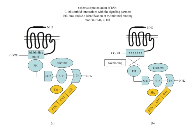
The functional consequences of the Etk/Bmx binding was further evaluated by inserting mutations to the “signal-binding” site. We prepared hPar 1 constructs with successive replacement of the designated seven residues (378-384; CQRYVYS) with A, termed as hPar 1-7A. This HA tagged mutant, HA-hPar 1-7A, completely failed to immunoprecipitate Etk/Bmx. In contrast, in the presence of HA-wt hPar 1, potent immunoprecipitation was obtained. We thus conclude that the critical region for Etk/Bmx binding to PAR1 C-tail resides in the vicinity of CQRYVYS. The physiological significance of PAR1-Etk/Bmx binding is emphasized by the following outcome. Activated MCF7 cells that express hPar 1-7A mutant failed to invade Matrigel-coated membranes. In-contrast, a potent invasion was obtained by activated wt hPar 1. This outcome highlights the fact that by preventing the binding of a key signaling partner to PAR1 C-tail, efficient inhibition of PAR1 pro-oncogenic functions, including the loss of epithelial cell polarity, migration, and invasion, is obtained (see Figures 1 and 2 for wt and mutated PAR1 C-tail and the ability to form a scaffold complexes with the signaling partners). Elucidation of the PAR1 C-tail binding domain may therefore provide a potent platform for future therapeutic vehicles in treating breast cancer.
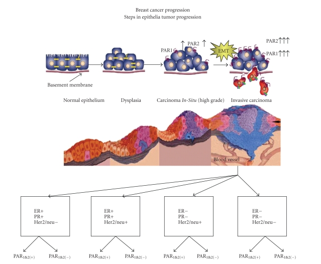
Figure 1.
Steps in breast cancer progression. Subtypes definition of breast cancer according to ER, PR, and Her2/neu status. Additional categorization is suggested including PARs status.
Figure 2.
Activation of PAR1 leads to the association of Etk/Bmx with PAR1 C-tail. This association is mediated through Etk/Bmx PH- domain enabling next the binding of Shc. The site of the “signal binding” domain (e.g., Etk/Bmx, as a prime signaling partner) in PAR1 has been identified. Insertion of successive replacement of A residues forming a PAR1 mutant incapable of binding Etk/Bmx showed impaired capabilities of PAR1 induced invasion and migration. This site provides therefore a platform for the development of future therapeutic medicaments in breast cancer.
The same approach may be utilized to identify a prime signaling partner for PAR2. This will eventually lead to characterization of a minimal PAR2 C-tail binding region. Generation of peptides that can enter the cells via adding Tat or penetratin, or alternatively, addition of either myristoylation, or another lipid moiety, will assist the peptides to cross the cell membrane. These peptides may prove as effective therapeutic inhibitors of PARs-induced breast cancer growth and development. Along this line of evidence, successful PAR1-derived peptides termed “pepducin” were developed by the group of Kuliopulos A [31]. This group has demonstrated that PAR1-induced breast tumor in a mouse model, in vivo, is blocked by the cell-penetrating lipopeptide “pepducin,” P1pal-7, which is a potent inhibitor of cell viability in breast carcinoma cells expressing PAR1. It has been shown that P1pal-7 is capable of promoting apoptosis in breast tumor xenografts and significantly inhibits metastasis to the lung.
In summary, PARs may provide a timely effective challenge for developing valuable prognostic vehicles and also critical targets for therapy in breast cancer. While the PAR prognostic vehicles stem from the extracelluar portion of the receptors, we offer the intracellular C-tail site as potential targets for therapy in breast cancer. What is the relative contribution of PAR1 versus PAR2 in breast cancer tumor growth and development is yet an open question and a subject of current evaluation.
Conflict of Interests
The authors have declared that no conflict of interests exists.
Acknowledgments
This work was supported by grants from the Israel Science Foundation (Grant no. 1313/07), Fritz-Thyssen Foundation, and Israel Cancer Research Fund (granted to R. B.). The funds had no role in the study design, data collection, analysis, decision to publish, or preparation of the paper.
References
- 1.Hanash S. Integrated global profiling of cancer. Nature Reviews Cancer. 2004;4(8):638–644. doi: 10.1038/nrc1414. [DOI] [PubMed] [Google Scholar]
- 2.Oakman C, Bessi S, Zafarana E, Galardi F, Biganzoli L, Di Leo A. Recent advances in systemic therapy: new diagnostics and biological predictors of outcome in early breast cancer. Breast Cancer Research. 2009;11(2):p. 205. doi: 10.1186/bcr2238. [DOI] [PMC free article] [PubMed] [Google Scholar]
- 3.Paik S, Shak S, Tang G, et al. A multigene assay to predict recurrence of tamoxifen-treated, node-negative breast cancer. New England Journal of Medicine. 2004;351(27):2817–2826. doi: 10.1056/NEJMoa041588. [DOI] [PubMed] [Google Scholar]
- 4.Coughlin SR. Thrombin signalling and protease-activated receptors. Nature. 2000;407(6801):258–264. doi: 10.1038/35025229. [DOI] [PubMed] [Google Scholar]
- 5.Arora P, Cuevas BD, Russo A, Johnson GL, Trejo J. Persistent transactivation of EGFR and ErbB2/HER2 by protease-activated receptor-1 promotes breast carcinoma cell invasion. Oncogene. 2008;27(32):4434–4445. doi: 10.1038/onc.2008.84. [DOI] [PMC free article] [PubMed] [Google Scholar]
- 6.Nierodzik ML, Karpatkin S. Thrombin induces tumor growth, metastasis, and angiogenesis: evidence for a thrombin-regulated dormant tumor phenotype. Cancer Cell. 2006;10(5):355–362. doi: 10.1016/j.ccr.2006.10.002. [DOI] [PubMed] [Google Scholar]
- 7.Rickles FR, Edwards RL. Activation of blood coagulation in cancer: trousseau’s syndrome revisited. Blood. 1983;62(1):14–31. [PubMed] [Google Scholar]
- 8.Camerer E, Duong DN, Hamilton JR, Coughlin SR. Combined deficiency of protease-activated receptor-4 and fibrinogen recapitulates the hemostatic defect but not the embryonic lethality of prothrombin deficiency. Blood. 2004;103(1):152–154. doi: 10.1182/blood-2003-08-2707. [DOI] [PubMed] [Google Scholar]
- 9.Palumbo JS, Kombrinck KW, Drew AF, et al. Fibrinogen is an important determinant of the metastatic potential of circulating tumor cells. Blood. 2000;96(10):3302–3309. [PubMed] [Google Scholar]
- 10.Riewald M, Kravchenko VV, Petrovan RJ, et al. Gene induction by coagulation factor Xa is mediated by activation of protease-activated receptor 1. Blood. 2001;97(10):3109–3116. doi: 10.1182/blood.v97.10.3109. [DOI] [PubMed] [Google Scholar]
- 11.Su S, Li Y, Luo Y, et al. Proteinase-activated receptor 2 expression in breast cancer and its role in breast cancer cell migration. Oncogene. 2009;28(34):3047–3057. doi: 10.1038/onc.2009.163. [DOI] [PMC free article] [PubMed] [Google Scholar]
- 12.Collier MEW, Li C, Ettelaie C. Influence of exogenous tissue factor on estrogen receptor alpha expression in breast cancer cells: involvement of beta1-integrin, PAR2, and mitogen-activated protein kinase activation. Molecular Cancer Research. 2008;6(12):1807–1818. doi: 10.1158/1541-7786.MCR-08-0109. [DOI] [PubMed] [Google Scholar]
- 13.Even-Ram S, Uziely B, Cohen P, et al. Thrombin receptor overexpression in malignant and physiological invasion processes. Nature Medicine. 1998;4(8):909–914. doi: 10.1038/nm0898-909. [DOI] [PubMed] [Google Scholar]
- 14.Grisaru-Granovsky S, Salah Z, Maoz M, Pruss D, Beller U, Bar-Shavit R. Differential expression of Protease activated receptor 1 (Par1) and pY397FAK in benign and malignant human ovarian tissue samples. International Journal of Cancer. 2005;113(3):372–378. doi: 10.1002/ijc.20607. [DOI] [PubMed] [Google Scholar]
- 15.Even-Ram SC, Grisaru-Granovsky S, Pruss D, et al. The pattern of expression of protease-activated receptors (PARs) during early trophoblast development. Journal of Pathology. 2003;200(1):47–52. doi: 10.1002/path.1338. [DOI] [PubMed] [Google Scholar]
- 16.Boire A, Covic L, Agarwal A, Jacques S, Sherifi S, Kuliopulos A. PAR1 is a matrix metalloprotease-1 receptor that promotes invasion and tumorigenesis of breast cancer cells. Cell. 2005;120(3):303–313. doi: 10.1016/j.cell.2004.12.018. [DOI] [PubMed] [Google Scholar]
- 17.Booden MA, Eckert LB, Der CJ, Trejo J. Persistent signaling by dysregulated thrombin receptor trafficking promotes breast carcinoma cell invasion. Molecular and Cellular Biology. 2004;24(5):1990–1999. doi: 10.1128/MCB.24.5.1990-1999.2004. [DOI] [PMC free article] [PubMed] [Google Scholar]
- 18.Tellez C, McCarty M, Ruiz M, Bar-Eli M. Loss of activator protein-2alpha results in overexpression of protease-activated receptor-1 and correlates with the malignant phenotype of human melanoma. Journal of Biological Chemistry. 2003;278(47):46632–46642. doi: 10.1074/jbc.M309159200. [DOI] [PubMed] [Google Scholar]
- 19.Salah Z, Maoz M, Pizov G, Bar-Shavit R. Transcriptional regulation of human protease-activated receptor 1: a role for the early growth response-1 protein in prostate cancer. Cancer Research. 2007;67(20):9835–9843. doi: 10.1158/0008-5472.CAN-07-1886. [DOI] [PubMed] [Google Scholar]
- 20.Salah Z, Haupt S, Maoz M, et al. p53 controls hPar1 function and expression. Oncogene. 2008;27(54):6866–6874. doi: 10.1038/onc.2008.324. [DOI] [PubMed] [Google Scholar]
- 21.Saban R, D’Andrea MR, Andrade-Gordon P, et al. Mandatory role of proteinase-activated receptor 1 in experimental bladder inflammation. BMC Physiology. 2007;7:p. 4. doi: 10.1186/1472-6793-7-4. [DOI] [PMC free article] [PubMed] [Google Scholar]
- 22.Su X, Camerer E, Hamilton JR, Coughlin SR, Matthay MA. Protease-activated receptor-2 activation induces acute lung inflammation by neuropeptide-dependent mechanisms. Journal of Immunology. 2005;175(4):2598–2605. doi: 10.4049/jimmunol.175.4.2598. [DOI] [PubMed] [Google Scholar]
- 23.Trejo J, Coughlin SR. The cytoplasmic tails of protease-activated receptor-1 and substance P receptor specify sorting to lysosomes versus recycling. Journal of Biological Chemistry. 1999;274(4):2216–2224. doi: 10.1074/jbc.274.4.2216. [DOI] [PubMed] [Google Scholar]
- 24.Hein L, Ishii K, Coughlin SR, Kobilka BK. Intracellular targeting and trafficking of thrombin receptors. A novel mechanism for resensitization of a G protein-coupled receptor. Journal of Biological Chemistry. 1994;269(44):27719–27726. [PubMed] [Google Scholar]
- 25.Cohen I, Maoz M, Turm H, et al. Etk/Bmx regulates proteinase-activated-receptor1 (PAR1) in breast cancer invasion: signaling partners, hierarchy and physiological significance. Plos One. 2010;5(6) doi: 10.1371/journal.pone.0011135. Article ID e11135. [DOI] [PMC free article] [PubMed] [Google Scholar]
- 26.Griffin CT, Srinivasan Y, Zheng YW, Huang W, Coughlin SR. A role for thrombin receptor signaling in endothelial cells during embryonic development. Science. 2001;293(5535):1666–1670. doi: 10.1126/science.1061259. [DOI] [PubMed] [Google Scholar]
- 27.Connolly AJ, Lshihara H, Kahn ML, Farese RV, Coughlin SR. Role of the thrombin receptor an development and evidence for a second receptor. Nature. 1996;381(6582):516–519. doi: 10.1038/381516a0. [DOI] [PubMed] [Google Scholar]
- 28.Qiu Y, Kung HJ. Signaling network of the Btk family kinases. Oncogene. 2000;19(49):5651–5661. doi: 10.1038/sj.onc.1203958. [DOI] [PubMed] [Google Scholar]
- 29.Qiu Y, Robinson D, Pretlow TG, Kung HJ. Etk/Bmx, a tyrosine kinase with a pleckstrin-homology domain, is an effector of phosphatidylinositol 3′-kinase and is involved in interleukin 6-induced neuroendocrine differentiation of prostate cancer cells. Proceedings of the National Academy of Sciences of the United States of America. 1998;95(7):3644–3649. doi: 10.1073/pnas.95.7.3644. [DOI] [PMC free article] [PubMed] [Google Scholar]
- 30.Tsai YT, Su YIH, Fang SS, et al. Etk, a Btk family tyrosine kinase, mediates cellular transformation by linking src to STAT3 activation. Molecular and Cellular Biology. 2000;20(6):2043–2054. doi: 10.1128/mcb.20.6.2043-2054.2000. [DOI] [PMC free article] [PubMed] [Google Scholar]
- 31.Yang E, Boire A, Agarwal A, et al. Blockade of PAR1 signaling with cell-penetrating pepducins inhibits Akt survival pathways in breast cancer cells and suppresses tumor survival and metastasis. Cancer Research. 2009;69(15):6223–6231. doi: 10.1158/0008-5472.CAN-09-0187. [DOI] [PMC free article] [PubMed] [Google Scholar]


