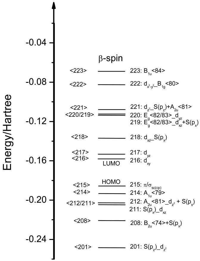
Figure 2.
Molecular orbital diagram (BP86/TZVP) of the Cyt P450cam model system shown in Scheme 1, as taken from the crystal structure (PDB code: 2CPP). The S(px), S(py), and S(pz) orbitals pertain to the 3p orbitals of sulfur. The π/σsc(cp) refers to a MO formed from π and σ bonding side chain (sc) orbitals from the Cys pocket (cp). The labels of the porphyrin core orbitals refer to free porphine, see Figure S1 in the Supporting Information. Molecular orbital labels a_b indicate that orbital a interacts with b and that a has a larger contribution in the resulting MO.

