Abnormal communication between arteries and veins are known as “arteriovenous malformations” (typically congenital) or “arteriovenous fistulae” (usually acquired). In the kidney, arteriovenous fistulae are more common than arteriovenous malformations and usually occur as a direct result of trauma or iatrogenic insult.
CASE REPORT
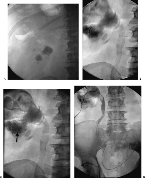
A 64-year-old man was referred to the Vascular and Interventional Radiology Section by the urology service in our hospital for percutaneous renal access prior to stone removal. After informed consent, conscious sedation was achieved with midazolam and fentanyl. The left flank was prepped and draped in usual fashion. Using both ultrasound and fluoroscopic guidance, a left lower-pole calyx containing a large renal calculus was punctured with a 21-gauge needle (Fig. 1A). A 0.018-inch guide wire (V18, Boston Scientific, Natick, MA) was advanced through the needle and multiple attempts were made to pass the wire around the stone into the renal pelvis (Fig. 1B). During one of these attempts, the tip of the wire was sheared off after being kinked between the needle tip and stone (Fig. 1C). Additional punctures were made and eventually, a guide wire was advanced around the stone into the renal pelvis. The needle was exchanged for transitional dilators and ultimately a 5-F end-hole catheter was inserted into the kidney down the ureter to terminate in the bladder (Fig. 1D). The patient then underwent operative stone extraction and ureteral stent insertion.
Figure 1.
Percutaneous renal access for stone extraction. (A) Fluoroscopic image shows large left renal calculi. (B) Fluoroscopic image shows needle puncture and attempted wire manipulation into the renal pelvis. (C) Fluoroscopic image shows continued wire manipulation. Note fractured wire tip (arrow). (D) Fluoroscopic image shows final catheter position from skin to bladder.
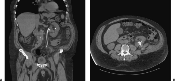
The patient was noted to have a slowly falling hematocrit on postoperative days 1 and 2, which prompted further evaluation with a computed tomography (CT) scan (Fig. 2). This revealed perinephric retroperitoneal hemorrhage, and the sheared guide wire tip was noted to be traversing the renal capsule. Because of a continuing slow drop in hematocrit, the patient was brought back to the interventional radiology section for further evaluation.
Figure 2.
CT scan after stone extraction. (A) Coronal CT reformatted image shows ureteral stent and small residual stone. (B) Axial CT image shows wire fragment and retroperitoneal hemorrhage.
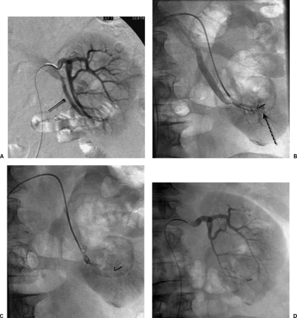
The right common femoral artery was catheterized and a 5-F sheath was inserted. An abdominal aortogram (not shown) was performed and revealed a solitary left renal artery and early filling vein, indicating an arteriovenous fistula. Selective catheterization was then performed using an RC-1 catheter (Boston Scientific). Selective renal angiography confirmed the abnormal connection between artery and vein, which appeared to originate at the site of the wire fragment (Figs. 3A,B). Three 4-mm coils were deployed at the site of the fistula (Fig. 3C). Postembolization renal arteriography demonstrated satisfactory occlusion of the arteriovenous fistula (Fig. 1D). The patient made an uneventful recovery.
Figure 3.
Embolization of arteriovenous fistula. (A) Selective renal angiogram shows arteriovenous fistula and early draining vein at lower pole of left kidney (small arrow). (B) Subselective digital subtraction angiogram shows arteriovenous fistula at site of wire fragment (dashed arrow). (C) Fluoroscopic image shows coil embolization of lower-pole renal artery. (D) Renal angiogram after embolization shows occlusion of fistula.
DISCUSSION
Renal calculi that are not suitable for lithotripsy or retrograde removal may be fragmented and extracted via a percutaneous route. To facilitate calculus removal, puncture is performed into the calyx containing the stone. This type of access is challenging, particularly when stones are large and obstructive. Procedures may be protracted, requiring repeated punctures with extensive wire manipulation. In this case, the sheared tip of a guide wire ultimately appeared to facilitate a communication between artery and vein, which led to continued perinephric hemorrhage. This injury was also likely exacerbated by stone extraction, which required insertion and manipulation of large catheters (30 French). Protracted bleeding after renal access and stone extraction is unusual and typically indicates arterial injury, either pseudoaneurysm or arteriovenous fistula.
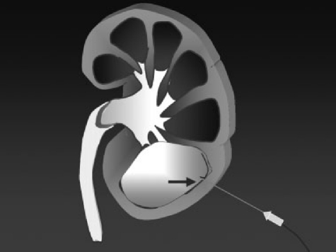
The V18 guide wire is a rigid 0.018-inch guide wire with a short hydrophilic tip. It is used preferentially in my hospital for nearly all visceral procedures when 21-gauge needles are used for access. The hydrophilic tip eases catheterization of bile ducts, renal calyces, and fluid collections. The relatively stiff body allows transitional 4- and 6-French dilators to easily pass over the wire to the intended target. One weakness of the wire is highlighted by the complication described herein. The junction point between the hydrophilic tip and body of the wire may occasionally kink, especially in lengthy, difficult access. (Fig. 4 When this occurs, the tip cannot be retracted through the needle; both the needle and wire must be removed simultaneously. If attempts are made to retract the wire into the needle, the tip will be sheared by the bevel of the needle.
Figure 4.
Illustration of complication. Wire is kinked between needle tip and stone.
Embolization is the treatment of choice for renal arterial hemorrhage. Renal arteries are end arteries. Therefore, embolization proximal to the site of bleeding effectively stops hemorrhage. This is in contrast to arteries of the bowel where collateral supply may require embolization on both sides of the bleeding site (i.e., “front door” and “back door” embolization). A variety of embolic agents may be successfully employed for arrest of hemorrhage. I prefer coils for arteriovenous fistula or pseudoaneurysm embolization due to ease of deployment and fluoroscopic visualization. Occasionally, it is helpful to combine coils with gelfoam to achieve optimal hemostasis.
SUGGESTED READING
Ray CE Jr. Renal embolization. Semin Intervent Radiol 2001;18:37–46


