Abstract
Language functional magnetic resonance imaging (fMRI) is a promising non-invasive technique for pre-surgical planning in patients whose lesions are adjacent to or within critical language areas. Most language fMRI studies in patients use blocked experimental design. In this study, we compared a blocked design and a rapid event-related design with a jittered inter-stimulus-interval (ISI) (or stochastic design) for language fMRI in six healthy controls, and eight brain tumor patients, using a vocalized antonym generation task. Comparisons were based on visual inspection of fMRI activation maps and degree of language lateralization, both of which were assessed at a constant statistical threshold for each design. The results indicated a relatively high degree of discordance between the two task designs. In general, the event-related design provided maps with more robust activations in the putative language areas than the blocked design, especially for brain tumor patients. Our results suggest that the rapid event-related design has potential for providing comparable or even higher detection power over the blocked design for localizing language function in brain tumor patients, and therefore may be able to generate more sensitive language maps. More patient studies, and further investigation and optimization of language fMRI paradigms will be needed to determine the utility and validity of this approach for pre-surgical planning.
Keywords: fMRI, Blocked design, Event-related design, Language, Pre-surgical planning
Introduction
The diagnostic aims of language functional magnetic resonance imaging (fMRI) for pre-surgical planning include identifying the language-dominant hemisphere (Binder et al., 1996), and localizing critical language areas in relation to brain lesions (Stippich et al., 2007b). Compared with the traditional invasive language mapping techniques (e.g., intracarotid amytal test (IAT, or Wada test), and electro-cortical stimulation (ECS)), language fMRI has the advantages of non-invasiveness, pre-surgical availability, repeatability, and less time and cost (Tharin and Golby, 2007). However, unlike somatosensory or motor systems that have generally predicable and consistent localization, the language system is far more complex and variable across individuals. Furthermore, it is difficult to compare the results from different laboratories due to variability in fMRI methodology, including language task paradigm, experimental design, stimulus presentation modality (visual or acoustic), behavior requirement (silent or vocalized response), and acquisition parameters (Stippich et al., 2007a). Therefore, the evaluation, optimization, and standardization of language fMRI methodology are very important for clinical applications in pre-surgical planning.
The objective of this work is to assess different experimental designs of language fMRI for pre-surgical planning purposes. There are two major types of experimental designs for fMRI studies: blocked and event-related designs. In a blocked design, a condition is presented continuously for an extended time interval (block) to maintain cognitive engagement, and different task conditions are usually alternating in time. Blocked design has the advantages of robustness (Brockway, 2000; Rombouts et al., 1997), relatively large blood-oxygen-level-dependent (BOLD) signal change relative to baseline (Buxton et al., 1998), and increased statistical power (Friston et al.,1999a). In an event-related design, discrete and short-duration events are presented with timing and order that may be randomized. Event-related design can detect transient variations in hemodynamic response, allow for analysis of individual response to trials (Schacter et al., 1997), reduce subject's expectation effects (D'Esposito et al., 1999), and is less sensitive to head motion (Birn et al., 1999). The inter-stimulus-interval (ISI) of an event-related design is a very important parameter. A slow event-related design refers to an ISI that is longer than the duration of the hemodynamic response function (HRF, 10–12 s) (Buckner et al., 1996), while a rapid event-related design refers to an ISI that is shorter than the duration of the HRF (Buckner et al.,1998). Compared to the slow design, a rapid event-related design allows for an ISI that is similar to those used in the classical neuropsychological experiments (e.g., event-related potentials (ERPs)), and also increases the statistical power by allowing for more stimuli per time unit (Friston et al., 1999a). Event-related designs can also be categorized as either periodic (with a constant ISI) or jittered (with a randomized ISI). A jittered ISI is commonly adopted in rapid event-related designs as a method for minimizing confounds from a subject's habituation and expectation (Liu et al., 2001). As events within a rapid presentation tend to overlap multiple consecutive hemodynamic responses, a linear model for the HRF summation is assumed (Burock et al., 1998). Such a rapid event-related design with a jittered ISI is also referred to as a “stochastic design” (Friston et al., 1999b). Finally, it has been demonstrated that a blocked design has high detection power, while a rapid event-related design is quite effective at estimating the hemodynamic impulse response (Birn, 2002).
Most of the reported pre-surgical language fMRI studies in patients use a blocked design (Binder et al., 1996; Brannen et al., 2001; Carpentier et al., 2001; Stippich et al., 2007b); this is likely due to its high detection power, simple design and implementation, and relative ease for patients to perform. A comparison between blocked and event-related designs has been reported for word-frequency effect (Chee et al., 2003) and semantic processing (Pilgrim et al., 2002) in healthy subject populations. They concluded that an event-related design can generate similar activations as a blocked design, and also has the capacity to overcome the confounding effects of habituation and expectation. However, to date there have been no reports directly comparing blocked and event-related fMRI designs in patients for the purpose of localizing language function. In this study, we compared the two experimental designs, both in healthy controls and brain tumor patients, using a vocalized antonym generation task. We focused on comparing their statistical maps, including activation detection in the putative language areas, language-specificity, and the effect of a brain tumor on language localization.
Materials and methods
Healthy controls and brain tumor patients
The protocol was approved by the Partner's Institutional Review Board. Six healthy controls (3 males, age = 29.0±6.2 years, range 22–36 years) and eight brain tumor patients (4 males, age =47.0±11.9 years, range 30–66 years) participated in the study and provided written informed consent. All subjects were native English speakers. Handedness was determined by the Edinburgh Handiness Inventory (EHI) (Oldfield, 1971). Patient #4 was left-handed; all other subjects were right-handed.
The patients' demographic and clinical data are listed in Table 1. The tumor volume was measured using the 3D Slicer software (http://www.slicer.org). One patient (#1) had a Wada test, and five patients (#2, #3, #5, #6, and #8) had intra-operative ECS testing. Pre- and post-operative language function and agreement between invasive language mapping and fMRI results are listed in Table 1.
Table 1.
Clinical information and agreement between invasive language mapping and fMRI results of brain tumor patients
| No. | Gender/ age |
EHI | Diagnosis | Tumor location/ size (cm3) |
Language | Impression of language from intra-operative language mapping and clinical findings |
Clinical findings more consistent with |
||
|---|---|---|---|---|---|---|---|---|---|
| Pre-op | Post-op | BL | ER | ||||||
| 1▴ | M/43 | 65 | Ganglioglioma WHO grade I–II | Left temporal/2.91 | Normal | Normal | Left hemisphere dominance for language | √ | √ |
| 2* | F/55 | 100 | Metastatic adenocarcinoma | Left temporal/7.58 | Normal | Normal | Left hemisphere involvement of language | √ | |
| 3* | F/30 | 100 | Glioblastoma WHO grade IV | Left temporal/38.42 | Speech difficulty | Better | Tumor surrounding areas involvement of language | √ | |
| 4 | M/43 | −100 | Glioblastoma WHO grade IV | Right temporal/40.83 | Normal | Normal | No language areas in the region of lesion | √ | |
| 5* | M/50 | 100 | Anaplastic oligoastrocytoma WHO grade III | Left temporal/7.27 | Occasional word-finding difficulty | Occasional word-finding difficulty → normal | Language areas were in the left hemisphere, and posterior to lesion | √ | |
| 6* | F/66 | 100 | Metastatic adenocarcinoma | Left parietal/1.67 left temporal/0.20 | Normal | Normal | No critical language areas immediately adjacent to lesion | √ | √ |
| 7 | M/35 | 100 | Oligoastrocytoma WHO grade II | Left frontal/19.62 | Normal | Normal | N/A | N/A | N/A |
| 8* | F/57 | 100 | Glioblastoma WHO grade IV | Left temporal/8.59 | Speech difficulty | N/A | No critical language areas in the region of lesion | √ | √ |
EHI: Edinburgh Handiness Inventory; positive EHI indicates right-handedness, negative EHI indicates left-handedness. BL: blocked design; ER: rapid event-related design.
Patient #1 had Wada test, which indicated left lateralization for language function.
Patients #2, #3, #5, #6, and #8 had intra-operative ECS testing.
Image acquisition
MR images were obtained using a 3.0 T GE Signa system (General Electric, Milwaukee, WI, USA). A single-shot gradient-echo echo-planar imaging (EPI) was used to acquire BOLD functional images (TR = 2000 ms, TE = 40 ms, flip angle = 90°, slice gap = 0 mm, FOV = 25.6 cm, dimension = 128 × 128 × 27, voxel size = 2 × 2 × 4 mm3) using a quadrature head coil. In each image volume, 27 axial slices were acquired using an ascending interleaved scanning sequence. Whole brain T1-weighted axial 3D-SPGR (spoiled gradient recalled) structural images (TR = 7500 ms, TE = 30 ms, flip angle = 20°, matrix = 256 × 256, voxel size = 0.5 × 0.5 × 1 mm3) were also acquired using an 8-channel head coil and ASSET (Array Spatial Sensitivity Encoding Technique, i.e., parallel imaging). Except for patient #8, all patients had gadolinium-enhanced SPGR images acquired (TR = 30 ms, TE = 5 ms, flip angle = 45°, matrix = 224 × 224, voxel size = 0.94 × 0.94 × 1.4 mm3).
Behavioral paradigm
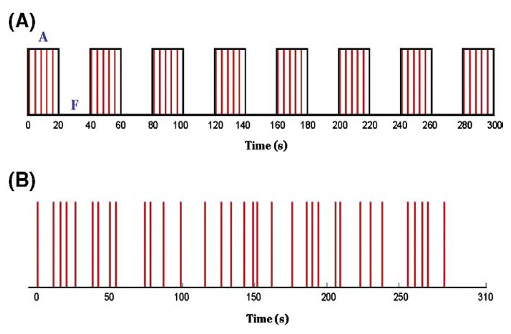
Subjects performed an antonym generation task with vocalized response (Suarez et al., 2008). The language task was implemented as blocked and rapid event-related designs in two runs. The two runs were administered in one scanning session, with the order pseudor-andomized across subjects. The time delay between the two runs was 3 min for the subject to rest in order to avoid fatigue; a different list of word stimuli was used in each design. The blocked design was 5 min 10 s (including 10 s pre-stimulus period to allow stabilization of the BOLD signal, excluded from analysis), consisting of interleaving 8 task blocks and 7 rest blocks with a 20 s block duration (Fig. 1A). In each task block, five words were presented sequentially, resulting in 40 word stimuli. Each word was shown for 2 s with a 2 s ISI. The rapid event-related design was 5 min 20 s (including 10 s pre-stimulus period), delivering 34 word stimuli (Fig. 1B). Each word was shown for 2 s with a jittered ISI (8.53±4.58 s, maximum ISI = 20.00 s, minimum ISI = 3.01 s). The order and exact timing for delivery of word stimuli was based on a stochastic design intended to maximize the statistical significance of the fMRI paradigm, diminish subject habituation, and minimize expectation effects, using the Optseq2 software package (NMR Center, Massachusetts General Hospital, Boston, MA, USA). Both stimulus paradigms were implemented using the Presentation software package (Version 9.70, Neurobehavioral Systems Inc., Davis, CA, USA), and stimuli were presented visually through MR-compatible video goggles (Resonance Technology, Los Angeles, CA, USA).
Fig. 1.
Schematic diagrams showing the two experimental designs of language fMRI. (A) Blocked design with alternating 8 antonym generation task blocks (indicated as “A”) and 7 fixation (control) blocks (indicated as “F”). (B) Rapid event-related design with jittered inter-stimulus-interval (ISI = 8.53±4.58 s). Red lines indicate the onsets of word stimuli.
The word stimuli were chosen from a pool of potential antonym pairs classified by our pilot group study of twenty native English speakers (9 males, average age = 28 years) (Suarez et al., 2008). Only those antonym pairs that generally elicited quick and accurate responses were used, such as UP–DOWN, LEFT–RIGHT, OFF–ON, OPEN–CLOSE, PUSH–PULL, or NORTH–SOUTH. During the tasks, subjects were instructed to verbalize the antonym of the presented word stimuli with minimal movement of their head, jaw, or lips. To further minimize head movement, foam padding was placed around the head, along with strips of tape spanning the video goggles and lightly adhered to the patient table. During the time period between the word stimuli, subjects were asked to relax and look at a crosshair shown in the center of the visual field.
Data pre-processing
Following functional image reconstruction, we used the Statistical Parametric Mapping software package (SPM2, Wellcome Department of Cognitive Neurology, London, UK) to perform motion correction by realigning the functional images to the first volume in each run. The displacement parameters in the x, y and z directions were recorded and used to assess gross head motion. The maximum net displacement was calculated as the norm of the vector determined by the maximum absolute displacement in each direction. Then we performed spatial smoothing on the realigned images using a 6 mm full-width-half-maximum (FWHM) Gaussian kernel. The structural SPGR image and gadolinium-enhanced SPGR image was coregistered to the mean functional image of the corresponding run for subsequent display of functional activations.
Data analysis
We performed the first-level single subject analysis based on the general linear model (GLM) (Friston et al., 1995) implemented in SPM2. For the blocked design, an estimate of the canonical HRF was used as the basis function, and only task conditions were explicitly modeled. For the event-related design, the basis function consisted of the canonical HRF model with temporal and dispersion derivatives. Run-specific responses were modeled in an event-related design (Friston et al., 1998) by convolving a series of Dirac's delta function, each representing a stimulus event onset, with the basis function. The statistical maps generated from each task design were thresholded at p<0.05 using a family-wise error (FWE) correction (Benjamini and Hochberg, 1995) for multiple comparisons.
We also performed a random effect paired t-test on the healthy control data that were spatially normalized to the Montreal Neurological Institute (MNI) space in order to test the difference between the two designs systematically.
Voxel count in the putative language areas
To facilitate the comparison between the two task designs' statistical maps in terms of the activation detection in the putative language areas, two region of interest (ROI) images were generated in the frontal and temporal lobes respectively, based on the Talairach Daemon database (Talairach and Tournoux, 1988), using the WFU Pick Atlas software (Version 1.04, Department of Radiology, Wake Forest University, Winston-Salem, NC, USA) (Lancaster et al., 1997; Lancaster et al., 2000; Maldjian et al., 2003). The first ROI consisted of the portion of the inferior frontal gyrus (IFG) overlapped by Brodmann areas (BAs) 44 and 45; the second ROI consisted of the posterior half of the superior temporal gyrus (STG). These ROIs are consistent with conventional description of Broca's and Wernicke's areas in the language dominant hemisphere and their homologues in the non-dominant hemisphere (Broca, 1861; Wernicke, 1874).
To obtain the corresponding ROI images in single subject's specific brain space, a “reverse” spatial normalization procedure was performed on the standard ROI images in the MNI space, using SPM2 and Brain Imaging Tools (BIT, http://web.mit.edu/swg/software.htm). First, the subject's SPGR image was segmented into gray matter, white matter, and cerebrospinal fluid. Then the gray matter image was spatially normalized to a common reference gray matter template brain. The inverse of the normalization matrix was used to transform the standard ROI images into the subject's specific space. The resulting specific ROI images were then overlaid onto the subject's SPGR image to confirm their structural accuracy by visual inspection. Then for each statistical map, the number of supra-threshold voxels located within the ROI images was counted. To indicate the hemispheric lateralization of language function, a laterality index (LI) was calculated using the following equation:
| (1) |
LH and RH are the numbers of supra-threshold voxels in the ROIs of the left and right hemispheres respectively. Positive LI values indicate left-dominant language function and negative values indicate right-dominant language function.
Results
Healthy controls
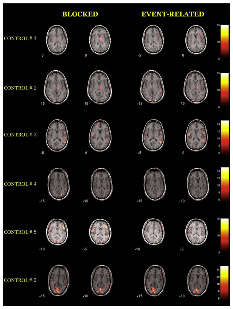
All healthy controls performed the tasks with head motion at acceptable levels. The maximum net displacement during each run was 0.91±0.55 mm for the blocked design, and 0.85±0.41 mm for the event-related design. There was no statistical difference between the gross head motion of the two designs (p = 0.85). The thresholded t-maps (p<0.05, FWE corrected) of the healthy controls' data are shown in Fig. 2. The voxel count in the language ROIs and LI results are listed in Table 2.
Fig. 2.
Thresholded t-maps (p<0.05, FWE corrected) of six healthy controls' data, comparing blocked and rapid event-related experimental designs. The background structural images are SPGR images coregistered to the mean functional image of the corresponding run. All images are in radiological convention (left side of the image is right side of the brain; right side of the image is left side of the brain).
Table 2.
Voxel count of the activations in the language ROIs and LI results
| No. | Frontal ROI (IFG overlapped by BAs 44 and 45) |
Temporal ROI (Posterior half of STG) |
|||||||
|---|---|---|---|---|---|---|---|---|---|
| BL |
ER |
BL |
ER |
||||||
| Left/right | LI | Left/right | LI | Left/right | LI | Left/right | LI | ||
| Healthy controls | 1 | 0/1 | −1 | 104/3 | 0.94 | 0/0 | N/A | 24/102 | −0.62 |
| 2 | 57/0 | 1 | 30/0 | 1 | 6/0 | 1 | 130/11 | 0.84 | |
| 3 | 18/25 | −0.16 | 38/0 | 1 | 476/164 | 0.49 | 514/166 | 0.51 | |
| 4 | 51/0 | 1 | 8/0 | 1 | 220/52 | 0.62 | 68/24 | 0.48 | |
| 5 | 141/17 | 0.78 | 14/0 | 1 | 318/180 | 0.28 | 18/14 | 0.13 | |
| 6 | 49/30 | 0.24 | 14/12 | 0.08 | 160/76 | 0.36 | 57/116 | −0.34 | |
| Brain tumor patients | 1 | 147/29 | 0.67 | 154/28 | 0.69 | 90/4 | 0.91 | 138/0 | 1 |
| 2 | 0/0 | N/A | 29/0 | 1 | 20/10 | 0.33 | 42/124 | −0.49 | |
| 3 | 0/0 | N/A | 113/19 | 0.71 | 22/40 | −0.29 | 17/400 | −0.92 | |
| 4 | 16/25 | −0.22 | 9/3 | 0.5 | 336/716 | −0.36 | 296/128 | 0.40 | |
| 5 | 6/0 | 1 | 6/5 | 0.09 | 128/272 | −0.36 | 250/580 | −0.40 | |
| 6 | 1/0 | 1 | 0/1 | −1 | 154/333 | −0.37 | 55/159 | −0.49 | |
| 7 | 145/17 | 0.79 | 46/0 | 1 | 121/45 | 0.46 | 44/15 | 0.49 | |
| 8 | 0/0 | N/A | 18/0 | 1 | 72/45 | 0.23 | 193/105 | 0.30 | |
For each subject's thresholded t-maps (p<0.05, FWE corrected), the number of supra-threshold voxels in the language ROIs was counted using a custom toolbox. BL: blocked design; ER: rapid event-related design; LI: laterality index.
For controls #1, #2, and #3, the event-related design showed more supra-threshold voxels in the putative language areas in the left frontal and temporal lobes than the blocked design. This observation was more pronounced for the activations in the left temporal lobe corresponding toWernicke's area. Note that for control #1 the blocked design showed almost no activation in the language ROIs at the chosen threshold.
For controls #4, #5 and #6, the blocked design seemed to indicate more robust activations in the language ROIs. Especially for control #5, the blocked design showed many more activated voxels in Wernicke's area than the event-related design, however with many more extraneous voxels in the non-language regions.
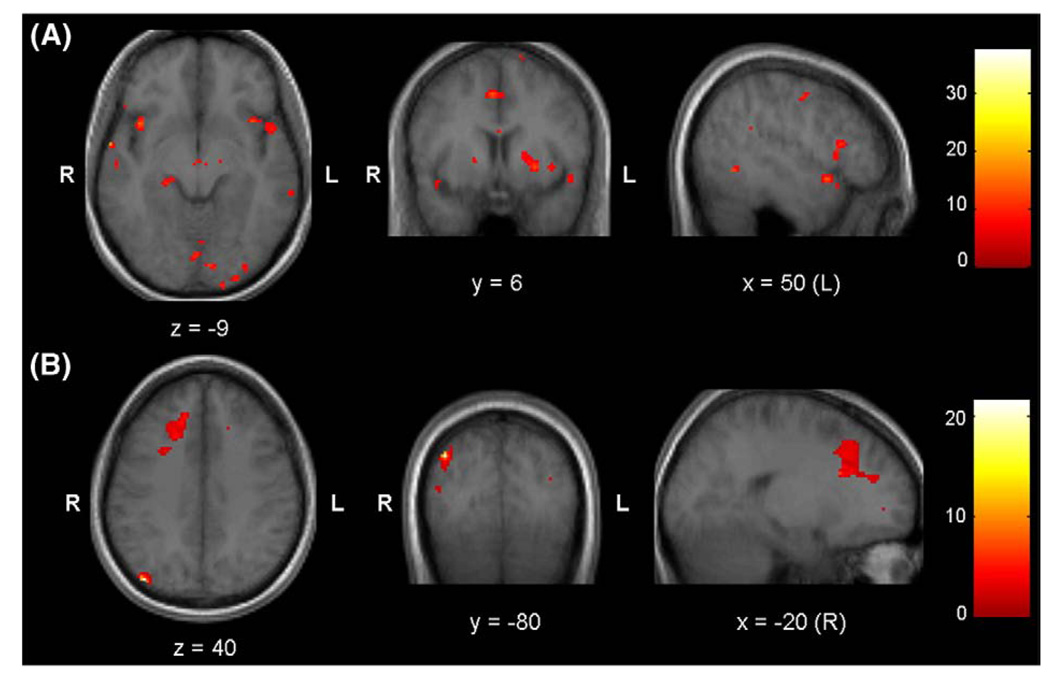
The thresholded t-maps of the random effect paired t-test are shown in Fig. 3. The results indicated that the event-related design had significantly higher activations in the putative language areas, as well as other areas, e.g., deep nuclei, thalamus, and visual areas (p<0.0005, uncorrected, df = 5, Fig. 3A). In comparison, the blocked design showed higher activations in the right frontal lobe and precuneus (p<0.05, uncorrected, df = 5, Fig. 3B).
Fig. 3.
Results of random effect paired t-test of healthy controls' data. (A) Event-related design>blocked design, thresholded at p<0.0005 (uncorrected, df = 5); (B) Blocked design>event-related design, thresholded at p<0.05 (uncorrected, df = 5).
Brain tumor patients
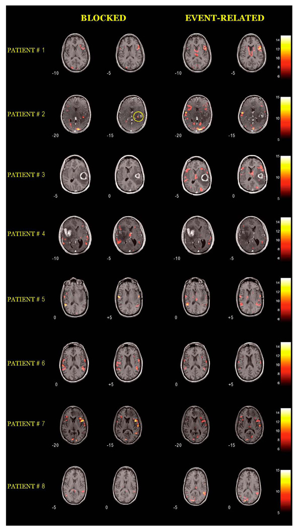
All brain tumor patients performed the tasks with limited gross head motion. The maximum net displacement during each run was 0.78±0.48 mm for the blocked design, and 0.79±0.64 mm for the event-related design. There was no statistical difference between the gross head motion of the two designs (p = 0.99). The thresholded t-maps (p<0.05, FWE corrected) of the patients' data are shown in Fig. 4. For a better view of the tumor and edema, we used gadolinium-enhanced SPGR images as the background structural images for the maps of patients #2, #3, and #4. The clinical impression, fMRI results, and consistency between these two from the two designs are summarized in Table 1. The voxel count in the language ROIs and LI results are listed in Table 2. Following is the observation of results for each patient.
Fig. 4.
Thresholded t-maps (p<0.05, FWE corrected) of eight brain tumor patients' data, comparing blocked and rapid event-related experimental designs. The background structural images are SPGR images (patients #1, #5, #6, #7, and #8) or gadolinium-enhanced SPGR images (patients #2, #3, and #4) coregistered to the mean functional image of the corresponding run. All images are in radiological convention.
Patient #1: This patient had a 1.5-year history of multiple daily seizures, and was diagnosed as medically refractory epilepsy with left temporal lobe lesion (not seen in the selected slices). Results from both experimental designs confirmed that the patient was left hemisphere dominant for language function, consistent with the Wada test findings. No intra-operative language mapping was performed. The event-related design identified more supra-threshold voxels in both Broca's and Wernicke's areas, and higher LIs. It also identified voxels around the ventricles (with lower t-scores than the main activations), which were likely due to noise.
Patient #2: This patient had a 4-year history of lung cancer, and a newly-found calcified mass in the left superior temporal lobe with significant surrounding edema and mild mass effect. Pathology confirmed metastatic adenocarcinoma. The intra-operative ECS proceeded along the left superior temporal gyrus and the inferior aspect of the left frontal lobe, and no evidence for speech arrest was identified in the region of the lesion. No post-operative language deficit was observed. The t-maps from the blocked design showed almost no supra-threshold voxels in the putative language areas, even when a more lenient threshold was tried (not shown in the figure). The blocked design identified activated voxels at the tumor margin (highlighted by yellow circle). In comparison, the event-related design identified activations in Broca's area, and more supra-threshold voxels in the right temporal lobe ROI than the left side. This observation was possibly due to the effect of edema on the left temporal lobe that reduced the BOLD signal. After this patient's second surgery for recurrent and extensive tumor her speech was slightly worse (occasional word-finding hesitation and paraphasia). This observation confirmed the left hemisphere involvement of language function. Therefore, in this patient the event-related design provided more sensitive language maps than the blocked design.
Patient #3: This patient's MRI indicated a large enhancing mass in the middle portion of the left temporal lobe, and considerable edema around the tumor. Similar to patient #2, the blocked design identified almost no activation in the putative language areas, but indicated extraneous voxels at the tumor margin or within the tumor (not shown in the selected slices). In comparison, the event-related design indicated robust activations in Broca's area and its homologue in the right hemisphere. For the activations in the temporal lobe, there were significantly more supra-threshold voxels in the right hemisphere than in the left side. This patient had pre-operative speech difficulty, and her speech was better post-operatively. Although the intra-operative ECS indicated that there were no critical speech areas in the immediate region of the tumor, this change in speech confirmed the involvement of the surrounding area in language function, which was consistent with the maps from the event-related design.
Patient #4: This patient had pre-operative language mapping because he was left-handed and his tumor was in the right temporal lobe. He did not have pre-operative or post-operative language deficit. No invasive language mapping was performed. The blocked design indicated more supra-threshold voxels in the right hemisphere ROIs than the left side, resulting in negative LIs. However, the activations in the right hemisphere seemed to be within the edematous region. In contrast, the event-related design indicated more supra-threshold voxels in the left hemisphere, resulting in positive LIs for both ROIs; and there were no extraneous activated voxels in the tumor or edema areas. There was no language deficit observed in this patient after 10 months, suggesting no language involvement in the lesion area, which was consistent with the findings of the event-related design.
Patient #5: This patient experienced several months of language and cognitive problems (word-finding difficulty, repetitive speech, short-term memory loss, and confusion). MRI indicated a large non-enhancing mass in the left temporal lobe. Results from both designs showed almost no supra-threshold voxels in Broca's area and more supra-threshold voxels in the Wernicke's ROI in the right temporal lobe than the left side, resulting in negative LIs. Compared with the blocked design, the event-related design indicated more activations outside the main language areas, posterior to the tumor in areas of edema and in the thalamus and deep nuclei. The intra-operative ECS did not result in any speech change. Following the surgery, this patient's speech was gradually getting better (from “occasionally word-finding difficulty” to “normal”). This observation confirmed that the language areas were in the left hemisphere and posterior to the lesion, which was more consistent with the maps from the event-related design.
Patient #6: This patient's MRI showed enhancing lesions in the left parietal and superior temporal lobes. Pathology returned as meta-static adenocarcinoma of non-small cell lung cancer. Similar to patient #5, both designs did not show activation in Broca's area, and indicated more supra-threshold voxels in the Wernicke's ROI of the right hemisphere than the left side. The blocked design indicated more activations in the temporal lobe than the event-related design, however with more extraneous voxels in the non-language regions. The intra-operative ECS indicated no critical language areas immediately adjacent to the lesion.
Patient #7: This patient had a 6-year history of oligoastrocytoma in the left frontal lobe. The two designs showed very similar activation patterns, with the blocked design indicating more supra-threshold voxels in the language ROIs. No intra-operative language mapping was performed.
Patient #8: This patient was diagnosed as glioblastoma in the left medial temporal lobe, with edema and mass effect. He had pre-operative speech difficulty. The blocked design did not indicate activated voxels in Broca's area and identified less activation in Wernicke's area than the event-related design. Intra-operative ECS did not result in difficulties with the patient's speech. However, during resection he began to have some speech and memory difficulties, which might indicate white matter disruption. This observation together with his pre-operative speech difficulties was consistent with fMRI-predicted left hemisphere lateralization of language function.
Discussion
As a promising non-invasive technique, pre-surgical language fMRI has been applied to determine language dominance and localize language areas for patients with lesions in or adjacent to critical language regions (Bookheimer, 2007). In this study, we compared analogous blocked and rapid event-related language fMRI in healthy controls and brain tumor patients in an effort to address the clinical utility of these two different experimental designs for pre-surgical language mapping.
Choice of language task and parameters of experimental design
For the language task, we chose an antonym generation task with overt responses as the language task. Antonym generation has been demonstrated to be reliable and robust in localizing Broca's area (Brannen et al., 2001). This behavioral task is expected to activate all the major aspects of language function: receptive decoding, expressive encoding, and vocalization.
We chose a vocalized language task (rather than a silent task) due to its advantages of higher fMRI signal strength and robustness of activations (Palmer et al., 2001), higher correlation with the ECS results (Petrovich et al., 2005), and the feasibility of monitoring task performance and comparing with the clinical gold-standard tests that use overt responses (Suarez et al., 2008). However, language fMRI studies have most often been restricted to covert responses due to the potential speech-induced motion artifact. Investigators have proposed different strategies for task design, data acquisition and analysis to address this problem (Abrahams et al., 2003; Birn et al.,1999, 2004). In this study, subjects were instructed to verbalize their responses in a way that minimized head movement. No motion artifacts were observed in the statistical maps from both designs, and realignment indicators demonstrated low levels of gross head motion during the task.
In order to make the blocked and event-related designs comparable to each other, the scan time, stimulus duration, and the number of words presented in each run were designed to be as similar as possible. The scan time was about 5 min for both designs, which is an acceptable acquisition time for patient studies. Furthermore, we used fixation as the control condition in order to provide a closer comparison between the two designs (Chee et al., 2003). However, the use of a low-level control condition such as fixation may introduce numerous non-language related activations in the results due to other cognitive processes involved in language processing (Price et al., 1997). One potential alternative is to use a higher-level baseline block (instead of fixation) in the blocked design, and use two events (e.g., language task event and baseline event) in the event-related design.
Comparison between experimental designs of language fMRI
We focused on comparing the language maps generated by the two experimental designs, by setting the same statistical level of the t-maps (p<0.05, FWE corrected, t-threshold = 4.99±0.07 for the blocked design, and 5.01±0.06 for the event-related design). It is worth mentioning that a direct comparison of the contrast-specific statistics should be avoided since any difference between the resulting statistics could be due to the efficiency of the designs (Mechelli et al., 2003b), and psychological effects of blocking stimuli (Pilgrim et al., 2002). In this study, we used a constant threshold for both designs and found large discrepancies in terms of laterality and activation patterns using blocked versus event-related designs. It is not clear however if a threshold at p<0.05 is entirely analogous for both designs. The event-related stochastic design may benefit from stricter thresholds in a manner that may not generalize to the blocked designs. For this reason we chose a relatively liberal threshold of p<0.05 in the comparisons. It should be emphasized that arbitrary threshold setting for fMRI maps may cause unstable results, and is particularly problematic for the interpretation of fMRI results for patient studies (Loring et al., 2002; Suarez et al., 2008). In pre-surgical planning, both sensitivity and specificity of language fMRI maps needs to be improved to provide the surgeon with a picture of critical language areas; while most importantly not missing the true language activations due to arbitrary threshold setting (Tharin and Golby, 2007).
a) Healthy control study
For the healthy control study, both designs were able to identify activations in the putative language areas at the chosen threshold, except for the blocked design of one control (#1), which did not reveal supra-threshold voxels in the Broca's and Wernicke's ROIs, and the event-related design of another control (#5), which showed few activated voxels in the Wernicke's ROI. In general, the event-related design generated maps with less extraneous activations in the non-language regions. The results of the paired t-test on the healthy control data indicated that the activations revealed by the event-related design were significantly higher in the putative language areas than the blocked design, suggesting that there was a systematic difference between the two designs, although the sample size was limited.
b) Brain tumor patient study
For the patient study, the event-related design in general identified more robust or at least similar activations compared with the blocked design, and the findings from the event-related design were more consistent with the clinical impression of the patients' language function. Although it is known that blocked design is less sensitive to the shape of HRF, our results suggest that for detection of language function in brain tumor patients, the stochastic design used in this study demonstrated detection power comparable or even higher than the blocked design. Thus, the stochastic design may pose a compromise between the conventional blocked and event-related designs, in terms of detection power and estimation efficiency (Liu et al., 2001). It has also been demonstrated that an event-related model may capture the form of HRF better than a blocked model, even for the blocked design fMRI, and therefore reducing the error variance and increasing the sensitivity (Mechelli et al., 2003a). Another possible advantage of the stochastic paradigm is that the randomness of stimuli delivery may elicit more attention from the subject.
In brain tumor patients, mass effect, edema, or neovascularization can alter blood flow or induce a neurovascular uncoupling effect, and therefore reduce activations in the functional areas (Bookheimer, 2007; Hou et al., 2006). It has also been reported that the BOLD effect is sensitive to the altered regional blood flow of high-grade brain tumor cases (Bogomolny et al., 2004). Consistent with these previous findings, our results showed considerably weaker language activations in the hemisphere where the lesion was located than we observed in the contralateral hemisphere, resulting in a LI value that may be misleading for language dominance. For example, many more supra-threshold voxels were identified in the right temporal lobe than the left side for patients #2, #3, and #5, resulting in negative LIs. This pattern is easily misinterpreted as right-hemispheric language dominance. However, in these adult-onset cases the activations in the homologous language areas in the right hemisphere are unlikely to reflect true lesion-induced functional reorganization. The additional activations in the cortex contralateral to the lesion (as seen in patients #2, #3, #4, and #5) have been proposed to reflect a compensatory response or disinhibition effect as the lesion hemisphere becomes compromised (Bookheimer, 2007). A study by (Ulmer et al., 2004) reported seven patients whose fMRI data were inconsistent with other functional localization methods, and two cases where the fMRI results incorrectly suggested right-hemispheric speech dominance. They postulated that the main reasons for these errors were abnormal blood flow response (neurovascular uncoupling), and tumor infiltration of the functional areas.
The blocked design of patients #2 and #3 identified supra-threshold voxels at the border or within the tumor, where the event-related design did not show any activation. In the case of a metastatic lesion as in patient #2, there cannot be functional tissue within the lesion itself, though there may be functional tissue immediately adjacent. The ECS testing indicated no speech arrest in the region of lesion. This observation indicated that the blocked design may generate false positives within the tumor. In contrast to metastatic lesions, some low-grade gliomas may contain functional tissue within the lesion due to tumor infiltration (Ojemann et al., 1996; Skirboll et al., 1996). In such cases, the surgeon may not be able to definitively interpret the significance of activations seen within the tumor on pre-operative fMRI and must proceed with caution by assuming that there is indeed functional tissue within the tumor and performing confirmatory testing at surgery prior to resection.
Accuracy of language ROIs
To facilitate a quantitative comparison between the language maps generated by the two designs, we counted the number of supra-threshold voxels located within the putative language ROIs (i.e., Broca's and Wernicke's areas and their homologues in the right hemisphere). We performed a “reverse normalization” on the standard ROI images to obtain the specific ROIs in each subject's own space, based on gray matter structure. This procedure is expected to be appropriate for a normal control study. For a patient study, one should proceed with caution, as the patient's brain is structurally abnormal, and the lesion may push and distort the cortex of the functional areas, leading to abnormal functional localization. Furthermore, the human language system is very complex and variable across individuals, and may be more so for the patients. As such, we inspected the accuracy of the specific ROIs by overlaying them onto each subject's structural image. The results indicated that the lesion-induced distortion of the brain structure had been well accounted for, and the functional localization appeared to be in the correct location and aligned very well to the cortex.
In this study, we compared two fMRI experimental designs, blocked and rapid event-related, of an antonym generation task for pre-surgical language mapping. Comparison based on the statistical maps generated by the two designs indicated a relatively high degree of discordance. In general, the rapid event-related design identified more activated voxels in the putative language areas and was more consistent with the clinical findings of language function for brain tumor patients, suggesting that it may generate more sensitive language maps. This study was not able to assess reproducibility of results; some studies suggest that there is greater intra-session variability for fMRI results in patients (Bosnell et al., 2008). Future work will be directed towards investigating reliability of fMRI pre-surgical language mapping using event-related design compared to blocked design.
Acknowledgments
This work is supported by NIH K08 NS048063, NIH-NCRR U41 RR019703, and The Brain Science Foundation. We thank Dr. Susan Whitfield-Gabrieli for her great help and suggestions on this work.
References
- Abrahams S, Goldstein LH, Simmons A, Brammer MJ, Williams SC, Giampietro VP, Andrew CM, Leigh PN. Functional magnetic resonance imaging of verbal fluency and confrontation naming using compressed image acquisition to permit overt responses. Hum. Brain Mapp. 2003;20:29–40. doi: 10.1002/hbm.10126. [DOI] [PMC free article] [PubMed] [Google Scholar]
- Benjamini Y, Hochberg Y. Controlling the false discovery rate: a practical and powerful approach to multiple testing. J. Roy. Stat. Soc. Ser. B, Methodol. 1995;57:289–300. [Google Scholar]
- Binder JR, Swanson SJ, Hammeke TA, Morris GL, Mueller WM, Fischer M, Benbadis S, Frost JA, Rao SM, Haughton VM. Determination of language dominance using functional MRI: a comparison with the Wada test. Neurology. 1996;46:978–984. doi: 10.1212/wnl.46.4.978. [DOI] [PubMed] [Google Scholar]
- Birn R. Detection versus estimation in event-related fMRI: choosing the optimal stimulus timing. NeuroImage. 2002;15:252–264. doi: 10.1006/nimg.2001.0964. [DOI] [PubMed] [Google Scholar]
- Birn RM, Bandettini PA, Cox RW, Shaker R. Event-related fMRI of tasks involving brief motion. Hum. Brain Mapp. 1999;7:106–114. doi: 10.1002/(SICI)1097-0193(1999)7:2<106::AID-HBM4>3.0.CO;2-O. [DOI] [PMC free article] [PubMed] [Google Scholar]
- Birn RM, Cox RW, Bandettini PA. Experimental designs and processing strategies for fMRI studies involving overt verbal responses. NeuroImage. 2004;23:1046–1058. doi: 10.1016/j.neuroimage.2004.07.039. [DOI] [PubMed] [Google Scholar]
- Bogomolny DL, Petrovich NM, Hou BL, Peck KK, Kim MJ, Holodny AI. Functional MRI in the brain tumor patient. Top. Magn. Reson. Imaging. 2004;15:325–335. doi: 10.1097/00002142-200410000-00005. [DOI] [PubMed] [Google Scholar]
- Bookheimer S. Pre-surgical language mapping with functional magnetic resonance imaging. Neuropsychol. Rev. 2007;17:145–155. doi: 10.1007/s11065-007-9026-x. [DOI] [PubMed] [Google Scholar]
- Bosnell R, Wegner C, Kincses ZT, Korteweg T, Agosta F, Ciccarelli O, De Stefano N, Gass A, Hirsch J, Johansen-Berg H, Kappos L, Barkhof F, Mancini L, Manfredonia F, Marino S, Miller DH, Montalban X, Palace J, Rocca M, Enzinger C, Ropele S, Rovira A, Smith S, Thompson A, Thornton J, Yousry T, Whitcher B, Filippi M, Matthews PM. Reproducibility of fMRI in the clinical setting: implications for trial designs. NeuroImage. 2008;42:603–610. doi: 10.1016/j.neuroimage.2008.05.005. [DOI] [PubMed] [Google Scholar]
- Brannen JH, Badie B, Moritz CH, Quigley M, Meyerand ME, Haughton VM. Reliability of functional MR imaging with word-generation tasks for mapping Broca's area. AJNR Am. J. Neuroradiol. 2001;22:1711–1718. [PMC free article] [PubMed] [Google Scholar]
- Broca PP. Perte de la parole ramolissement chronique et destruction partielle du lobel anterieur gauche de cerveau. Bull. Mem. Soc. Anthropol. Paris. 1861;2:249–262. [Google Scholar]
- Brockway JP. Two functional magnetic resonance imaging f(MRI) tasks that may replace the gold standard, Wada testing, for language lateralization while giving additional localization information. Brain Cogn. 2000;43:57–59. [PubMed] [Google Scholar]
- Buckner RL, Bandettini PA, O'Craven KM, Savoy RL, Petersen SE, Raichle ME, Rosen BR. Detection of cortical activation during averaged single trials of a cognitive task using functional magnetic resonance imaging. Proc. Natl. Acad. Sci. U. S. A. 1996;93:14878–14883. doi: 10.1073/pnas.93.25.14878. [DOI] [PMC free article] [PubMed] [Google Scholar]
- Buckner RL, Goodman J, Burock M, Rotte M, Koutstaal W, Schacter D, Rosen B, Dale AM. Functional-anatomic correlates of object priming in humans revealed by rapid presentation event-related fMRI. Neuron. 1998;20:285–296. doi: 10.1016/s0896-6273(00)80456-0. [DOI] [PubMed] [Google Scholar]
- Burock MA, Buckner RL, Woldorff MG, Rosen BR, Dale AM. Randomized event-related experimental designs allow for extremely rapid presentation rates using functional MRI. NeuroReport. 1998;9:3735–3739. doi: 10.1097/00001756-199811160-00030. [DOI] [PubMed] [Google Scholar]
- Buxton RB, Wong EC, Frank LR. Dynamics of blood flow and oxygenation changes during brain activation: the balloon model. Magn. Reson. Med. 1998;39:855–864. doi: 10.1002/mrm.1910390602. [DOI] [PubMed] [Google Scholar]
- Carpentier A, Pugh KR, Westerveld M, Studholme C, Skrinjar O, Thompson JL, Spencer DD, Constable RT. Functional MRI of language processing: dependence on input modality and temporal lobe epilepsy. Epilepsia. 2001;42:1241–1254. doi: 10.1046/j.1528-1157.2001.35500.x. [DOI] [PubMed] [Google Scholar]
- Chee MW, Venkatraman V, Westphal C, Siong SC. Comparison of block and event-related fMRI designs in evaluating the word-frequency effect. Hum. Brain Mapp. 2003;18:186–193. doi: 10.1002/hbm.10092. [DOI] [PMC free article] [PubMed] [Google Scholar]
- D'Esposito M, Zarahn E, Aguirre GK. Event-related functional MRI: implications for cognitive psychology. Psychol. Bull. 1999;125:155–164. doi: 10.1037/0033-2909.125.1.155. [DOI] [PubMed] [Google Scholar]
- Friston KJ, Holmes AP, Worsley KJ, Poline J-P, Frith CD, Frackowiak RSJ. Statistical parametric maps in functional imaging: a general linear approach. Hum. Brain Mapp. 1995;2:189–210. [Google Scholar]
- Friston KJ, Fletcher P, Josephs O, Holmes A, Rugg MD, Turner R. Event-related fMRI: characterizing differential responses. NeuroImage. 1998;7:30–40. doi: 10.1006/nimg.1997.0306. [DOI] [PubMed] [Google Scholar]
- Friston KJ, Holmes AP, Price CJ, Buchel C, Worsley KJ. Multisubject fMRI studies and conjunction analyses. NeuroImage. 1999a;10:385–396. doi: 10.1006/nimg.1999.0484. [DOI] [PubMed] [Google Scholar]
- Friston KJ, Zarahn E, Josephs O, Henson RN, Dale AM. Stochastic designs in event-related fMRI. NeuroImage. 1999b;10:607–619. doi: 10.1006/nimg.1999.0498. [DOI] [PubMed] [Google Scholar]
- Hou BL, Bradbury M, Peck KK, Petrovich NM, Gutin PH, Holodny AI. Effect of brain tumor neovasculature defined by rCBV on BOLD fMRI activation volume in the primary motor cortex. NeuroImage. 2006;32:489–497. doi: 10.1016/j.neuroimage.2006.04.188. [DOI] [PubMed] [Google Scholar]
- Lancaster JL, Summerln JL, Rainey L, Freitas CS, Fox PT. The Talairach Daemon, a database server for Talairach Atlas Labels. NeuroImage. 1997;5:S633. [Google Scholar]
- Lancaster JL, Woldorff MG, Parsons LM, Liotti M, Freitas CS, Rainey L, Kochunov PV, Nickerson D, Mikiten SA, Fox PT. Automated Talairach atlas labels for functional brain mapping. Hum. Brain Mapp. 2000;10:120–131. doi: 10.1002/1097-0193(200007)10:3<120::AID-HBM30>3.0.CO;2-8. [DOI] [PMC free article] [PubMed] [Google Scholar]
- Liu TT, Frank LR, Wong EC, Buxton RB. Detection power, estimation efficiency, and predictability in event-related fMRI. NeuroImage. 2001;13:759–773. doi: 10.1006/nimg.2000.0728. [DOI] [PubMed] [Google Scholar]
- Loring DW, Meador KJ, Allison JD, Pillai JJ, Lavin T, Lee GP, Balan A, Dave V. Now you see it, now you don't: statistical and methodological considerations in fMRI. Epilepsy Behav. 2002;3:539–547. doi: 10.1016/s1525-5050(02)00558-9. [DOI] [PubMed] [Google Scholar]
- Maldjian JA, Laurienti PJ, Kraft RA, Burdette JH. An automated method for neuroanatomic and cytoarchitectonic atlas-based interrogation of fMRI data sets. NeuroImage. 2003;19:1233–1239. doi: 10.1016/s1053-8119(03)00169-1. [DOI] [PubMed] [Google Scholar]
- Mechelli A, Henson RN, Price CJ, Friston KJ. Comparing event-related and epoch analysis in blocked design fMRI. NeuroImage. 2003a;18:806–810. doi: 10.1016/s1053-8119(02)00027-7. [DOI] [PubMed] [Google Scholar]
- Mechelli A, Price CJ, Henson RN, Friston KJ. Estimating efficiency a priori: a comparison of blocked and randomized designs. NeuroImage. 2003b;18:798–805. doi: 10.1016/s1053-8119(02)00040-x. [DOI] [PubMed] [Google Scholar]
- Ojemann JG, Miller JW, Silbergeld DL. Preserved function in brain invaded by tumor. Neurosurgery. 1996;39:253–258. doi: 10.1097/00006123-199608000-00003. discussion 258–259. [DOI] [PubMed] [Google Scholar]
- Oldfield RC. The assessment and analysis of handedness: the Edinburgh inventory. Neuropsychologia. 1971;9:97–113. doi: 10.1016/0028-3932(71)90067-4. [DOI] [PubMed] [Google Scholar]
- Palmer ED, Rosen HJ, Ojemann JG, Buckner RL, Kelley WM, Petersen SE. An event-related fMRI study of overt and covert word stem completion. Neuro-Image. 2001;14:182–193. doi: 10.1006/nimg.2001.0779. [DOI] [PubMed] [Google Scholar]
- Petrovich N, Holodny AI, Tabar V, Correa DD, Hirsch J, Gutin PH, Brennan CW. Discordance between functional magnetic resonance imaging during silent speech tasks and intraoperative speech arrest. J. Neurosurg. 2005;103:267–274. doi: 10.3171/jns.2005.103.2.0267. [DOI] [PubMed] [Google Scholar]
- Pilgrim LK, Fadili J, Fletcher P, Tyler LK. Overcoming confounds of stimulus blocking: an event-related fMRI design of semantic processing. NeuroImage. 2002;16:713–723. doi: 10.1006/nimg.2002.1105. [DOI] [PubMed] [Google Scholar]
- Price CJ, Moore CJ, Friston KJ. Subtractions, conjunctions, and interactions in experimental design of activation studies. Hum. Brain Mapp. 1997;5:264–272. doi: 10.1002/(SICI)1097-0193(1997)5:4<264::AID-HBM11>3.0.CO;2-E. [DOI] [PubMed] [Google Scholar]
- Rombouts SA, Barkhof F, Hoogenraad FG, Sprenger M, Valk J, Scheltens P. Test-retest analysis with functional MR of the activated area in the human visual cortex. AJNR Am. J. Neuroradiol. 1997;18:1317–1322. [PMC free article] [PubMed] [Google Scholar]
- Schacter DL, Buckner RL, Koutstaal W, Dale AM, Rosen BR. Late onset of anterior prefrontal activity during true and false recognition: an event-related fMRI study. NeuroImage. 1997;6:259–269. doi: 10.1006/nimg.1997.0305. [DOI] [PubMed] [Google Scholar]
- Skirboll SS, Ojemann GA, Berger MS, Lettich E, Winn HR. Functional cortex and subcortical white matter located within gliomas. Neurosurgery. 1996;38:678–684. discussion 684–685. [PubMed] [Google Scholar]
- Stippich C, Blatow M, Krakow K. Presurgical fMRI of language function. In: Stippich C, editor. Clinical Functional MRI: Presurgical Functional Neuroimaging. Heidelberg: Springer; 2007a. pp. 115–134. [Google Scholar]
- Stippich C, Rapps N, Dreyhaupt J, Durst A, Kress B, Nennig E, Tronnier VM, Sartor K. Localizing and lateralizing language in patients with brain tumors: feasibility of routine preoperative functional MR imaging in 81 consecutive patients. Radiology. 2007b;243:828–836. doi: 10.1148/radiol.2433060068. [DOI] [PubMed] [Google Scholar]
- Suarez RO, Whalen S, O'Shea JP, Golby AJ. A surgical planning method for functional MRI assessment of language dominance: influences from threshold, region-of-interest, and stimulus mode. Brain Imag. Behav. 2008;2:59–73. [Google Scholar]
- Talairach J, Tournoux P. Co-Planar Stereotactic Atlas Of The Human Brain. Stuttgart: Thieme; 1988. [Google Scholar]
- Tharin S, Golby A. Functional brain mapping and its applications to neurosurgery. Neurosurgery. 2007;60:185–201. doi: 10.1227/01.NEU.0000255386.95464.52. discussion 201–202. [DOI] [PubMed] [Google Scholar]
- Ulmer JL, Hacein-Bey L, Mathews VP, Mueller WM, DeYoe EA, Prost RW, Meyer GA, Krouwer HG, Schmainda KM. Lesion-induced pseudo-dominance at functional magnetic resonance imaging: implications for preoperative assessments. Neurosurgery. 2004;55:569–579. doi: 10.1227/01.neu.0000134384.94749.b2. discussion 580–581. [DOI] [PubMed] [Google Scholar]
- Wernicke K. Eine psychologische Studie auf anatomischer Basis. Breslau: M. Crohn und Weigert; 1874. Der aphasische Sympotomencomplex. [Google Scholar]


