



Abstract
Increased sensitivity to light-induced melatonin suppression characterizes some, but not all, patients with bipolar illness or seasonal affective disorder. The aim of this study was to test the hypothesis that patients with premenstrual dysphoric disorder (PMDD), categorized as a depressive disorder in DSM-IV, have altered sensitivity to 200 lux light during mid-follicular (MF) and late-luteal (LL) menstrual cycle phases compared with normal control (NC) women. As an extension of a pilot study in which we administered 500 lux to 8 PMDD and 5 NC subjects, in the present study we administered 200 lux to 10 PMDD and 13 NC subjects during MF and LL menstrual cycle phases. We admitted subjects to the General Clinical Research Center (GCRC) in dim light (< 50 lux) to dark (during sleep) conditions at 16:00 h where nurses inserted an intravenous catheter at 17:00 h and collected plasma samples for melatonin at 30-min intervals from 18:00 to 10:00 h, including between 00:00 and 01:00 h for baseline values, between 01:30 and 03:00 h during the 200 lux light exposure administered from 01:00-03:00 h, and at 03:30 and 04:00 h after the light exposure. Median % melatonin suppression was significantly greater in PMDD (30.8%) vs. NC (−0.2%) women (p = 0.040), and was significantly greater in PMDD in the MF (30.8%) than in the LL (−0.15%) phase (p = 0.047). Additionally, in the LL (but not the MF) phase, % suppression after 200 lux light was significantly positively correlated with serum estradiol level (p = 0.007) in PMDD patients, but not in NC subjects (p > .05).
Keywords: light suppression, melatonin, menstrual cycle, depression, women
INTRODUCTION
Increased sensitivity to light-induced melatonin suppression characterizes some (Lewy et al., 1981; Nathan et al., 1999a; Nurnberger et al., 1988) but not all (Nurnberger et al., 2000; Whalley et al., 1991) patients with, or at risk for, bipolar illness, some (Gaddy et al., 1990; Nathan et al., 1999a; Thompson et al., 1990), but not all (Murphy et al., 1993; Partonen et al., 1997; Thalen et al., 1995) patients with seasonal affective disorder (SAD), but not patients with unipolar depressive illness (Cummings et al., 1989; Lam et al., 1990; Nathan et al., 1999a; Thalen et al., 1995). Some of the inconsistencies of these findings may be attributable to differences in the methods employed and, more specifically, to the light intensities used to test light suppression of melatonin. As McIntyre et al. (1989a) observed, light of lesser intensity (e.g., 200-350 lux) may be more suitable to dichotomize patient and control groups, although the authors did not indicate in this study that subjects had dilated pupils, so they likely were undilated. Many of these studies used light of 500 lux or greater. In previous reports, investigators reported either no (Nathan et al., 1997, 2000) or some (40% greater in women) (Monteleone et al., 1995) significant sex differences in melatonin suppression by light. Nathan et al. (1999b) also observed no significant differences in % melatonin suppression across stages of the menstrual cycle to 200 lux of light in healthy control subjects. In previous work (Parry et al., 1997), we found no statistically significant differences between women with premenstrual dysphoric disorder (PMDD) and normal control (NC) women in plasma melatonin suppression to 500 lux light during the mid-follicular (MF) and late-luteal (LL) menstrual cycle phases. The aim of the present study was to test the hypothesis that diagnostic group or menstrual cycle phase differences would become evident with use of 200, rather than with 500, lux light, as McIntyre (1989a) suggested.
SUBJECTS AND METHODS
Subjects
We recruited potential patients with PMDD and NC subjects primarily by advertisement for participation in mood, sleep, and light studies during the menstrual cycle. Screening procedures consisted of a structured menstrual assessment questionnaire (adapted by Parry and Mostofi from the Menstrual Assessment Form as described in Roy-Byrne et al. (1986)), the Structured Clinical Interview for DSM-IV (SCID) (First et al., 1995), psychiatric interview, physical examination, and laboratory tests, including chemistry panel, complete blood count, urinalysis, and measurements of thyroid indices. If the subject did not have other major medical, gynecologic, or psychiatric illness, had regular (26-32 day) menstrual cycles, reported recurrent premenstrual affective symptoms severe enough to disrupt social or occupational functioning (PMDD only), and was willing to endure the rigors of a research study over several months, she was admitted for a 2 to 3 month prospective evaluation for diagnostic assessment. A past, but not recent (within the last year), history of affective illness was permitted for PMDD, but not NC subjects. NC subjects had to be without a lifetime history of psychiatric illness (including alcohol dependence) and to have no active medical illnesses.
During the 2-month evaluation, both potential PMDD and NC subjects completed twice-daily (morning and evening) mood ratings (100-mm visual analogue scales of depression, anxiety, irritability, fatigue, withdrawal, physical symptoms, and appetite) and visited the clinic weekly for interview-based (21-item Hamilton Rating Scale for Depression) (HRSD) (Hamilton, 1967) and self-report (Beck Depression Inventory) (BDI) (Beck et al., 1961) depression ratings. In addition, an addendum to the HRSD to assess atypical items of depression (Structured Interview Guide for the HRSD-Seasonal Affective Disorder Version (Williams et al., 1994)) and a hypomania rating scale to determine if light interventions induced manic or hypomanic symptoms were used in the evaluation (Rosenthal & Heffernan, 1986). On the basis of this examination, PMDD subjects had to meet DSM-IV criteria for Premenstrual Dysphoric Disorder (APA, 1994) to be selected for the study. To meet impairment criteria, PMDD subjects had to have a mean score of ≥ 14 on the HRSD, ≥ 10 on the BDI, and a 30% increase in daily ratings in the late-luteal phase (1 wk before the onset of menses), and demonstrate a reduction in mean scores to ≤ 7 on the HRSD, ≤ 5 on the BDI, and < 50 mm on daily ratings by the week after the cessation of menses. All the PMDD subjects had debilitating affective symptoms that occurred during the late-luteal phase of each menstrual cycle throughout the year, i.e., they did not have seasonal premenstrual symptoms. To be included in this study, NC subjects had to have mean HRSD scores < 7 and BDI ratings < 5 at all menstrual cycle phases, and their daily ratings needed to show < 30% clinical variation in association with the menstrual cycle. PMDD versus NC diagnosis was not determined until the end of the 2-month evaluation.
Subjects were free of psychoactive medication for at least 2 months before study entry (during the diagnostic evaluation) and for the duration of the study. The use of natural remedies and herbs were excluded on the basis of interview and questionnaires. Substance abuse and recent prescription medication use was ruled out by obtaining urine toxicology screens prior to admissions. Subjects needed to be off oral contraceptives, and to have not been smoking for 3 months prior to entering the evaluation phase of the study, but their use before this time was not exclusionary.
We asked subjects to maintain their habitual sleep and wake times for at least 1 wk before entering the study, as documented by sleep logs. In the screening forms and in the entrance interview, we asked subjects not to increase or decrease caffeine intake by more than one to two beverages/day so as not to induce caffeine excess or withdrawal, and not to take NSAIDs (Tylenol was OK) as per the recommendations of the Associated Professional Sleep Societies workshop on measuring melatonin in humans (Benloucif et al., 2008).
The protocol was approved by the Human Subjects Committee of the University of California, San Diego (UCSD) and meets the ethical standards of the journal (Portaluppi et al., 2008). All subjects gave written informed consent after the procedures had been explained fully.
METHODS
Melatonin Suppression by Light
Subjects who met criteria described above were studied during MF and LL menstrual cycle phases as determined by urine luteinizing hormone (LH) and serum estradiol and progesterone levels (see “Assays” described below). Admissions to the General Clinical Research Center (GCRC) of the University of California, San Diego Medical Center were scheduled 8 ± 2 days after the onset of menses for the MF phase (when both PMDD and NC subjects were asymptomatic) and 2 to 4 days before the next predicted onset of menses (determined by the mid-cycle LH surge) for the LL phase (when PMDD, but not NC, subjects were symptomatic). We chose to study subjects in both the MF and LL menstrual cycle phases to determine whether abnormal sensitivity of melatonin to suppression by light, if present in PMDD, was a trait or a state marker.
Subjects were admitted to the GCRC at16:00 h and placed in dim (< 50 lux) light at bed rest; lavatory facilities were provided in the room. Polysomnography (PSG) recordings were also obtained in conjunction with other measurements. Subjects were allowed to sleep at their usual bedtimes, which had been maintained for the week prior to admission. To allow time for adaptation, nurses inserted an intravenous catheter at 17:00 h, and threaded the catheter through a porthole in the wall to an adjoining room to allow for blood sample collection while subjects were asleep. Overnight blood samples were collected every 30 min, from 18:00-10:00h. Lights were turned out at 22:30 h. Plasma melatonin was measured from samples taken before (24:00, 24:30, 01:00 h), during (01:30, 02:00, 02:30, 03:00 h) and after (03:30, 04:00 h) the light pulse administered from 01:00-03:00 h. At 01:00 h, the subjects were awakened and exposed to 200 lux of light, administered using Apollo Brite Lite III (24 × 13 × 4-inch in size producing 10,000 lux, 4,100 Kelvin, with median irradiance of 3.82 × 10⁻³ w/cm2; or 7.59 × 10⁻⁵ w/cm2 at 169 inches for 200 lux) portable illumination boxes comprised of full spectrum cool white fluorescent light bulbs with intensity adjusted and ultraviolet light blocked by a neutral density filter (Apollo Light, Orem, UT). Light boxes were calibrated and exposure intensity was documented by a photometer (United Detector, Orlando, FL) set at 200 lux.
Light boxes were placed at eye level, and subjects were asked to gaze at the light box for 1 min every 3 min. A sleep technician monitored sleep by polysomnography to ensure that the subjects were kept awake during the time of the light exposure. The light exposure time was scheduled an hour earlier than the light suppression studies of Lewy et al. (1981) and Nurnberger et al. (1988). We selected this earlier time because we previously observed earlier offset times of melatonin secretion throughout the menstrual cycle in PMDD patients (Parry et al., 1990), and we wanted to prevent confounding of the effects of melatonin suppression to light with spontaneous offset times. During light exposure, plasma samples for melatonin were obtained every 30 min. Lights were turned off at 03:00 h after the plasma melatonin samples were obtained and subjects were allowed to return to sleep. Two additional plasma melatonin samples were obtained at 03:30 and 04:00 h in the dark to measure any possible rebound of melatonin secretion following light exposure. Subjects were discharged home after 10:00 h. Later during that same month, the process was repeated in the LL phase.
Assays
Blood samples for melatonin were placed in ethylenediaminetetracetic acid-containing plastic tubes, centrifuged, the plasma extracted and frozen immediately, and stored at −70° C until assayed. All samples for the same subject were run in duplicate in the same assay. Initial assays for plasma melatonin are described previously (Anderson et al., 1976; Brzezinski et al., 1988). With the exception of two subjects, we assayed plasma melatonin concentrations by radioimmunoassay (RIA) with kits manufactured by IBL Immuno-Biological Laboratories, Hamburg, Germany. As the manufacturer changed this kit, plasma samples for the last two NC subjects were assayed with Direct Melatonin RIA kits manufactured by Bühlmann Laboratories (ALPCO Diagnostics, Windham NH). This widely used RIA kit uses calibrators ranging from 1 - 81 pg/ml and reports intra- and inter-assay CVs of 6.7 % and 10.4 %, respectively. The standard range is from 1.0 - 81 pg/ml, with an analytical sensitivity of 0.8 pg/ml. For melatonin statistical analyses, assay type (IBL vs. Bühlmann) was included as a covariate to correct for differences between assays; no significant effect of the different assay methods was found.
Estimation of dim light melatonin onset (DLMO): Using a visual inspection method, we defined the DLMO as the time corresponding to the first elevated point when the slope (dy/dt) of the log-transformed melatonin concentration curve became steeply positive for at least three consecutive timepoints relative to the slope of the points immediately preceding it (Parry et al., 2008).
Reproductive Hormonal Assays: The timing of the mid-cycle luteinizing hormone (LH) surge as determined by a colorimetric urinary immunoassay (Unipath Limited, Bedford, UK) was used to document ovulation. Menstrual cycle phase was documented by serum estradiol and progesterone levels drawn at 06:00 and 18:00 h during GCRC admissions. Estradiol and progesterone assays are described previously (Anderson et al., 1976).
Statistical Analyses: Mean absolute (“raw”) melatonin levels in the dark, during baseline (00:00-01:00 h), during light exposure (01:30-0:300 h), and during recovery from light exposure in the dark (03:30-04:00 h) were calculated. The initial analyses on plasma melatonin concentrations were done using “mixed,” one between (NC vs. PMDD), one within (Baseline vs. Light Exposure) analyses of variance (ANOVA) to test the effects of Diagnosis, Light Exposure, and the Diagnosis x Light Exposure interaction. Similar tests were used to compare baseline vs. recovery melatonin levels. Separate analyses were done on the MF and luteal data. Follow-up tests on the Diagnosis x Light Exposure interaction were performed with simple-effects analyses (one-factor ANOVAs). We also calculated % suppression during light exposure (01:30-03:00 h) and recovery (03:30-04:00 h) as the % change from baseline melatonin concentrations (mean of 00:00 - 01:00 h). When % suppression scores proved to be non-normally distributed, we used non-parametric (Wilcoxen Signed-Rank, Mann-Whitney U) tests to compare groups. We used Pearson correlations to measure the relationships of sleep onset time and DLMO to estradiol and progesterone measures.
RESULTS
Clinical Demographics
We posted advertisements in local papers, clinic flyers and on the internet, and asked for physician referrals to find motivated volunteers meeting strict entrance criteria for the study of the suppressive effects of light on PMDD vs. NC subjects. In response to these notices, we received 2,457 calls from interested subjects; screened 456 women by telephone or mailed them screening packets, and scheduled 99 women for weekly visits over a 2-month diagnostic evaluation period. The primary reasons for excluding subjects were that they did not meet diagnostic criteria or that they were noncompliant during the screening process. Several subjects dropped from the study. Reasons for their dropping included: schedule conflicts, relocation, childcare needs, work or school requirements, and loss of interest. We reported the results of the effects of 500 lux of light on melatonin suppression in the first 8 PMDD and 5 NC subjects previously (Parry et al., 1997). We obtained complete data sets on 23 subjects (13 NC, 10 PMDD) who completed the light suppression studies with 200 lux.
The mean age (± SD) of NC subjects was 37.73 ± 5.14 yrs (range 23 to 44 yrs) and for the PMDD subjects, 37.83 ± 6.16 yrs (range 30 to 44 yrs). There were no statistically significant group differences in age, parity, or body mass index. Thirty-three percent of PMDD subjects had a previous history of a major depressive disorder (MDD) as determined by SCID interviews. One NC subject was African-American (4.3%) and one was Asian (4.3%); one PMDD patient was Hispanic (4.3%), and the remaining 20 subjects (87.1%) were Caucasian.
Raw melatonin levels before and after 200 lux light
As shown in Table 1, baseline plasma melatonin concentrations from 00:00 to 01:00 h, prior to 200 lux light exposure, were somewhat (but not significantly) higher in the PMDD than in the NC group in both the MF (mean = 74.1 vs. 61.3 pg/ml) and LL phases (mean = 82.2 vs. 60.4 pg/ml). ANOVA revealed that after exposure to 200 lux light in the MF phase, there was a significant main effect of Light Exposure (Baseline vs. Light) [F(1,21) = 10.53, p = 0.004] and a non-significant main effect of Diagnosis [F(1,21) = 0.00, p = 0.993]; however, the Diagnosis x Light Exposure interaction also approached significance [F(1,21) = 3.80, p = 0.065]. Simple-effects analyses of this interaction showed plasma melatonin declined significantly in the PMDD [F(1,9) = 6.89, p = 0.028] but not the NC group [F(1,12) = 2.14, p = 0.169]. Exposure to 200 lux light in the LL phase did not significantly alter plasma melatonin in either PMDD [F(1,9) = p = 0.756) or NC (p = 0.251) groups. Melatonin levels during the hour after light exposure (03:30, 04:00 h, “recovery”) did not differ significantly from those during light exposure in both groups, in either MF or LL phases (all p > 0.05).
Table 1.
Mean (±SD) plasma melatonin levels (pg /ml) in normal controls (NC) and women with premenstrual dysphoric disorder (PMDD) at baseline (00:00-01:00 h), during exposure to 200 lux light (01:30 – 03:00 h), and after light exposure (03:30-04:00 h) in mid-follicular and late-luteal phases
| TIME INTERVALS | |||
|---|---|---|---|
| 00:00-01:00 h (Baseline) |
01:30-03:00 h (Light Exposure) |
03:30-04:00 h (Recovery) |
|
| MID-FOLLICULAR PHASE | |||
| PMDD NC |
74.1 (±59.0) 61.3 (±53.8) |
50.6 (±35.2)* 58.0 (±31.8) |
54.3 (±40.4) 53.6 (±36.9) |
| LATE-LUTEAL PHASE | |||
| PMDD NC |
82.2 (±66.5) 60.4 (±60.7) |
65.6 (±56.3) 62.7 (±39.6) |
61.3 (±44.5) 61.3 (±40.6) |
p = 0.028, Baseline vs. Light Exposure, PMDD group.
% Melatonin Suppression After 200 Lux Light: Initially, we analyzed the % suppression data with ANOVA, as described above. These analyses showed no significant differences between groups or light exposure conditions. However, further inspection of the data and tests of the distributions of the %-transformed scores (Shapiro-Wilk test) confirmed that % suppression scores were not normally distributed (p = 0.001). Therefore, we carried out tests on % suppression scores using non-parametric (Wilcoxen Signed-Rank and Mann-Whitney U) tests. Consistent with the raw data, analyses of group differences showed the % suppression from baseline was greater in PMDD vs. NC (30.8% vs. −0.2%, z = −2.057, p = 0.040) in the MF phase. The median % melatonin suppression in response to 200 lux light from 01:30 – 03:00 h was significantly greater in the MF than in the LL phase in the PMDD group (30.8% vs. – 0.15%, z = 1.99, p = 0.047; negative value denotes non-suppression, with small increase in melatonin secretion); this menstrual phase difference was non-significant in the NC group (10.5% vs. −6.8%, z = 0.314, p = 0.754).
Reproductive Hormones
The NC and PMDD groups did not differ significantly in baseline mean serum estradiol or progesterone during the light sensitivity measurements, nor were any significant interactions between diagnosis and phase (all p > 0.05). As expected in ovulating women, mean serum progesterone was significantly higher during the LL (mean = 6.43 ng/ml) than the MF phase (mean = 0.86 ng/ml), F(1,28) = 47.1, p = 0.0000006; see Table 2. In the LL phase, % suppression was significantly correlated with serum estradiol in NC+PMDD subjects, combined (r = 0.622, p = 0.023). The correlation was non-significant during the MF phase (both p > 0.05). Examination of group differences showed LL phase % suppression was significantly correlated with serum estradiol in PMDD subjects (r = 0.892, p = 0.007), but the correlation was non-significant in NC subjects (r = 0.598, p = 0.210); see Figure 2. In LL, the correlation between % suppression and serum progesterone approached, but did not attain, statistical significance in NC + PMDD combined (r = 0.552 p = 0.051). Within groups, correlations were moderate but non-significant in both groups (NC: r = 0.613, p = 0.196; PMDD: r = 0.646, p = 0.117). The % suppression during the MF phase was not significantly correlated with serum progesterone level (r = 0.095, p = 0.757), with the correlations being in opposite directions in the NC and PMDD groups (NC: r = 0.455, p = 0.369; PMDD: r = −0.192, p = 0.681). Small group N’s (NC = 6, PMDD = 7) may have obscured significant group effects.
Table 2.
Mean (±SD) serum estradiol and progesterone levels, sleep onsettimes, and dim light melatonin onset (DLMO) times in normal controls (NC) and women with premenstrual dysphoric disorder (PMDD) in mid-follicular and late-luteal phases. Results of independent t-tests and p-values are presented
| Estradiol (pg/ml) |
Progesterone (ng/ml) |
Sleep Onset Time (hh:mm) |
DLMO (hh:mm) |
|
|---|---|---|---|---|
| MID-FOLLICULAR PHASE | ||||
| PMDD NC |
73.18 (±62.70) 112.05 (±185.08) |
0.47 (±0.25) 0.40 (±0.17) |
22:38 (±0:37) 23:12 (±0:46) |
20:33 (±0:44) 20:21 (±0:46) |
| t | .53 | .57 | 1.42 | .64 |
| p | .610 | .579 | .181 | .530 |
| LATE-LUTEAL PHASE | ||||
| PMDD NC |
76.15 (±24.29) 82.71 (±45.79) |
7.78 (±4.83)} 5.76 (±4.83)*** |
22:48 (±0:41) 23:19 (±0:41) |
20:42 (±0:32) 20:28 (±0:57 ) |
| t | 0.35 | 0.77 | 1.63 | 0.76 |
| p | 0.734 | 0.454 | 0.125 | 0.455 |
denotes significant difference (p = 0.000006) in progesterone levels between follicular and luteal phases (in NC + PMDD combined).
Figure 2.

Percent melatonin suppression with 200 lux light as a function of serum estradiol concentration on the night of luteal phase suppression testing.
Relation to Potentially Modifying Variables
DLMO, sleep onset time (SOT), and season: The NC vs. PMDD groups did not differ with respect to baseline DLMO or SOT; see Table 2. Testing of subjects took place across all seasons, with fewest in the fall (NC = 2, PMDD = 1), most in the winter (NC = 6, PMDD = 4), and the remainder in spring and summer (NC = 5, PMDD = 5). A chi-square analysis confirmed that NC and PMDD did not differ significantly in their seasonal distributions (X2 = 0.56, p = .91). Further, the main effect of season and the Season x Diagnosis interaction effect on % suppression were not significant in both the MF and LL phases (all p > .05).
DISCUSSION
Confirming our hypothesis, the findings from this study suggest, in contrast to our earlier finding with 500 lux light (Parry et al., 1997), that PMDD patients are more sensitive than NC subjects to the acute suppressive effects of 200 lux light on melatonin secretion in the asymptomatic MF phase. As McIntyre et al. (1989a) observed, patient groups can be better differentiated in regard to light-induced melatonin suppression with a 200 as compared with a 500–lux light stimulus. Our results showing increased sensitivity to light suppression at 200 lux in patients with a depressive disorder compared with healthy control subjects is similar to several published studies. In particular, the initial findings of Lewy et al. (1985, 1981) in bipolar patients, Nurnberger et al. (1988) in young people at risk for major affective illness, and Nathan et al. (1999a) in symptomatic bipolar patients on medication are similar in this respect. These findings were not replicated, however, by Whalley et al. (1991) in recovered, drug-free bipolar patients or by Nurnberger et al. (2000) in euthymic bipolar patients. In SAD, increased sensitivity of melatonin to light suppression was reported by Gaddy et al. (1990), Nathan et al. (1999a), and Thompson et al. (1990) in the symptomatic winter, but not the asymptomatic summer months. In fact SAD patients showed subsensitivity to light in the summer. The increased sensitivity to light in SAD, however, was not replicated by Murphy et al. (1993), Thalen et al. (1995), or by Partonen et al. (1997), who although they did not find altered melatonin suppression in response to light in SAD, did find that exposure to light reduced the level of subjective sleepiness more in SAD than in control subjects. Our findings are in contrast to studies of patients with unipolar illness in which altered sensitivity to light-induced melatonin suppression was not found (Cummings et al., 1989; Lam et al., 1990; Nathan et al., 1999a; Nurnberger et al., 2000; Thalen et al., 1995), although only the Nathan et al. study (1999a) used 200, rather than 500, lux of light to differentiate groups. Thus, our findings confirm the work of McIntyre et al. (1989a), that 200 lux is a better intensity to use in light suppression studies to differentiate diagnostic groups. The inconsistencies in the studies cited above most likely are attributable to the different intensities of light used in these studies.
The fact that NC subjects did not suppress to 200 lux light in our study maybe a function of them having freely moving, undilated pupils, their photoperiodic history (more bright light exposure prior to hospital admissions may have made them less sensitive to light suppression), differences in ambient light exposure, characteristics of the light stimulus, its intensity or duration, or their age and hormonal status. Thus, we suspect that the freely moving pupils used in our study, along with other characteristics of the light used, resulted in a reduced magnitude of response. Nevertheless, at least one carefully controlled earlier study by McIntyre et al. (1989b), reported non-significant melatonin suppression with 200 lux light, as we found in subjects with undilated pupils.
The pattern of light exposure also may account for reduced responses. Participants were instructed to gaze at the lights for 1 min every 3 min or so during the 120 min light pulse; thus, they would have received only 30 min of 200 lux light. Allowing subjects to gaze away from the light for what could have been up to 2/3 of the exposure time may have reduced melatonin suppression. Further, although subjects lay mostly supine with heads on a pillow throughout testing, we did not institute rigorous postural control, as would have been ideal.
The sensitivity to light-induced melatonin suppression was significantly greater in the MFthan the LL menstrual cycle phase. Lewy et al. (1985) also found increased sensitivity to 500 lux light occurred in euthymic bipolar patients, suggesting a possible trait marker for the illness; however, Whalley et al. (1991), administering 500 lux light in recovered, drug-free bipolar patients, found no differences compared with control subjects in the extent of melatonin suppression, thereby not supporting the hypothesis that melatonin suppression to light is a reliable trait marker for bipolar illness. Although Nurnberger et al. (1988) initially found young people (aged 15-25 yrs) at high risk for affective disorder were more likely to show increased sensitivity to suppression of melatonin by light compared with healthy controls, in a subsequent study (Nurnberger et al., 2000), the authors found no group differences in light suppression in euthymic bipolar, unipolar, or control groups. Also in contrast to our findings, increased light-induced melatonin suppression in SAD was found when patients were symptomatic in the winter months (Gaddy et al., 1990; Thompson et al., 1990), suggesting the response is a state, rather than trait, marker in SAD.
Our findings of increased suppression to light in the follicular compared with the luteal phase in PMDD, but not NC subjects, represents a partial confirmation of the finding of Nathan et al. (1999b), who found no significant differences in the % suppression of melatonin during menses, follicular, and luteal phases in 6 healthy control women. That estradiol levels in the luteal, but not MF, phase correlated with the magnitude of % suppression more in PMDD than in NC subjects, also suggests the role of hormonal influences of the menstrual cycle on sensitivity to light in patients with depressive disorders. As we did not study men, we could not address the potential sex differences reported in light-induced melatonin suppression in some (Monteleone et al., 1995), but not all (Nathan et al., 1997), studies. Overall, the findings of this and other studies also argue for the value of employing dimmer light intensities in searching for potential differences in light suppression effects in mood disorder patients.
We did not find significant influences of the DLMO, SOT, or season of the year tested, variables that potentially can modify melatonin responses to light suppression (Higuchi et al., 2005, 2007 Thompson et al., 1990), particularly as the spectral composition of ambient light exposure changes throughout the day and seasons (Thorne et al., 2009). Photoperiodic history and ambient light also can modify melatonin responses to light (Hebert et al., 2002; Smith & Eastman, 2009) as we observed in relation to phase-shift-responses (Parry et al., 1997).
This study does have limitations. One is that we measured ambient light and photoperiodic history by Actillume during follicular and luteal menstrual cycle phases in a separate month rather than at the time of the light suppression tests. Another potential limitation is that we did not use specific wavelengths to test light suppression. Brainard et al. (2008) found that 460-nm light was significantly more effective than 420-nm in suppressing melatonin, while Figueiro et al. (2009) observed significant suppression of plasma and salivary melatonin when comparing low-level (±10 lux) and high-level (±50 lux) 470-nm light, though the suppression was faster and the effects more prolonged with the higher intensity, suggesting a habituation of the circadian system to the effects of low-intensity light levels. Polychromatic light, however, may be more effective in suppressing nocturnal melatonin than monochromatic blue light (Revell & Skene, 2007) and may vary at different times of the night (Lockley, 2009). Furthermore, melatonin suppression is maximal when exposure occurs on the nasal and inferior part of the retina (Glickman et al., 2003; Visser et al., 1999).
Further work with attention to these modifying variables is warranted in future studies. Increased sensitivity to light as measured by melatonin suppression may be related to altered entrainment as we observed with regard to light-induced phase-shift responses (Parry et al., 1997) and as Evans et al. (2009) observed in an animal model with regard to time zone travel. There may be some therapeutic implication of these findings in that the mood stabilizer lithium carbonate can reduce the sensitivity of melatonin to light (Hallam et al., 2005; Nurnberger et al., 2000; Seggie et al., 1989), so lithium might correct this abnormality among PMDD patients.
Figure 1.
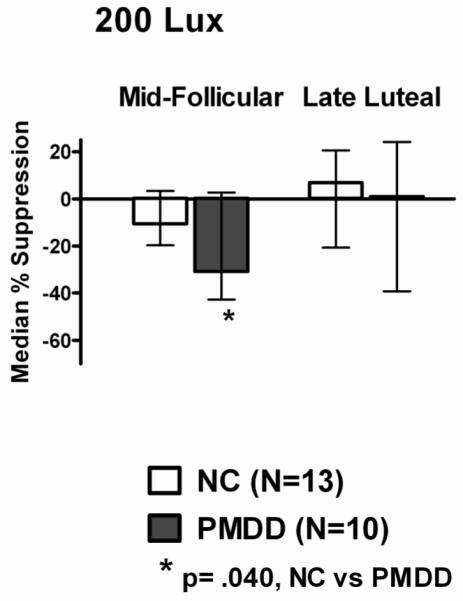
Median % suppression from baseline (± 25th and 75th percentiles) in plasma melatonin after two consecutive hours of exposure to 200 lux light in mid-follicular (MF) and late-luteal (LL) phases of the menstrual cycle, in normal controls (NC) and women with premenstrual dysphoric disorder (PMDD). The p-value denotes significance of difference between NC and PMDD during the follicular phase.
Acknowledgements
This work was supported by NIH grant R01 MH063462 and NIH Clinical Research Center (CRC) grant M01 RR00827. We thank Alan Turken, B.S., for his excellent work in performing the melatonin assays. Daniel F. Kripke, M.D. provided scientific consultation.
Footnotes
DECLARATION OF INTEREST
The authors report no conflicts of interest. The authors alone are responsible for the content and writing of the paper.
REFERENCES
- Anderson DC, Hopper BR, Lasley BL, Yen SS. A simple method for the assay of eight steroids in small volumes of plasma. Steroids. 1976;28:179–196. doi: 10.1016/0039-128x(76)90108-2. [DOI] [PubMed] [Google Scholar]
- APA . Diagnostic and Statistical Manual of Mental Disorders. 4 edition American Psychiatric Association; Washington, D. C.: 1994. [Google Scholar]
- Beck AT, Ward CH, Mendelson M, Mock J, Erbaugh J. An inventory for measuring depression. Arch. Gen. Psychiatry. 1961;4:561–571. doi: 10.1001/archpsyc.1961.01710120031004. [DOI] [PubMed] [Google Scholar]
- Benloucif S, Burgess HJ, Klerman EB, Lewy AJ, Middleton B, Murphy PJ, Parry BL, Revell VL. Measuring melatonin in humans. J. Clin. Sleep Med. 2008;4:66–69. [PMC free article] [PubMed] [Google Scholar]
- Brainard GC, Sliney D, Hanifin JP, Glickman G, Byrne B, Greeson JM, Jasser S, Gerner E, Rollag MD. Sensitivity of the human circadian system to short-wavelength (420-nm) light. J. Biol. Rhythms. 2008;23:379–386. doi: 10.1177/0748730408323089. [DOI] [PubMed] [Google Scholar]
- Brzezinski A, Lynch HJ, Seibel MM, Deng MH, Nader TM, Wurtman RJ. The circadian rhythm of plasma melatonin during the normal menstrual cycle and in amenorrheic women. J. Clin. Endocrinol. Metab. 1988;66:891–895. doi: 10.1210/jcem-66-5-891. [DOI] [PubMed] [Google Scholar]
- Cummings MA, Berga SL, Cummings KL, Kripke DF, Haviland MG, Golshan S, Gillin JC. Light suppression of melatonin in unipolar depressed patients. Psychiatry Res. 1989;27:351–355. doi: 10.1016/0165-1781(89)90149-2. [DOI] [PubMed] [Google Scholar]
- Evans JA, Elliott JA, Gorman MR. Dim nighttime illumination accelerates adjustment to timezone travel in an animal model. Curr. Biol. 2009;19:R156–157. doi: 10.1016/j.cub.2009.01.023. [DOI] [PubMed] [Google Scholar]
- Figueiro MG, Bierman A, Bullough JD, Rea MS. A personal light-treatment device for improving sleep quality in the elderly: dynamics of nocturnal melatonin suppression at two exposure levels. Chronobiol. Int. 2009;26:726–739. doi: 10.1080/07420520902927809. [DOI] [PMC free article] [PubMed] [Google Scholar]
- First MB, Gibbon M, Spitzer RL, Williams JBW. Structured Clinical Interview for DSM-IV Axis I Disorders- research version. Biometerics Research Dept., New York State Psychiatric Institute; New York: 1995. [Google Scholar]
- Gaddy JR, Stewart KT, Byrne B, Doghramji K, Rollag MD, Brainard GC. Light-induced plasma melatonin suppression in seasonal affective disorder. Prog. Neuropsychopharmacol. Biol. Psychiatry. 1990;14:563–568. doi: 10.1016/0278-5846(90)90007-4. [DOI] [PubMed] [Google Scholar]
- Glickman G, Hanifin JP, Rollag MD, Wang J, Cooper H, Brainard GC. Inferior retinal light exposure is more effective than superior retinal exposure in suppressing melatonin in humans. J. Biol. Rhythms. 2003;18:71–79. doi: 10.1177/0748730402239678. [DOI] [PubMed] [Google Scholar]
- Hallam KT, Olver JS, Horgan JE, McGrath C, Norman TR. Low doses of lithium carbonate reduce melatonin light sensitivity in healthy volunteers. Int. J. Neuropsychopharmacol. 2005;8:255–259. doi: 10.1017/S1461145704004894. [DOI] [PubMed] [Google Scholar]
- Hamilton M. Development of a rating scale for primary depressive illness. Br. J. Soc. Clin. Psychol. 1967;6:278–296. doi: 10.1111/j.2044-8260.1967.tb00530.x. [DOI] [PubMed] [Google Scholar]
- Hebert M, Martin SK, Lee C, Eastman CI. The effects of prior light history on the suppression of melatonin by light in humans. J. Pineal Res. 2002;33:198–203. doi: 10.1034/j.1600-079x.2002.01885.x. [DOI] [PMC free article] [PubMed] [Google Scholar]
- Higuchi S, Motohashi Y, Maeda T, Ishibashi K. Relationship between individual difference in melatonin suppression by light and habitual bedtime. J. Physiol. Anthropol. Appl. Human Sci. 2005;24:419–423. doi: 10.2114/jpa.24.419. [DOI] [PubMed] [Google Scholar]
- Higuchi S, Motohashi Y, Ishibashi K, Maeda T. Less exposure to daily ambient light in winter increases sensitivity of melatonin to light suppression. Chronobiol. Int. 2007;24:31–43. doi: 10.1080/07420520601139805. [DOI] [PubMed] [Google Scholar]
- Lam RW, Berkowitz AL, Berga SL, Clark CM, Kripke DF, Gillin JC. Melatonin suppression in bipolar and unipolar mood disorders. Psychiatry Res. 1990;33:129–134. doi: 10.1016/0165-1781(90)90066-e. [DOI] [PubMed] [Google Scholar]
- Lewy AJ, Wehr TA, Goodwin FK, Newsome DA, Rosenthal NE. Manic-depressive patients may be supersensitive to light. Lancet. 1981;1:383–384. doi: 10.1016/s0140-6736(81)91697-4. [DOI] [PubMed] [Google Scholar]
- Lewy AJ, Nurnberger JI, Jr., Wehr TA, Pack D, Becker LE, Powell RL, Newsome DA. Supersensitivity to light: possible trait marker for manic-depressive illness. Am. J.Psychiatry. 1985;142:725–7. doi: 10.1176/ajp.142.6.725. [DOI] [PubMed] [Google Scholar]
- Lockley SW. Spectral sensitivity and spectral adaptation of the human circadian timing system; 21st Annual Scientific Meeting of the Australasian Sleep Association; Melbourne, Victoria, Australia. 2009. [Google Scholar]
- McIntyre IM, Norman TR, Burrows GD, Armstrong SM. Human melatonin suppression by light is intensity dependent. J. Pineal Res. 1989a;6:149–156. doi: 10.1111/j.1600-079x.1989.tb00412.x. [DOI] [PubMed] [Google Scholar]
- McIntyre IM, Norman TR, Burrows GD, Armstrong SM. Quantal melatonin suppression by exposure to low intensity light in man. Life Sci. 1989b;45:327–332. doi: 10.1016/0024-3205(89)90142-2. [DOI] [PubMed] [Google Scholar]
- Monteleone P, Esposito G, La Rocca A, Maj M. Does bright light suppress nocturnal melatonin secretion more in women than men? J. Neural Transm. Gen. Sect. 1995;102:75–80. doi: 10.1007/BF01276567. [DOI] [PubMed] [Google Scholar]
- Murphy DG, Murphy DM, Abbas M, Palazidou E, Binnie C, Arendt J, Campos Costa D, Checkley SA. Seasonal affective disorder: response to light as measured by electroencephalogram, melatonin suppression, and cerebral blood flow. Br. J. Psychiatry. 1993;163:327–331. 335–337. doi: 10.1192/bjp.163.3.327. [DOI] [PubMed] [Google Scholar]
- Nathan PJ, Burrows GD, Norman TR. The effect of dim light on suppression of nocturnal melatonin in healthy women and men. J. Neural Transm. 1997;104:643–648. doi: 10.1007/BF01291882. [DOI] [PubMed] [Google Scholar]
- Nathan PJ, Burrows GD, Norman TR. Melatonin sensitivity to dim white light in affective disorders. Neuropsychopharmacology. 1999a;21:408–413. doi: 10.1016/S0893-133X(99)00018-4. [DOI] [PubMed] [Google Scholar]
- Nathan PJ, Norman TR, Burrows GD. Effect of the menstrual cycle stage on the melatonin suppression by dim white light. Psychoneuroendocrinology. 1999b;24:193–200. doi: 10.1016/s0306-4530(98)00075-4. [DOI] [PubMed] [Google Scholar]
- Nathan PJ, Wyndham EL, Burrows GD, Norman TR. The effect of gender on the melatonin suppression by light: a dose response relationship. J. Neural Transm. 2000;107:271–279. doi: 10.1007/s007020050022. [DOI] [PubMed] [Google Scholar]
- Nurnberger JI, Jr., Adkins S, Lahiri DK, Mayeda A, Hu K, Lewy A, Miller A, Bowman ES, Miller MJ, Rau L, Smiley C, Davis-Singh D. Melatonin suppression by light in euthymic bipolar and unipolar patients. Arch. Gen. Psychiatr. 2000;57:572–579. doi: 10.1001/archpsyc.57.6.572. [DOI] [PubMed] [Google Scholar]
- Nurnberger JI, Jr., Berrettini W, Tamarkin L, Hamovit J, Norton J, Gershon E. Supersensitivity to melatonin suppression by light in young people at high risk for affective disorder. A preliminary report. Neuropsychopharmacology. 1988;1:217–223. doi: 10.1016/0893-133x(88)90020-6. [DOI] [PubMed] [Google Scholar]
- Parry BL, Berga SL, Kripke DF, Klauber MR, Laughlin GA, Yen SS, Gillin JC. Altered waveform of plasma nocturnal melatonin secretion in premenstrual depression. Arch. Gen. Psychiatry. 1990;47:1139–1146. doi: 10.1001/archpsyc.1990.01810240059010. [DOI] [PubMed] [Google Scholar]
- Parry BL, Udell C, Elliott JA, Berga SL, Klauber MR, Mostofi N, LeVeau B, Gillin JC. Blunted phase-shift responses to morning bright light in premenstrual dysphoric disorder. J. Biol. Rhythms. 1997;12:443–456. doi: 10.1177/074873049701200506. [DOI] [PubMed] [Google Scholar]
- Parry BL, Meliska CJ, Sorenson DL, Lopez AM, Martinez LF, Nowakowski S, Hauger RL, Elliott JA. Increased melatonin and delayed offset in menopausal depression: role of years past menopause, follicle-stimulating hormone, sleep end time, and body mass index. J. Clin. Endocrinol. Metab. 2008;93:54–60. doi: 10.1210/jc.2006-2853. [DOI] [PMC free article] [PubMed] [Google Scholar]
- Partonen T, Vakkuri O, Lonnqvist J. Suppression of melatonin secretion by bright light in seasonal affective disorder. Biol. Psychiatry. 1997;42:509–513. doi: 10.1016/S0006-3223(96)00376-9. [DOI] [PubMed] [Google Scholar]
- Portaluppi F, Touitou Y, Smolensky MH. Ethical and methodological standards for laboratory and medical biological rhythm research. Chronobiol. Int. 2008;25:999–1016. doi: 10.1080/07420520802544530. [DOI] [PubMed] [Google Scholar]
- Revell VL, Skene DJ. Light-induced melatonin suppression in humans with polychromatic and monochromatic light. Chronobiol. Int. 2007;24:1125–1137. doi: 10.1080/07420520701800652. [DOI] [PubMed] [Google Scholar]
- Rosenthal NE, Heffernan MM. Bulimia, carbohydrate craving and depression: a central connection? In: Wurtman RJ, Wurtman JJ, editors. Nutrition and the Brain. Raven Press; New York: 1986. [Google Scholar]
- Roy-Byrne PP, Rubinow DR, Hoban MC, Parry BL, Rosenthal NE, Nurnberger JI, Byrnes S. Premenstrual changes: a comparison of five populations. Psychiatry Res. 1986;17:77–85. doi: 10.1016/0165-1781(86)90062-4. [DOI] [PubMed] [Google Scholar]
- Seggie J, Carney PA, Parker J, Grof E, Grof P. Effect of chronic lithium on sensitivity to light in male and female bipolar patients. Prog. Neuropsychopharmacol. Biol. Psychiatry. 1989;13:543–549. doi: 10.1016/0278-5846(89)90144-9. [DOI] [PubMed] [Google Scholar]
- Smith MR, Eastman CI. Phase delaying the human circadian clock with blue-enriched polychromatic light. Chronobiol. Int. 2009;26:709–725. doi: 10.1080/07420520902927742. [DOI] [PMC free article] [PubMed] [Google Scholar]
- Thalen BE, Kjellman BF, Morkrid L, Wetterberg L. Melatonin in light treatment of patients with seasonal and nonseasonal depression. Acta Psychiatr. Scand. 1995;92:274–284. doi: 10.1111/j.1600-0447.1995.tb09583.x. [DOI] [PubMed] [Google Scholar]
- Thompson C, Stinson D, Smith A. Seasonal affective disorder and season-dependent abnormalities of melatonin suppression by light. Lancet. 1990;336:703–706. doi: 10.1016/0140-6736(90)92202-s. [DOI] [PubMed] [Google Scholar]
- Thorne HC, Jones KH, Peters SP, Archer SN, Dijk DJ. Daily and seasonal variation in the spectral composition of light exposure in humans. Chronobiol. Int. 2009;26:854–866. doi: 10.1080/07420520903044315. [DOI] [PubMed] [Google Scholar]
- Visser EK, Beersma DG, Daan S. Melatonin suppression by light in humans is maximal when the nasal part of the retina is illuminated. J. Biol. Rhythms. 1999;14:116–121. doi: 10.1177/074873099129000498. [DOI] [PubMed] [Google Scholar]
- Whalley LJ, Perini T, Shering A, Bennie J. Melatonin response to bright light in recovered, drug-free, bipolar patients. Psychiatry Res. 1991;38:13–9. doi: 10.1016/0165-1781(91)90048-t. [DOI] [PubMed] [Google Scholar]
- Williams JB, Link MJ, Rosenthal NE, Amira L, Terman M. Structured Interview Guide for the Hamilton Depression Rating Scale, Seasonal Affective Disorders Version (SIGH-SAD) New York Psychiatric Institute; New York: 1994. revised edition. [Google Scholar]