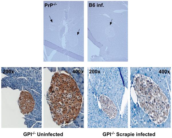
Fig. 3.
Deposition of PrP in the islets of GPI−/− transgenic mice. Pancreas isolated from PrP knockout mice (PrP−/−), B6 mice, or GPI−/− transgenic mice infected with 2% scrapie intracranially was sectioned and stained for PrP as described in Materials and Methods. PrP was detected only in the islets of the pancreas, not in the acinar cells, in the GPI−/− PrP tg mice. No PrP was detected in the pancreas of PrP−/− mice or in B6 mice infected with scrapie at 160 days post-infection. Arrows indicate the Islets of Langerhans in the tissues. Images of the islets from GPI−/− PrP tg mice were taken at 200x and 400x magnification. Control tissues are shown at 50x magnification.

