Abstract
This review focuses on how the study of anatomy in India has evolved through the centuries. Anatomical knowledge in ancient India was derived principally from animal sacrifice, chance observations of improperly buried human bodies, and examinations of patients made by doctors during treatment. The Vedic philosophies form the basis of the Ayurvedic tradition, which is considered to be one of the oldest known systems of medicine. Two sets of texts form the foundation of Ayurvedic medicine, the Susruta Samhita and the Charaka Samhita. The Susruta Samhita provided important surgical and anatomical information of the understanding of anatomy by Indians in the 6th century BCE. Here we review the anatomical knowledge known to this society.
Keywords: anatomy, ancient India, Charaka Samhita, Susruta Samhita
Anatomy in ancient India
Healing traditions and medical practices are inextricably tied to human history. The oldest known civilizations have healing traditions associated with them and have added to our current knowledge of the medical sciences, particularly anatomy. Areas such as Greece, Mesopotamia, Egypt and China have shaped the study of medicine and human anatomy. As one of the oldest civilizations, India is rich in such history and tradition, which includes significant contributions to our understanding of human morphology.
The foundation for modern Indian Ayurvedic medicine can be found in ancient texts, some of which predate the Christian era by 4000 years (Persaud, 1997). The developmental history of ancient India can be divided into three periods: the Vedic (c. 1500–500 BCE), Brahmanic (600 BCE–1000 CE) and finally the Mughal (1000 CE until the 18th century) (Persaud, 1997). The ancient Indian name of the ‘science’ of medicine is ‘Ayurveda,’ the Veda for (lengthening of) the span of life, which is considered an upanga (subsidiary) to the Atharvaveda. The science of medicine is also called ‘Vaidyasastra’ and the physician is called vaidya, ‘possessing knowledge’ (vidya) (Winternitz & Jha, 1986). The Vedic philosophies form the basis of the Ayurvedic tradition, which is considered to be one of the oldest known systems of medicine and was compiled during the Vedic period (Bhagvat Sing Jee, 1978). The four Vedas are considered the oldest Sanskrit literature and are the main religious texts that form the basis of the Hindu religion. The Vedas contain rituals, hymns and incantations. The Ayurveda scripture focuses on health and medicinal practices (Micozzi, 2006). According to tradition, the Ayurveda originally consisted of eight parts (astranga), in which major surgery (salya), minor surgery (salakya), treatment of diseases of the body (kayaacikitsa), demonology (teachings on the diseases caused by demons) (bhutavidya), healing of diseases of children (Kaumarabhrtya), toxicology (agadatantra), elixir (rasayana) and aphrodisiaca (vajikarana) were included (Winternitz & Jha, 1986). Two main sets of texts form the foundation of Ayurvedic medicine, the Susruta Samhita and the Charaka Samhita. The Susruta Samhita was written by the famous physician and surgeon Susruta in the 6th century BCE who taught at the University of Benares (alternatively Kasi or Varanasi) on the Ganges River. He is best known for his tome of surgical wisdom, practices and tools. In Susruta's work, it is evident that considerable thought was given to anatomical structure and function, as Susruta was a proponent of human dissection (Persaud, 1984); his texts include a systematic method for the dissection of the human cadaver. Charaka lived in the mid 2nd century and was associated with the north-western part of India and the ancient university of Taksasila. Charaka Samhita contains 120 chapters arranged in five books. The Sarira-sthaka discusses mainly anatomy, embryology and technique of dissection. The original date of the Charaka Samhita is not known but some estimate its composition to have occurred early in the 4th century BCE (Porter, 1998). The Charaka Samhita is often philosophical and ethical in its considerations and includes an Oath of Initiation that is akin to the Hippocratic Oath. The teaching of medicine in ancient India followed a hereditary model, with the knowledge being passed from ‘Guru’ to ‘Sisya’. These ancient Indian texts were written solely in Sanskrit and were inaccessible to anyone who was not a direct disciple of that Guru or that particular school (Nagaratnam, 1989). Anatomical knowledge in ancient India was derived principally from the sacrifice of animals, by chance observations of improperly buried bodies, and examinations of patients by physcians (Zysk, 1985).
Susruta
Susruta was believed to have been born in the Eastern part of India near Bihar. Known as the father of Indian surgery, Susruta was the first to practise rhinoplasty in India. When he lived has long been a controversial subject among many medical historians. Susruta's famous work, the Susruta Samhita, has not survived and its only existence is in the form of revisions and copies (Ruthkow, 1961). Late Vedic hymns ascribed to Susruta suggested that he must have flourished during the latter part of the Vedic age, which would place him around 1000 BCE.
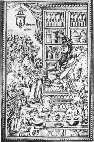
Susruta's Samhita emphasized surgical matters, including the use of specific instruments and types of operations. It is in his work that one finds significant anatomical considerations of the ancient Hindu. There is also compelling evidence suggesting that the knowledge of human anatomy was revealed by both inspection of the surface of the human body and through human dissection, as he believed that students aspiring to be surgeons should acquire a good knowledge of the structure of the human body (Hoernle, 1907; Keswani, 1970) (Fig. 1). Interestingly, in neither the writings of Susruta or of Charaka is there any indication that animal dissection was practised. Their anatomical knowledge, therefore, appears to have been gleaned from human dissection. Moreover, their writings show a considerable familiarity with the bones of the human body (Banerjee, 2006; Hoernle, 1907).
Fig. 1.

A picture of Susruta examining a patient. Note the palpation of the radial pulse.
The advancement of surgery during ancient Indian medical history is significant when considering the obstacles that deterred the study of anatomy. According to Hindu tenets, the human body is sacred in death. Hindu law (Shastras) states that no body may be violated by the knife and that persons older than 2 years of age must be cremated in their original condition (Ruthkow, 1961). Susruta was, however, able to bypass this decree and achieve his remarkable knowledge of human anatomy by using a brush-type broom, which scrapped off skin and flesh without the dissector having to actually touch the corpse.
Susruta's description of anatomical specimens included over 300 bones, as well as types of joints, ligaments and muscles from various parts of the body (Hoernle, 1907). Critics suggest that Susruta's overestimate of the number of bones contained in the human body may be due to the large number of child cadavers he observed (i.e. it is very possible that Susruta accounted for individual parts of bones that had not yet fused.) Despite his erroneous accounts of the skeleton, Susruta offered an in-depth understanding of bones, muscles, joints and vessels that far exceeded the knowledge of the time (Persaud, 1997).
The Susruta Samhita
Arguably the oldest surgical textbook is the Susruta Samhita. The literal translation of this Susruta is ‘that which is well heard’ or ‘one who has thoroughly learned by hearing’ (Chari, 2003). The first translation of this book from Sanskrit was the Arabic translation of the late 8th century. It was later translated into Latin, German and English (Mukhopadhaya, 1929). The most recent English translation was by Kaviraj Bhishagharatan, published in 1910; a later edition was released in 1963 (Bhishagratna, 1963). The Susruta Samhita is divided into two parts, the Purva-tantra and the Uttara-tantra. The Purva-tantra is subdivided into five books, the Sutrasthana, Nidana, Sarirasthana, Chikitasathanam and the Kalpastham, totalling 120 chapters, collectively (Mukhopadhaya, 1929). At the approximate time of the Susruta Samhita, the healing arts were divided into five parts, which included the Rogaharas (physicians), Shaylyaharas (surgeons), Vishaharas (poison healers), Krityaharas (demon doctors), and Bhisagatharvans (magic doctors) (Chari, 2003).
The Sutrasthana deals with basic medical science and pharmacology; Nidana, addresses disease processes; Chikitsasthanam is the bulk of the text, 34 chapters on surgical procedures and post-operative management; and the Kalpasthanam is composed of eight chapters on toxicology.
Anatomy in the Susruta Samhita
It was Susruta's belief that for one to be a skilful and erudite surgeon, one must first be an anatomist. The Sarirasthana is made up of 10 chapters regarding the study of human anatomy.
Susruta –Samhita said:
‘The different parts or members of the body as mentioned before including the skin, cannot be correctly described by one who is not well versed in anatomy. Hence, any one desirous of acquiring a thorough knowledge of anatomy should prepare a dead body and carefully, observe, by dissecting it, and examine its different parts.
[tasmat nihsamsayam jnanam harta salyasya vanchata/
Sodhayitva mrtam samyag drastavyah anga-vinisccayah//
Pratyaksatah hi yat drstam sastra-drstam ca yat bhavet/
Samasatah tat ubhayam bhuayh jnana-vivardhanam// (Chattopadhyaya, 1933)]’
Dissection preparation
As previously discussed, the issue of using humans for dissection was in opposition to the religious law of the time; however, it was an essential tool for the true understanding of human anatomy. The following is the method that Susruta developed that enabled him to work within the confines of these laws.
‘Therefore for dissecting purposes, a cadaver should be selected which has all of whose parts of the body present, of a person who had not died due to poisoning, but not suffered from a chronic disease (before death), had not attained a 100 years of age and from which the fecal contents of the intestines have been removed. Such a cadaver, whose all parts are wrapped by any one of “munja” (bush or grass), bark, “kusa” and flax, etc. and kept inside a cage, should be put in a slowly flowing river and allowed to decompose in an unlighted area. After proper decomposition for seven nights, the cadaver should be removed (from the cage) and then dissected slowly by rubbing it with the brushes made out of any of usira (fragrant roots of plant), hair, bamboo or “balvaja” (coarse grass). In this way, as previously described, skin, etc. and all the internal and external parts with their subdivisions should be visually examined’ (Singhal & Guru, 1973).
Interestingly, the Susruta Samhita mentions the role of a student in the dissection: ‘A pupil, otherwise well-read, but uninitiated, in the practice (of medicine or surgery) is not competent to take in hand the medical and surgical treatment of disease.’ According to the Susruta Samhita, medical students should be taught the art of making cuts in the body of a puspaphala (a kind of gourd), alavu (bottle-gourd) or ervaruka (cucumber) prior to dissection of human cadavers (Chattopadhyaya, 1933).
Head and neck
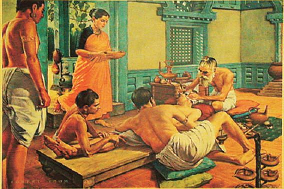
The Hindu Laws of Manu that governed much of life in ancient India dictated that the punitive measure for the crime of adultery was to have the offender's nose cut from the face (Persaud, 1997). It is in the Susruta Samhita that a procedure for repairing such damage is discussed, and this represents the equivalent of a modern ‘free flap’ used in reconstructive surgical techniques and thus implies a good knowledge of human facial anatomy (Fig. 2).
Fig. 2.

Susruta is depicted performing an otoplastic operation. The patient, drugged with wine, is steadied by friends and relatives as the great surgeon sets about fashioning an artificial ear lobe. He will use a section of flesh to be cut from the patient's cheek; it will be attached to the stump of the mutilated organ, treated with homeostatic powders, and bandaged.
‘First the leaf of a creeper, long and broad enough to fully cover the whole of the severed or clipped part, should be gathered; and a patch of living flesh, equal in dimension to the receding leaf, should be sliced off [from down upward] from the region of the cheek and, after scarifying it with a knife, swiftly adhered to the severed nose. Then the cool-headed physician should steadily tie it up with a bandage decent to look at and perfectly suited to the end for which it has been employed. The physician should make sure that the adhesion of the severed parts has been fully affected and then insert two small pipes into the nostrils to facilitate respiration, and to prevent the adhered flesh from hanging down. After that, the adhered part should be dusted with [hemostatic] powders; and the nose should be enveloped in Karpasa cotton and several times sprinkled over with the refined oil of pure seasmum’ (Mahabir, 2001).
In ancient India, piercing of the earlobes and subsequent enlarging of the hole was a widely practised method for warding off evil. However, this often resulted in the tearing of the earlobes. Susruta instructed that a pedicle flap reconstructive procedure be done where the graft of skin is taken from an adjacent area, carefully leaving its vascular supply intact. The graft is then rotated to the area of the defect and reattached.
‘A surgeon well versed in the knowledge of surgery should slice off a patch of living flesh from the cheek of a person so as to have on its ends attached to its former seat (cheek). Then the part, where the artificial ear lobe is to be made, should be slightly scarified (with a knife) and the living flesh, full of blood and sliced off as previously directed, should be adhered to it (so as to resemble a natural ear lobe in shape). The flap should then be covered with honey and butter and bandaged with cotton and linen and dusted with the power of baking clay.’ Again, such a procedure would necessitate a good working knowledge of human anatomy of the facial region including blood supply.
Susruta gave considerable time to ophthalmic study, as conditions such as cataracts were common in his region of the world. His description of the eye included five basic elements: earth (Bhu), fire/heat (Agni), air (Vayu), fluid (Jala) and void (Akasa). The extraocular muscles are the solid earth; heat/fire is the blood flowing through the vessels. Air forms the iris and pupil; fittingly, the vitreous part is attributed to the fluid element. Finally, the lacrimal ducts are derived from the void. Susruta delineated five anatomical divisions (Madalas) of the eye: eyelashes, eyelid, sclera, choroid and the pupil (Raju, 2003). The following is the procedure for the removal of cataracts:
‘…This procedure is auspiciously performed primarily in the warm season… [Preoperatively] the skin is rubbed with a pledget of cotton saturated with an oily medicine followed by a heated bath. The patient is given a light refreshment. The sick room is fumigated with vapors of white mustard, bdellium, nimva leaves, and the resinous gums of shala trees (in order to rid the area of insects and the diseases they harbor)… Incense of cannabis is used in addition to wine for sedation… [Technique] The patient sits on a high stool with the surgeon facing him. The hands are secured with proper fastenings. The patient is asked to look at his own nose while the surgeon rests his little finger on the (bony margin of the outer angle of the orbit), holding a Yava Vaktra Salaka between his thumb, index, and middle fingers. The left eye should be pierced with the right hand, and vice versa. The eye is entered at the junction of the medial and lateral two-thirds of the outer portion of the sclera. If a sound is produced following the gushing of a watery fluid, the needle is in the correct place, but if the puncture is followed by bleeding, the needle is misplaced. The eye is then sprinkled with breast milk. Care is taken to avoid blood vessels in the region. The tip of the needle is then used to incise the anterior capsule of the lens. With the needle in this position, the patient is asked to blow down the nostril, while closing the opposite naris. After this, lens material (Kapha) is seen coming alongside the needle. When the patient is able to perceive objects, the needle is removed… [Postoperatively] indigenous roots, leaves, and ghee are applied with a bandage. The patient then lies flat and is asked not to sneeze, cough or move. The eye is examined every fourth day for 10 days. If the whitish material recurs, the same procedure is repeated….’ (Raju, 2003).
Pelvis and perineum
The Hindu religion places a great deal of emphasis on reproduction and sexual energy. Male urogenital issues received a great deal of attention in the Susruta Samhita, though the health of women was also addressed. Susruta advocated the use of dilators, irrigating syringes and catheters. The following is a method of management of a urethral stricture via dilation and urethroplasty:
‘In the case of Niruddhaprakasha (stricture of the urethra), a tube open at both ends made of iron, wood, or shellac should be lubricated with clarified butter and gently introduced into the urethra. Thicker and thicker tubes should be made to dilate in this manner and emollient food should be given to the patient. As an alternative, an incision should be made into the lower part of the penis avoiding the sevani (raphe) and it should be treated as an incidental ulcer’ (Das, 1983).
Conclusions
Susruta's seminal work, the Susruta Samhita, forms the basis for the Ayurvedic tradition, which is still widely practised today. The contributions of ancient civilizations to our modern understanding are well appreciated, with ancient India being no exception. An appreciation of the evolution of anatomical knowledge can be gleaned from reviewing such ancient texts.
References
- Banerjee GN. Hellenism in Ancient India. New Delhi: Red Books; 2006. [Google Scholar]
- Bhagvat Sing Jee HH. A Short History of Aryan Medical Science. Delhi: New Asian Publishers; 1978. [Google Scholar]
- Bhishagratna KL. An English Translation of the Susruta Samhita. Varanasi: Chowkhamba Sanskrit Series Office; 1963. [Google Scholar]
- Chari PS. Susruta and our heritage. Indian J Plast Surg. 2003;36:4–13. [Google Scholar]
- Chattopadhyaya DP. Science and Society in Ancient India. Calcutta: Research India Publications; 1933. [Google Scholar]
- Das S. Susruta of India, the pioneer in the treatment of urethral structure. Surg Gynecol Obstet. 1983;157:581–582. [PubMed] [Google Scholar]
- Hoernle AF. Studies in the Medicine of Ancient India. Part I. Osteology of the Bones of the Human Body. Oxford: Claredon Press; 1907. [Google Scholar]
- Keswani NH. Medical education in India since ancient times. In: O’Malley CD, editor. The History of Medical Education. Berkeley: University of California Press; 1970. pp. 329–366. [Google Scholar]
- Mahabir RC. Ancient Indian civilization: ahead by a nose. J Invest Surg. 2001;14:3–5. [PubMed] [Google Scholar]
- Micozzi MS. Fundamentals of Complementary and Integrative Medicine. 3rd edn. St. Louis: Saunders Elsevier; 2006. [Google Scholar]
- Mukhopadhaya G. History of Indian Medicine. Calcutta: Calcutta University Press; 1929. [Google Scholar]
- Nagaratnam AMA. New Light on Ancient Indian Anatomy. Vol. 19. Hyderabad, India: B. Rao for the Central Council for Research; 1989. pp. 1–20. [PubMed] [Google Scholar]
- Persaud TVN. Early History of Human Anatomy. Springfield, IL: Charles C Thomas; 1984. [Google Scholar]
- Persaud TVN. A History of Anatomy. The Post-Vesalian Era. 1st edn. Springfield, IL: Charles C Thomas; 1997. [Google Scholar]
- Porter R. The Greatest Benefit to Mankind. A Medical History of Humanity. 1st edn. New York: W.W. Norton & Company; 1998. [Google Scholar]
- Raju VK. Susruta of ancient India. Indian J Ophthalmol. 2003;51:119–122. [PubMed] [Google Scholar]
- Ruthkow IM. Great Ideas in the History of Surgery. Baltimore: The Williams & Wilkins Company; 1961. [Google Scholar]
- Singhal GD, Guru LV. Anatomical & Obstetric Considerations in Ancient Indian Surgery. Banaras: Banaras Hindu University Press; 1973. [Google Scholar]
- Winternitz M, Jha S. History of Indian Literature. 2nd edn. Vol. 2. New Delhi: Motilal Banarsidass; 1986. [Google Scholar]
- Zysk KG. Religious Medicine. The History of India Medicine and Revolution. part 7. Vol. 75. Philadelphia: Transactions of the American Philosophical Society; 1985. [Google Scholar]


