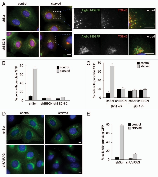
Figure 7.
Bif-1 together with the PI3KC3 complex is required for Atg9 redistribution during starvation. (A) HeLa cells stably expressing Atg9L1-EGFP were infected with lentivirus expressing Beclin 1 shRNA (shBEC N or shBEC N-2) or scrambled shRNA (shScr) and subjected to selection with 1 µg/ml puromycin for 5 days. The cells were then incubated in complete or starvation medium for 1.5 h and subjected to immunofluorescent staining with an anti-TGN46 sheep polyclonal antibody followed by a secondary antibody conjugated with Alexa Fluor 568. Magnified images are shown in the right part. (B) The percentage of cells with cytoplasmic Atg9 foci in (A) was quantified (mean ± SD; n = 4 × 211 for shScr and shBEC N1, n = 219 for shBEC N-2). (C) Bif-1+/+ and Bif-1−/− MEFs stably expressing shBEC N or control shScr were transfected with Atg9L1-EGFP. Twenty-four hours after transfection, the cells were cultured in complete or starvation medium and the percentage of cells with GFP foci was quantified (mean ± SD; n = 3 × 67). (D) HeLa cells stably expressing Atg9L1-EGFP were infected with lentivirus expressing UVRAG shRNA (shUVRAG) or control shScr. After selection with puromycin for 5 days, the cells were incubated in complete or starvation medium for 1.5 h and subjected to fluorescent microscopic analysis. (E) The percentage of cells with cytoplasmic Atg9 foci in (D) was calculated (mean ± SD; n = 3 × 256). The scale bars represent 10 µm.

