Mesenteric chylous cysts are rare. This study suggests that even large mesenteric chylous cysts may be managed with minimally invasive means.
Keywords: Chylous cyst, Mesenteric cyst, Laparoscopic enucleation
Abstract
Mesenteric chylous cysts are rare pathologic entities that often present with unspecific symptoms. The preoperative diagnosis requires all the common abdominal imaging techniques, but usually the correct diagnosis may be made only at the operation stage or during the histological examination. The treatment of choice is the complete surgical excision that may be safely performed by laparoscopy. A 58-year-old man underwent laparoscopic excision of a huge mesenteric chylous cyst. The technique entails the perfect control of the major abdominal vessels running near the tumor and the complete sealing of the chylous and blood vessels to and from the cyst.
INTRODUCTION
Mesenteric cysts are rare pathologic entities. First reported in 1507 by Beneviene or Benevenni,1 now they are identified in about 1 out of 100 000 adult hospital admissions.2–5 About half of them are chylous cysts, first described at necropsy by Rokitansky in 1842.6 Although often asymptomatic, they can sometimes reach such a volume as to cause pain, bowel obstruction, and other aspecific symptoms. In addition, they may mimic other abdominal diseases, such as tumors, benign masses, and congenital anomalies. The mainstay of therapy is the complete surgical removal of the cyst. This report deals with the case of a mesenteric chylous cyst treated by laparoscopy.
CASE REPORT
A 58-year-old man came to our observation for chronic abdominal pain in the right lower quadrant, where a rounded swelling could be noticed and palpated, resembling the shape and dimensions of the head of a large fetus. The patient did not complain of vomiting or constipation or change in bowel habits and did not report any other abdominal or systemic symptom. He reported just mild essential hypertension that had required treatment for a couple of years.
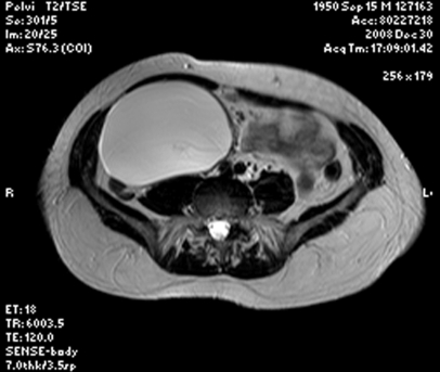
Abdominal ultrasonography, computed tomography, and a magnetic resonance scan revealed a circumscribed cyst measuring about 14cm × 12cm × 9cm, with a thick capsule, containing a dense fluid with a high-fat content (Figure 1). The cyst was located in the right lower quadrant, within the mesenterium of the small bowel, strictly adherent to the superior mesenteric vessels. Blood tests did not reveal any abnormality. Serologic tests for hydatidosis, amebiasis, and other parasitic diseases were negative.
Figure 1.
T2/TSE sequence magnetic resonance imaging showing a huge mesenteric cyst in the right lower quadrant of the abdomen.
With the informed consent of the patient, we decided to try to remove this mass by laparoscopy.
After antibiotic short-term intravenous prophylaxis, general anesthesia was induced and maintained as usual (with the “totally intravenous anesthesia” technique we use for most of our laparoscopic operations). Myoresolution was obtained by the administration of bolus doses of vecuronium bromide.
Three trocars were used, placed according to our technique of laparoscopic right colectomy, aiming to obtain perfect control of the superior mesenteric pedicle and the right lower quadrant. The first 10-mm to 12-mm trocar was placed with the “open” technique on the transverse umbilical line, just laterally to the lateral margin of the left rectus abdominis muscle, for the introduction of a 30° laparoscope. The second 10-mm trocar was placed in the left upper quadrant, on the same vertical line of the first cannula. The third 5-mm trocar had a suprapubic location. The initial exploration of the abdomen revealed a huge tumor arising within the mesenterium of the small bowel, leaving the small bowel at its left side and the right colon at its right side. The peritoneal layer of the right side of the ileal mesenterium covered the lump. The dissection started with an incision of the peritoneal covering in the cephalic aspect of the mass at its superior margin. The mass was then peeled away from the underlying fat tissue by ultrasonic dissection, while the small vessels to and from the cyst were progressively sealed and divided. Particular care was taken during the detachment of the cyst from the superior mesenteric vessels, which were particularly attached to the right side of the cyst itself. During the dissection, albeit delicate, the cyst was damaged and a dense white milk-like fluid spilled. The opening of the cyst was therefore enlarged to permit complete draining of the fluid. The cyst was completely dissected free and removed in an endoscopic bag. Abundant irrigation of the peritoneal cavity was performed, to remove all the residual cystic fluid. Fearing of a chylous fistula, a drain was inserted and the trocars removed. Total operative time was 90 minutes. The postoperative period was uneventful, and the patient resumed oral food intake after 24 hours (liquid intake after 8 hours). On postoperative day 3, the drain was removed (no chylous fistula), and the patient was discharged. The patient did not experience any postoperative early or late complications. His recovery was complete, and he returned to his normal social activities one week after the operation.
The results of the laboratory tests on the cyst and its content confirmed it to be a chylous cyst. Bacteriologic examination of the cystic fluid yielded negative findings.
DISCUSSION
Mesenteric cysts are rare abdominal pathologic entities, discovered in about 0.001% of all adult hospital admissions in the USA.2–5 About half of them are chylous cysts. According to dePerrot et al,4 other mesenteric cysts may be of mesothelial, enteric, dermoid, urogenital, traumatic, or infectious genesis. Malignant changes occur in less than 3% of cases.7 Chylous cysts are often congenital but may be related to previous abdominal surgery, pelvic diseases, and trauma.2–5,8,9 The first description of a chylous cyst dates back to 1842,6 and since then relatively few cases have been reported in writing.
The diagnosis may be challenging because chylous cysts may mimic other pathologies, such as pancreatic pseudocysts or cystic tumors,10 pelvic diseases,11 and aortic aneurysms.12
Symptoms are usually poor and quite unspecific.2,10,11,13 Rarely, the clinical presentation may be dramatic, with acute severe abdominal pain and symptoms of intestinal obstruction,14 or simulating the rupture of an aortic aneurysm.12 In the presented case, the only symptom was the progressive distension of the abdomen with a swelling in the right lower quadrant, but the patient did not complain of any other abdominal troubles.
The preoperative diagnosis may be achieved with the common imaging examinations of the abdomen (ultrasonography, computed tomography, nuclear magnetic resonance). Ultrasonography usually demonstrates a cystic tumor, whose content may form a fluid-fluid level.15 Computed tomography scans show a cystic mass with a thick wall and a fluid content with a low CT number.10,15 Abdominal imaging is particularly useful to demonstrate the relation of the tumor to the major abdominal vessels and with the bowel loops, to adequately plan the surgical approach.
The treatment of choice is the complete surgical excision of the cyst, which may need to be associated with bowel or pancreatic resection or splenectomy, depending on the location of the cyst. The first effective surgical treatment of a mesenteric cyst was performed in 1880 by Tillaux.16 In 1897, O'Conor17 described the marsupialization of a chylous mesenteric cyst by suturing its edges to the skin, although initially he tried to enucleate it but gave up fearing an eventual uncontrollable hemorrhage. Wiesen et al18 reported the complete enteroscopic excision of a mesenteric chylous cyst developing into the lumen of the proximal jejunum. Some authors performed laparotomic or laparoscopic fenestration of the cyst, if located near a major abdominal vessel.9,12 In our opinion, such a surgical option must be avoided due to the risk of malignancy.
In this case, we performed the complete excision of the cyst by laparoscopy. The position of the trocars must be judged with respect to the location of the tumor. Because the tumor was located between the right colon and the small bowel, we placed the cannulas according to our technique of right colectomy, aiming to achieve good control of the superior mesenteric vessels, the last small bowel loops, and the right colon with the underlying ureter, should it have been necessary to mobilize the cecum, the right colon, and the terminal ileus. A second concern of the technique is the relation between the cyst and the major abdominal vessels. In this case, the progressive dissection of the cyst was initiated at its cephalic edge, near the angle between the transverse mesocolon and the mesentery, where the superior mesenteric vessels have been accurately detached from the mass, paying attention to its branches to the small bowel and to the cecum and ascending colon. To avoid any risk of chylous fistula, every vessel must be accurately sealed and divided by the harmonic dissector. Obviously, it is not always possible to enucleate the cyst without opening it, in particular, if its content is under pressure. However, the chyle leakage from the cyst does not represent any particular concern, as long as the surgeon can accurately wash the surgical field and eventually insert the drain.
CONCLUSION
Even if it is a rare disease, surgeons must consider the diagnosis of a mesenteric chylous cyst each time they face a cystic abdominal tumor. The complete removal of the cyst must be regarded as the first-choice therapy, but this operation needs to be accurately planned on the basis of the preoperative diagnostic workup. Depending on the surgeon's experience, this operation may also be performed safely by laparoscopy, the only concern being the risk of iatrogenic injuries to the major abdominal vessels and bowel loops.
Acknowledgments
We wish to thank Mr. Matt Llewellin, Director of the EF Bournemouth School of English, for his helpful assistance in reviewing and checking the manuscript.
Contributor Information
Giovanni Domenico Tebala, Digestive and Laparoendoscopic Surgery Unit, Aurelia Hospital, Rome, Italy..
Ida Camperchioli, Digestive and Laparoendoscopic Surgery Unit, Aurelia Hospital, Rome, Italy.; Department of Surgery, University of Rome, Tor Vergata, Rome, Italy.
Valeria Tognoni, Digestive and Laparoendoscopic Surgery Unit, Aurelia Hospital, Rome, Italy.; Department of Surgery, University of Rome, Tor Vergata, Rome, Italy.
Michele Noia, Department of Surgery, Città di Roma Hospital, Rome, Italy..
Achille Lucio Gaspari, Department of Surgery, University of Rome, Tor Vergata, Rome, Italy..
References:
- 1. Martin WL, Grotzinger PJ, Zaydon TJ. Chylous cyst of the mesentery. Ann Surg. 1954;140:132–135 [DOI] [PMC free article] [PubMed] [Google Scholar]
- 2. O'Brien MF, Winter DC, Lee G, Fitzgerald EJ, O'Sullivan GC. Mesenteric cysts. A series of six cases with a review of the literature. Irish J Med Sci. 1999;168:233–236 [DOI] [PubMed] [Google Scholar]
- 3. Kurtz RJ, Heimann TM, Holt J, Beck AR. Mesenteric and retroperitoneal cysts. Ann Surg. 1986;203:109–112 [DOI] [PMC free article] [PubMed] [Google Scholar]
- 4. dePerrot M, Brundler MA, Totsch M, Mentha G, Morel P. Mesenteric cysts. Towards less confusion? Dig Surg. 2000;17:323–328 [DOI] [PubMed] [Google Scholar]
- 5. Kwan E, Lau H, Yuen WK. Laparoscopic resection of a mesenteric cyst. Gastrointest Endosc. 2004;59:154–156 [DOI] [PubMed] [Google Scholar]
- 6. Levison CG, Wolfssohn M. A mesenteric chylous cyst. Cal West Med. 1929;24:480–482 [PMC free article] [PubMed] [Google Scholar]
- 7. Liew SC, Glenn DC, Storey DW. Mesenteric cyst. Aust N Z J Surg. 1994;64:741–744 [DOI] [PubMed] [Google Scholar]
- 8. Wykypiel HF, Margreiter R. Chylous cyst formation following laparoscopic fundoplication. Wien Klin Wochenschr. 2007;119(23-24):729–732 [DOI] [PubMed] [Google Scholar]
- 9. Sarli L, Cortellini P, Pavlidis C, Simonazzi M, Sebastio N. Successful management of para-aortic lymphocyst with laparoscopic fenestration. Surg Endosc. 2000;14:373. [DOI] [PubMed] [Google Scholar]
- 10. Yasoshima T, Mukaiya M, Hirata K, et al. A chylous cyst of the mesentery: report of a case. Surg Today. 2000;30:185–187 [DOI] [PubMed] [Google Scholar]
- 11. Covarelli P, Arena S, Badolato M, et al. Mesenteric chylous cysts simulating a pelvic disease: a case report. Chir Ital. 2008;60:319–322 [PubMed] [Google Scholar]
- 12. Ho TP, Bhattacharya V, Wyatt MG. Chylous cyst of the small bowel mesentery presenting as a contained rupture of an abdominal aortic aneurysm. Eur J VascEndovasc Surg. 2002;23:82–83 [DOI] [PubMed] [Google Scholar]
- 13. Miljkovic D, Gmijovic D, Radojkovic M, Gligorijevic J, Radovanovic Z. Mesenteric cyst. Arch Oncol. 2007;15:91–93 [Google Scholar]
- 14. Kamat MM, Bahal NK, Prabhu SR, Pai MV. Multiple chylous cysts of abdomen causing intestinal obstruction. J Postgrad Med. 1992;38:206–207 [PubMed] [Google Scholar]
- 15. Sahin DA, Akbulut G, Saycol V, San O, Tokyol C, Dilek ON. Laparoscopic enucleation of mesenteric cyst: a case report. Mt Sinai J Med. 2006;73(7):1019–1020 [PubMed] [Google Scholar]
- 16. Tillaux PJ. Cyste du mesentere un homme: ablation par la gastromie: quersion. Revue de Therapeutiques Medico-Chirurgicale Paris. 1880:47:479 [Google Scholar]
- 17. O'Conor J. Chylous cyst of mesentery: operation: recovery. Br Med J. 1897;February 13:391. [DOI] [PMC free article] [PubMed] [Google Scholar]
- 18. Wiesen A, Sideridis K, Stark B, Bank S. Mesenteric chylous cyst. Gastroint Endosc. 2006;63:502. [DOI] [PubMed] [Google Scholar]


