Abstract
Electronic health record (EHR) systems serving pediatric populations typically incorporate growth charts to help healthcare providers monitor children’s growth. Currently, easily implementable growth charts are not available for subpopulations having growth that differs from the population as a whole, such as children with Down syndrome. This manuscript describes an approach for generating subpopulation-specific growth charts meeting requirements for implementation into EHR systems, using as an example weights for children with Down syndrome. Gender-specific growth curves were generated from 2358 weight values obtained from 331 patients with Down syndrome from July 2001 until March 2005. The project generated printable curves and computable data tables formatted according to growth chart standards set forth by the Centers for Disease Control and Prevention to facilitate implementation into EHR systems. This approach will help developers implementing growth charts and provides actual data tables for monitoring growth in children with Down syndrome.
Background and Rationale
Pediatric growth charts provide a valuable function of Electronic Health Record (EHR) systems supporting healthcare delivery to children.1–4 Growth charts help healthcare providers identify growth abnormalities that herald incident social, psychological and physical diseases, and their use is a standard of care around the world.5 The United States Centers for Disease Control and Prevention (CDC) has published publicly available pediatric growth charts for healthy, normally developing children.6–8 The CDC growth charts are available as data files that can be easily integrated into EHR systems, with normative reference curves covering the 3rd, 5th, 10th, 25th, 50th, 75th, 90th, 95th, and 97th percentiles of growth for both genders from ages 0–21 years.6–8 When implemented into an EHR system, growth charts should allow healthcare providers to record height, weight and head circumference measurements, and to compare these data to normative population-based curves. The American Academy of Pediatrics Task Force on Medical Informatics’ “Special Requirements for Electronic Medical Record Systems in Pediatrics” recommended that EHR systems be able to adjust normative growth curves based on diagnostic features that may affect growth, such as the presence of Down syndrome.1, 2 Currently, growth charts for pediatric subpopulations having growth that differs from the unaffected population are unavailable in a format that is easily implemented into EHR systems. The absence of readily available and standardized subpopulation-specific growth charts poses a challenge to EHR system developers and to healthcare providers caring for these children.
Down syndrome is the most common congenital disease affecting children’s growth, occurring in one out of every 732 live births in the United States.9 The features of Down syndrome, initially described by J. Langdon Down, include intellectual impairment, congenital heart disease, and characteristic dysmorphisms.10 These patients are at increased risk for a number of medical complications affecting nearly every organ system and grow differently from the unaffected population. Growth charts for children with Down syndrome have been developed in paper-based formats. The most commonly used Down syndrome growth charts in the US are based on work done by Cronk et al., published in 1988.11, 12 These represent a broad cohort of children with Down syndrome, including those with comorbid conditions such as congenital heart disease. The growth data underlying these charts was obtained before modern advances in the treatments for prematurity, congenital heart disease and nutritional deficiencies, and may not reflect the growth of children with Down syndrome in the US today. Additionally, the data underlying the Cronk charts are not available for direct implementation into EHR systems. Other studies by Myrelid in Sweden13 and Styles in Ireland14, both published in 2002, report more modern estimates of growth. However, these European studies are restricted to relatively small geographic settings, and neither provide data that can support conversion to EHR system growth charts.
The goal of the current work was to develop updated growth curves representing the weight growth of children with Down syndrome, that support easy implementation into EHR systems, and that account for recent advances in the care for diseases common to children with Down syndrome, including prematurity, congenital cardiac disease, and nutritional challenges in infancy.
Methods
Setting and Subjects
Vanderbilt University Medical Center (VUMC) includes the Monroe Carell Jr. Children’s Hospital at Vanderbilt (VCH) and the outpatient Doctors’ Office Tower (DOT), a primary and tertiary care facility with large local and regional primary referral bases for children. The VCH has 222 inpatient beds, including 114 intensive care beds, and an additional 25 emergency department beds. In fiscal year 2009, there were 235,849 pediatric visits at Children’s Hospital, and more than 171,000 children were seen in Children’s clinics. The VUMC includes a multidisciplinary clinic that addresses the multiple medical, social and nutritional needs of children and families with Down syndrome. This Vanderbilt Down Syndrome Clinic includes practitioners in developmental medicine, cardiology, and genetics as well as a nutritionist, physical, occupational, and speech therapists, and a social worker.
Source of Data
We obtained all data from the institutional EHR system and demographic databases. The recruitment interval for this study was from July 1, 2001 through March 1, 2005. We queried the EHR system to obtain anthropomorphic measurements and demographic information for all patients aged 0–21 during the recruitment interval. From this data, we derived tables containing the height, weight, head circumference, gender, birth date and ICD-9 encoded diagnosis codes for all patients. These data were then filtered to identify patients with Down syndrome as follows. We identified patients as having Down syndrome if they: 1) were included in the Down Syndrome Clinic’s patient census, and 2) had an ICD-9 code 758.0 (Down syndrome) diagnosis made during at least one clinical visit. The final Down syndrome dataset contained height, weight, head circumference, gender and birth date for all patients meeting the study criteria for having Down syndrome
Statistical Considerations
Before generating growth charts from the raw Down syndrome dataset, we first reviewed tables and scatter plots of the growth data to identify potential errors and outliers. The scatter plots were visually inspected, and all outliers were manually reviewed by two investigators (AQA, QC) with oversight provided by two others (STR, TLM). Data errors and outliers fell into three categories. First, values were recorded using the wrong units of measurement. For example, weights in pounds were encoded as being in kilograms. Second, values were substantially different from other values recorded from the same patient in a short time period. Third, values were not biologically plausible. For example, with weight values were considered implausible if they were over 150 kilograms at any age, and over 30 kilograms between the ages of 0–36 months. It is possible that some values considered implausible were valid; however, these were likely due to the co-presence of significant obesity that is more than would be expected when accounting for the presence of Down syndrome. In addition, data points that were duplicated were trimmed.
Following the standards of Centers for Disease Control and Prevention, we created the growth charts at the 5th, 10th, 25th, 50th, 75th, and 95th percentiles. Due to the limited size of our Down syndrome dataset, the 3rd and 97th percentiles of weight are not reliable, and are not presented here. To construct the final set of Down syndrome reference growth charts representing both genders and covering ages 0–19 years, we used nonparametric quantile regression with quadratic B-splines based on age. We selected this approach over the LMS method of Cole and Green15 because quantile regressions do not impose any parametric assumptions on the response distributions, and can incorporate covariates.16 For example, because LMS methods model the conditional mean of the weight based upon the power transformation family of Box and Cox, the resulting growth charts will depend heavily on the Gaussian assumption, especially for the extreme percentiles curves. On the other hand, quantile regressions make no assumption on the distribution of the weights. In creating the B-spline functions, we selected knots at the minimum and maximum boundaries, as well as at the 33.3% and 66.7% percentiles for both boys and girls. This resulted in knots at: 0, 11, 76, and 225 months for the growth chart of boys; and 0, 8, 51, and 227 months for the growth chart of girls. These knots are the age points chosen to construct B-spline functions.16 We then generated the basis B-spline functions using these knots.
Because children with Down syndrome may have any number of comorbid conditions requiring frequent clinical visits, it is possible that a selection bias was present in the data. This occurs because comorbid conditions may correlate with both children’s growth patterns and the frequency with which they receive clinical care. For example, a child with Down syndrome who also has congenital heart disease may grow more slowly and have more clinic visits relative to a child with Down syndrome who does not have congenital heart disease. As a result of this correlation, children with Down syndrome who have comorbidities likely contributed more data points to the study’s database than children who do not have comorbidities. To accommodate this selection bias, weighted quantile regression was used, with the weights defined as the reciprocals of the number of records from each unique patient identifier code.
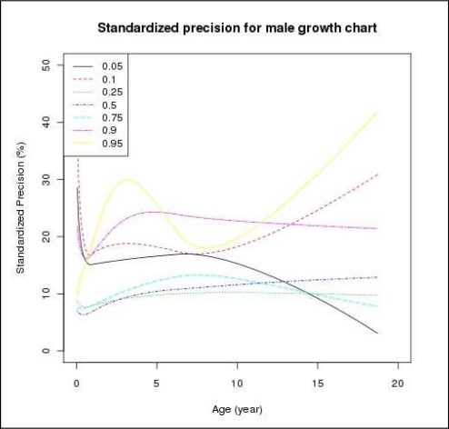
To illustrate the precision of our estimates, we created standardized precision plots. Standardized precision corrects the precision measurement for the increasing value for the estimate that would be expected in a growth chart. Standardized precision is calculated as the half-width of the confidence interval (i.e., the precision), divided by the estimate itself. For example, the median weight for a boy of 11.5 months was estimated to be 8.5 kilograms, with a 95% confidence interval of 7.9 – 9.1 kilograms. In this case, the precision of this estimate is 0.6 kilograms, and the standardized precision is 7.0%.
The current project focused on creating Down syndrome growth curves representing weight growth. All analyses, as well as graphs and tables, were generated using the quanreg and splines packages of R 2.10.1 (http://www.r-project.org/). The VUMC Institutional Review Board approved this project as compliant with institutional and national ethical standards for research involving human subjects.
Results
During the study period, a total of 532,808 weights from 151,254 patients aged 0–21 years were identified in the VUMC EHR system. Among these, 331 patients (181 males and 150 females) met criteria for having Down syndrome, and contributed 2570 weight observations. Of these 2570 observations, 186 were exact duplicates and were removed. With visual inspection, we identified fifteen outliers. Six were plausibly recorded in pounds instead of in kilograms; two were plausibly recorded correctly initially, but were then mis-converted to pounds. These values were all manually corrected and retained in the Down syndrome database. The other seven observations were drastically different from other measurements taken from the same patient at nearby time points and were eliminated. For example, subject ID 524 had one weight of 132.34 kg at age 33 months, while the 13 other weights for this subject fell within the range 12.5 at 26 months to 16.9 kg at 60 months. We deleted her 132.34 kg weight because we inferred from her other weights that this outlier was incorrect. Due to the sparseness of the observations after 19 years old, we limited our data observations from males and females to between the age of 0 and 19 years old. As a result, the final working dataset contained 1303 observations from 179 boys and 1055 observations from 147 girls, between the age of 0 and 19 years old. The median ages were 27 months for males and 25 months for females.
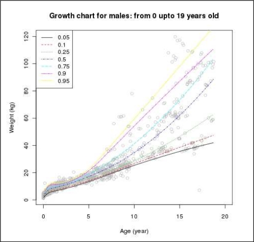
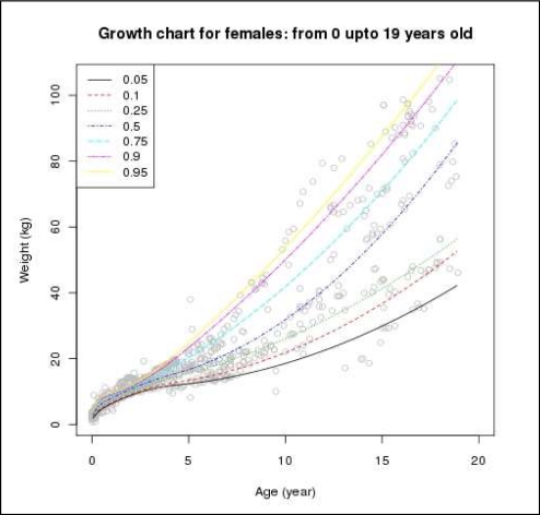
From this dataset, we developed growth charts for boys and girls from 0 to 19 years old (i.e., 0 to 228 months of age), as presented in Figures 1 and 2. Data tables containing weights representing each percentile for month age and formatted like the CDC growth chart data tables were also generated. An extract from the data table for boys is presented in the Table. Standardized precision plots were also generated for both genders. These plots indicate the relative accuracy of our percentile plots as a percentage of the patient’s predicted weight at the corresponding percentile. Figure 3 gives an example of such plots for growth curves for boys with Down syndrome. This graph indicates that the relative accuracy of these curves is fairly constant for the 25th through the 90th percentiles, with better accuracy achieved for the 25th through the 75th percentiles. Relative accuracy decreases with age at the 95th and 10th percentile and increases with age at the 5th percentile. These changes are due to the increase in both the dispersion and skewness of weight with age that is illustrated in Figure 1.
Figure 1.
Weight percentile growth curve for boys with Down syndrome, ages 0–19 years
Figure 2.
Weight percentile growth curve for girls with Down syndrome, ages 0–19 years
Table.
Sample extract from the Down syndrome growth data table for boys; monthage is age in months. τ (tau) is the percentile value in kilograms
| monthage | τ = 0.05 | τ = 0.10 | τ = 0.25 | τ = 0.50 |
|---|---|---|---|---|
| 0.5 | 1.79711 | 2.40166 | 2.92434 | 3.42588 |
| 1.5 | 2.45099 | 3.08119 | 3.63104 | 4.22726 |
| 2.5 | 3.05106 | 3.70483 | 4.28176 | 4.96145 |
| 3.5 | 3.59733 | 4.27260 | 4.87649 | 5.62846 |
| 4.5 | 4.08979 | 4.78448 | 5.41524 | 6.22829 |
| 5.5 | 4.52844 | 5.24049 | 5.89801 | 6.76093 |
Figure 3.
Example of standardized precision plots for boys with Down syndrome, showing the relative accuracy of the percentile grown curves as a percentage of the child’s weight.
Discussion
Major goals of implementing EHR systems into clinical practice include improving and standardizing healthcare delivery. These goals are consistent with integrating standard population-based pediatric growth charts into EHR systems. However, there currently do not exist appropriate standardized EHR-compatible growth curves representing the subpopulations for whom they would be most valuable, such as for children with Down syndrome. Without a consistent approach or a standardized set of normative curves, each EHR system would have to implement specialized growth curves according to its own methods. If various EHR systems use different approaches for monitoring growth in subpopulations, the curves will invariably differ across systems. Such a lack of standardization will hamper screening for growth abnormalities and cloud communication between healthcare providers and families.
In addition to healthcare providers and families, researchers may be affected by inconsistencies among measurement interpretations. In November, 2007, the CDC and the National Down Syndrome Society sponsored a conference, “Setting a Public Health Research Agenda for Down Syndrome.”17 Priority areas included identification of risk and preventive factors for physical health and improved understanding of comorbid conditions.17 Because multiple researchers will be involved in fulfilling this research agenda, it is essential to adopt standardized methods for evaluating and reporting growth among children with Down syndrome.
This paper reports the results of a project to generate a set of growth curves representing children with Down syndrome seen at a multidisciplinary clinic serving middle Tennessee. The Down syndrome growth curves include the 5th, 10th, 25th, 50th, 75th, 90th, 95th, percentiles for both genders, from ages 0–19 years. Curves representing the 3rd and 97th percentiles of growth and covering ages from 19–21 years are not provided due to sparseness of data in the dataset. This work generated curves in the form of a graphical chart that can be printed, and as a computable data table. To facilitate implementation into EHR systems, the project formatted data table to have the same structure as the CDC’s growth curves data tables for normally developing children. The approach used in this project (i.e., modeling growth curves using a quantile regression model) allows us to include factors that covary with growth, such as the presence of congenital heart disease and of premature birth. This approach will permit future work evaluating the impact of these factors on growth of children with Down syndrome.
Limitations
This study should be interpreted in light of its limitations. First, the data obtained to generate the Down syndrome-specific growth charts were obtained from a single tertiary institution. These specific charts may not accurately represent the growth of children in the general Down syndrome population who do not require referral for specialized care or those who live in other geographic regions. Second, the growth charts generated in this work included all children identified as having Down syndrome, regardless of comorbid conditions that may also have affected growth.18–20 For example, about 40%–60% of children with Down syndrome are born with congenital heart defects.18 Children with Down syndrome are two to three times more likely than other newborns to be born prematurely or with a low birthweight.19 They also exhibit increased rates of leukemia, respiratory problems, celiac disease, and hypothyroidism.20 These limitations can be overcome through future work pooling data from children with Down syndrome receiving care at sites from different geographic locations and stratifying by the presence of comorbidities. Third, the current work focused on weight only. Children with Down syndrome also grow differently than the general population in terms of height, body mass index and head circumference. A complete set of growth charts representing a special population would need to include normative data for all these anthropomorphic measures.
Acknowledgments
This work was supported in part by a grant from the Vanderbilt Institute for Clinical and Translational Research (Rosenbloom, VR512).
References
- 1.American Academy of Pediatrics: Task Force on Medical Informatics Special requirements for electronic medical record systems in pediatrics. Pediatrics. 2001;108:513–5. doi: 10.1542/peds.108.2.513. [DOI] [PubMed] [Google Scholar]
- 2.Spooner SA. Special requirements of electronic health record systems in pediatrics. Pediatrics. 2007;119:631–7. doi: 10.1542/peds.2006-3527. [DOI] [PubMed] [Google Scholar]
- 3.Rosenbloom ST, Qi X, Riddle WR, et al. Implementing pediatric growth charts into an electronic health record system. J Am Med Inform Assoc. 2006;13:302–8. doi: 10.1197/jamia.M1944. [DOI] [PMC free article] [PubMed] [Google Scholar]
- 4.Shiffman RN, Spooner SA, Kwiatkowski K, Brennan PF. Information technology for children’s health and health care. J Am Med Inform Assoc. 2001;8:546–51. doi: 10.1136/jamia.2001.0080546. [DOI] [PMC free article] [PubMed] [Google Scholar]
- 5.de Onis M, Wijnhoven TM, Onyango AW. Worldwide practices in child growth monitoring. J Pediatr. 2004;144:461–5. doi: 10.1016/j.jpeds.2003.12.034. [DOI] [PubMed] [Google Scholar]
- 6.Clinical Growth Charts US Center for Disease Control and Prevention, National Center for Health Statistics. (Accessed March 11, 2010, 2010, at http://www.cdc.gov/growthcharts/.)
- 7.Kuczmarski RJ, Ogden CL, Grummer-Strawn LM, et al. CDC growth charts: United States. Adv Data. 2000:1–27. [PubMed] [Google Scholar]
- 8.Kuczmarski RJ, Ogden CL, Guo SS, et al. Vital Health Stat. 2002. 2000 CDC Growth Charts for the United States; pp. 1–190. [PubMed] [Google Scholar]
- 9.Canfield MA, Honein MA, Yuskiv N, et al. National estimates and race/ethnic-specific variation of selected birth defects in the United States, 1999–2001. Birth Defects Res A Clin Mol Teratol. 2006;76:747–56. doi: 10.1002/bdra.20294. [DOI] [PubMed] [Google Scholar]
- 10.Down JL. Observations on an ethnic classification of idiots. 1866. Ment Retard. 1866;33:54–6. [PubMed] [Google Scholar]
- 11.Cronk C, Crocker AC, Pueschel SM, et al. Growth charts for children with Down syndrome: 1 month to 18 years of age. Pediatrics. 1988;81:102–10. [PubMed] [Google Scholar]
- 12.American Academy of Pediatrics: Health supervision for children with Down syndrome. Pediatrics. 2001;107:442–9. doi: 10.1542/peds.107.2.442. [DOI] [PubMed] [Google Scholar]
- 13.Myrelid A, Gustafsson J, Ollars B, Anneren G. Growth charts for Down’s syndrome from birth to 18 years of age. Arch Dis Child. 2002;87:97–103. doi: 10.1136/adc.87.2.97. [DOI] [PMC free article] [PubMed] [Google Scholar]
- 14.Styles ME, Cole TJ, Dennis J, Preece MA. New cross sectional stature, weight, and head circumference references for Down’s syndrome in the UK and Republic of Ireland. Arch Dis Child. 2002;87:104–8. doi: 10.1136/adc.87.2.104. [DOI] [PMC free article] [PubMed] [Google Scholar]
- 15.Cole TJ, Green PJ. Smoothing reference centile curves. Stat Med. 1992;11:1305–19. doi: 10.1002/sim.4780111005. [DOI] [PubMed] [Google Scholar]
- 16.Wei Y, Pere A, Koenker R, He X. Quantile regression methods for reference growth charts. Stat Med. 2006;25:1369–82. doi: 10.1002/sim.2271. [DOI] [PubMed] [Google Scholar]
- 17.Rasmussen SA, Whitehead N, Collier SA, Frias JL. Setting a public health research agenda for Down syndrome. Am J Med Genet A. 2008;146A:2998–3010. doi: 10.1002/ajmg.a.32581. [DOI] [PubMed] [Google Scholar]
- 18.Freeman SB, Taft LF, Dooley KJ, et al. Population-based study of congenital heart defects in Down syndrome. Am J Med Genet. 1998;80:213–7. [PubMed] [Google Scholar]
- 19.Frid C, Drott P, Otterblad Olausson P, Sundelin C, Anneren G. Maternal and neonatal factors and mortality in children with Down syndrome born in 1973–1980 and 1995–1998. Acta Paediatr. 2004;93:106–12. doi: 10.1080/08035250310007303. [DOI] [PubMed] [Google Scholar]
- 20.Van Riper M, Cohen WI. Caring for children with Down syndrome and their families. J Pediatr Health Care. 2001;15:123–31. doi: 10.1067/mph.2001.110627. [DOI] [PubMed] [Google Scholar]


