Abstract
Nervous lipofibromatous hamartoma is a rare tumor-like condition involving the peripheral nerves, whereby the epineurium and perineurium are enlarged and distorted by excess of fatty and fibrous tissues that infiltrate between and around nerve boundaries. The median nerve is much more likely to develop a hamartoma than other nerves with a predilection for the carpal tunnel. We present a case of carpal tunnel syndrome in an adult caused by fibrolipomatous hamartoma of the median nerve, successfully removed by excision of the fibrolipomatous tissue and decompression.
Keywords: Nervous lipofibromatous, Hamartoma, Median nerve, Carpal tunnel
Introduction
Neural fibrolipoma, also called fibrolipomatous hamartoma (FLH), is a rare, slow-growing, and benign tumor of a peripheral nerve, most often occurring in the median nerve of younger patients [8]. The tumor is often noticeable as a mass during manual palpation. Although frequently reported in patients under the age of 40 years, there have been a few reports of FLH in older patients [17]. In lipofibromatous hamartoma (LH), the epineurium and perineurium are enlarged and distorted by excess mature fat and fibrous tissue that infiltrate between and around nerve boundaries. LH of a nerve has also been reported as lipomatosis, fibrolipomatous nerve enlargement, lipofibroma, fibrofatty overgrowth, fatty infiltration of the nerve, and neurolipoma [6, 22]. According to the new World Health Organization Classification of tumors, it is designated as nervous lipomatosis [9]. The median nerve is much more likely to be involved by hamartoma than other nerves, with a predilection for the carpal tunnel. Involvement of the ulnar [1, 10], radial [11], plantar [22], and sciatic nerves [24] has also been described. It occurs more frequently in infants, and less commonly in children and young adults [7]. Many authors consider it as a congenital tumor [12, 22].
Case Report
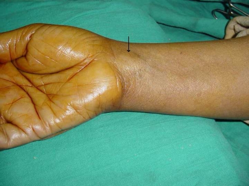
A 37-year-old right-handed lady was admitted at our department for right hand paresthesias in the area of distribution of the median nerve. On physical examination, 2-cm soft-tissue masses were noted on the distal volar aspect of her forearm, with no atrophy of the thenar eminence (Fig. 1). Tinel’s sign and Phalen’s test were positive all over the mass. Neurological deficits consist of paresthesia of the middle and ring finger at the time of compression of the mass, and the two-point discrimination test was about 4 mm in the median territory (at the pulp). No lymph node tumefaction was detected. General somatic exploration did not show any signs of neurofibromatosis. Electromyographic studies suggested a reduction in sensory conduction at the wrist, with a 50% sensory axonal loss and a slight increase in distal motor latency.
Fig. 1.
Soft-tissue masses in the distal volar aspect of her forearm
X-rays of the forearm and the hand did not reveal any ectopic calcification.
Ultrasound examination at that time demonstrated a carpal tunnel mass and magnetic resonance imaging (MRI) showed a signal of fat intensity without postgadolinium enhancement. The mass involved the median nerve on its length in the distal third of the right forearm, dissociating the nerve fibers, which were seen as low-intensity signal on T1- and T2-weighted images (Figs. 2 and 3).
Fig. 2.
Magnetic resonance imaging. One arrow, fibrolipoma; two arrows, medial nerve
Fig. 3.
Magnetic resonance imaging. One arrow, fibrolipoma; two arrows, medial nerve
These features were consistent with fibrolipomatous hamartoma of the right median nerve.
To protect the nerve, we started with the neurolysis and decompression. The fibrolipomatous hamartoma was distinguishable from the median nerve itself; a reasonable dissection allowed total excision of the tumor (Fig. 4).
Fig. 4.
Preoperative aspect
At 3-month follow-up, the patient was cured and became asymptomatic. The median nerve got its normal functional status.
Discussion
Intraneural lipoma, also known as neural fibrolipoma, lipofibromatous hamartoma, or perineural lipoma, is a benign mass composed of hypertrophied fibrofatty tissue intermixed with nerve tissue. The condition is best described by the term intraneural lipoma, first used by Morley in 1964 [19].
The median nerve and its branches are the main sites of involvement, followed by the radial nerve, ulnar nerve, nerves at the dorsal aspect of the foot, brachial plexus, and cranial nerves [16, 22]. Adults younger than 30 years of age are selectively affected [23].
There has been inconsistency in the nomenclature of this lesion. The accepted terminology, which most accurately reflects the hamartomatous nature of the tumor, is fibrolipoma of the median nerve [13, 22]. The etiology is poorly understood [2, 4], and the majority of cases occur in children or young adults [4, 13].
Clinical features consist of sensory and motor symptoms with or without macrodactyly. The neural symptoms are often slowly progressive with pain, paresthesia, and weakness. Carpal and cubital tunnel syndromes, caused by fibrolipomatous infiltration of median and ulnar nerves, respectively, have been described previously [18, 21]. The condition may remain static or demonstrate slow and progressive loss of nerve function [14].
Macrodactyly is approximately seen in to two thirds of the cases [2, 22].
Pathologically, the tumor is characterized by fibrofatty enlargement of the median nerve, usually confined by the nerve sheath [22]. There is massive epineural and perineural fibrosis surrounding and compressing individual nerve bundles [22]. Individual nerve fibers themselves are usually normal [13, 22]. The nerves and surrounding fibrosis are interspersed in hamartomatous fatty tissue, usually confined to the nerve sheath [2, 4].
MRI imaging is an important tool for preoperative evaluation, especially for differentiating lipoma and neurofibroma, from malignant tumors [3, 20].
MRI characteristics reflect the histology of the tumor.
The individual nerve fascicles and surrounding fibrosis result in long cylindrical bands of low T1- and T2-weighted signal.
The differential diagnosis of this tumor includes intraneural lipoma, ganglionar cyst, traumatic neuroma, and vascular malformations (in which flow void can mimic the cylindrical low-signal voids of lipofibromatous hamartoma) [5].
Knowledge of the extent of nerve involvement by fibrolipoma is important for presurgical planning.
Most authors, however, report progression of symptoms over time with or without carpal tunnel release. In both of these cases, there was progression of symptoms, despite the history of previous carpal tunnel release. These progressive symptoms are most likely related to direct compression of individual nerve bundles by massive perineural fibrosis rather than by compression of the enlarged median nerve in the confines of the carpal tunnel.
The current treatment practice is to provide initial decompression by carpal tunnel release. Dissection and microsurgical excision is usually reserved for those cases with progressive and disabling median nerve compromise despite previous carpal tunnel release. However, this technique can result in permanent loss of motor and sensory function and has only had modest success rates reported [2, 15, 22].
Ultimately, the decision to use additional preoperative tests, or to proceed to the operating room for direct inspection and biopsy or excision, hinges on the individual surgeon’s judgment.
Acknowledgments
Conflict of interest The authors declare that they have no conflict of interest.
References
- 1.Amadio PC, Reiman HM, Dobyns JH. Lipofibromatous hamartoma of nerve. J Hand Surg [Am] 1988;13:67–75. doi: 10.1016/0363-5023(88)90203-1. [DOI] [PubMed] [Google Scholar]
- 2.Amadio PC, Reiman HM, Dobyns JH. Lipofibromatous hamartoma of nerve. J Hand Surg. 1988;13A:67–75. doi: 10.1016/0363-5023(88)90203-1. [DOI] [PubMed] [Google Scholar]
- 3.Boren WL, Henry RE, Jr, Wintch K. MR diagnosis of fibrolipomatous hamartoma of nerve: association with nerve territoryoriented macrodactyly (macrodystrophia lipomatosa) Skeletal Radiol. 1995;24:296–297. doi: 10.1007/BF00198419. [DOI] [PubMed] [Google Scholar]
- 4.Cavallaro MC, Taylor JA, Gorman JD, et al. Imaging findings in a patient with fibrohpomatous hamartoma of the median nerve. Am J Roentgenol. 1993;161:837–838. doi: 10.2214/ajr.161.4.8372770. [DOI] [PubMed] [Google Scholar]
- 5.Cavallaro MC, Taylor JA, Gorman JD, et al. Imaging findings in a patient with fibrolipomatous hamartoma of the median nerve. AJR Am J Roentgenol. 1993;161:837–838. doi: 10.2214/ajr.161.4.8372770. [DOI] [PubMed] [Google Scholar]
- 6.Maeseneer M, Jaovisidha S, Lenchik L, et al. Fibrolipomatous hamartoma: MR imaging findings. Skeletal Radiol. 1997;26:155–160. doi: 10.1007/s002560050212. [DOI] [PubMed] [Google Scholar]
- 7.Enzinger FW, Weise SW. Soft tissue tumours. St Louis: Mosby; 1988. pp. 332–334. [Google Scholar]
- 8.Evans HA, Donnelly LF, Johnson ND, et al. Fibrolipoma of the median nerve: MRI. Clin Radiol. 1997;52:304–307. doi: 10.1016/S0009-9260(97)80060-8. [DOI] [PubMed] [Google Scholar]
- 9.Fletcher CDM, Unni K, Mertens K, editors. WHO classification of tumours. Pathology and genetics of tumours of soft tissue and bone. Lyon: IARC; 2002. pp. 24–25. [Google Scholar]
- 10.Frykman GK, Wood VE. Peripheral nerve hamartoma with macrodactyly in the hand: report of three cases and review of the literature. J Hand Surg [Am] 1978;3:307–312. doi: 10.1016/s0363-5023(78)80029-x. [DOI] [PubMed] [Google Scholar]
- 11.Herrick RT, Godsil RD, Jr, Widener JH. Lipofibromatous hamartoma of the radial nerve: a case report. J Hand Surg [Am] 1980;5:211–213. doi: 10.1016/s0363-5023(80)80003-7. [DOI] [PubMed] [Google Scholar]
- 12.Langa V, Posner MA, Steiner GE. Lipofibroma of the median nerve: a report of two cases. J Hand Surg [Br] 1987;12:221–223. doi: 10.1016/0266-7681_87_90017-9. [DOI] [PubMed] [Google Scholar]
- 13.Langa V, Possner MA, Striner GE. Lipofibroma of the median nerve: a report of two cases. J Hand Surg. 1987;12B:221–223. doi: 10.1016/0266-7681_87_90017-9. [DOI] [PubMed] [Google Scholar]
- 14.Louis DS, Hankin FM, Greene TL, et al. Lipofibromas of the median nerve: long-term follow up of four cases. Am J Hand Surg. 1985;10a(3):403–407. doi: 10.1016/s0363-5023(85)80044-7. [DOI] [PubMed] [Google Scholar]
- 15.Louis DS, Hankin FM, Greene TL, et al. Lipofibromas of the median nerve: long-term follow-up of four cases. J Hand Surg. 1985;10A:403–408. doi: 10.1016/s0363-5023(85)80044-7. [DOI] [PubMed] [Google Scholar]
- 16.Ly JQ, Bui-Mansfield LT, SanDiego JW, Beaman NA, Ficke JR. Neural fibrolipoma of the foot. J Comput Assist Tomogr. 2003;27:639–640. doi: 10.1097/00004728-200307000-00035. [DOI] [PubMed] [Google Scholar]
- 17.Marom EM, Helms CA. Fibrolipomatous hamartoma: pathognomonic on MR imaging. Skeletal Radiol. 1999;28:260–264. doi: 10.1007/s002560050512. [DOI] [PubMed] [Google Scholar]
- 18.Meyer BU, Roricht S. Fibrolipomatous hamartoma of the proximal ulnar nerve associated with macrodactyly and macrodystrophia lipomatosa as an unusual cause of cubital tunnel syndrome. J Neurol Neurosurg Psychiat. 1997;63(6):808–810. doi: 10.1136/jnnp.63.6.808a. [DOI] [PMC free article] [PubMed] [Google Scholar]
- 19.Morley GH. Intraneural lipoma of the median nerve in the carpal tunnel. Report of a case. J Bone Joint Surg Br. 1964;46:734–735. [PubMed] [Google Scholar]
- 20.Nogueira A, Pena C, Martinez MJ, et al. Hyperostotic macrodactyly and lipofibromatous hamartoma of the median nerve associated with carpal tunnel syndrome [review] Chir Main. 1999;18:261–271. doi: 10.1016/s0753-9053(99)80039-8. [DOI] [PubMed] [Google Scholar]
- 21.Ranawat CS, Arora MM, Singh RG. Macrodystrophia lipomatosa with carpal-tunnel syndrome. A case report. Am J Bone Joint Surg Sep. 1968;50(6):1242–1244. [PubMed] [Google Scholar]
- 22.Silverman TA, Enzinger FM. Fibrolipomatous hamartoma of nerve. A clinicopathologic analysis of 26 cases. Am J Surg Pathol. 1985;9:7–14. doi: 10.1097/00000478-198501000-00004. [DOI] [PubMed] [Google Scholar]
- 23.Breuseghem I, Sciot R, Pans S, Geusens E, Brys P, Wever I. Fibrolipomatous hamartoma in the foot: atypical MR imaging findings. Skeletal Radiol. 2003;32:651–655. doi: 10.1007/s00256-003-0684-3. [DOI] [PubMed] [Google Scholar]
- 24.Wong BZ, Amrami KK, Wenger DE, et al. Lipomatosis of the sciaticnerve: typical and atypical MRI features. Skeletal Radiol. 2006;35:180–184. doi: 10.1007/s00256-005-0034-8. [DOI] [PubMed] [Google Scholar]


