Abstract
A 45-year-old female presented with complaint of a lump in the right breast for the last 6 months. Excisional biopsy had been performed outside our institution, which was suggestive of a pure mammary chondrosarcoma. Modified radical mastectomy of the right breast was performed and histopathology was suggestive of a pure mammary chondrosarcoma of the right breast. One axillary lymph node was positive for metastasis, the rest of the lymph nodes were free of metastasis. Immunohistochemistry was suggestive of a metaplastic carcinoma with a predominant chondrosarcoma in the right breast. Postoperative radiotherapy was given to the patient with a further plan of chemotherapy.
Key Words: Breast carcinoma, Metaplastic carcinoma, Predominant chondrosarcoma
Introduction
Metaplastic carcinoma with predominant chondrosarcoma of the breast is exceedingly rare. Most benign and malignant tumors of the breast arise from the glandular epithelium. However, in some cases, the glandular epithelium differentiates into nonglandular mesenchymal tissue, a process called metaplasia [1]. Metaplastic changes, including squamous cell, spindle cell, and heterologous mesenchymal growth, occur in fewer than 5% of breast carcinomas [5]. Sarcomas of the breast are relatively rare, accounting for less than 1% of all primary malignant tumors of the female breast [2]. Carcinosarcomas, a subgroup of metaplastic carcinomas, are the rarest primary malignancies of the breast (found in <0.1% of cases) [3]. Metaplastic carcinomas are usually seen in women over 50 years of age. Here, we present a case of metaplastic carcinoma with predominant chondrosarcoma of the right breast with local single axillary lymph node metastasis. We report this case because of its rarity.
Case Report
A 45-year-old female presented with complaint of a lump in the right breast for the last 6 months. On examination, there was a painless, freely mobile, hard lump of 7 × 5 cm in the right breast. The opposite breast and the axilla were normal. Hematological investigations were within normal limits, chest X-ray, ECG and heart and lung functions were normal. A surgical scar of a previous biopsy was present in the superolateral quadrant of the right breast. Excisional biopsy of the right breast lesion delivered the diagnosis of a pure mammary chondrosarcoma of the right breast, and fine needle aspiration cytology of the ipsilateral axillary lymph node was positive for malignancy. Modified radical mastectomy of the right breast with axillary dissection was done. The postoperative period was uneventful.
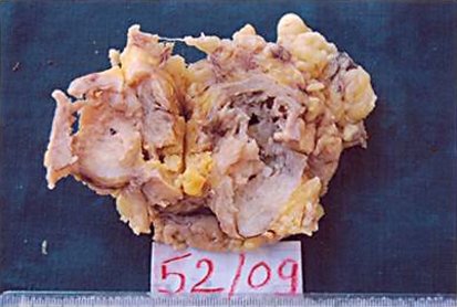
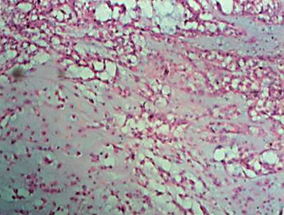
On gross examination, the lesion was grayish white with areas of necrosis. Histopathology was suggestive of a pure mammary chondrosarcoma with all surgical margins free of tumor (fig. 1, fig. 2). One axillary lymph node mass (2.5 cm) was positive for metastasis. The remaining 4 axillary lymph nodes were free of tumor. Immunohistochemistry was suggestive of a poorly differentiated infiltrating duct carcinoma of the right breast with a dominant chondrosarcomatous component. Estrogen receptor (ER), progesterone receptor (PR), and c-erbB2 were negative. S-100 was positive in the metaplastic cartilaginous area, while tumor cells showed strong immunoreactivity for cytokeratins and epithelial membrane antigen in epithelial areas, which confirms the presence of metaplastic carcinoma with predominant chondrosarcoma with high-grade infiltrating duct carcinoma. Postoperative radiotherapy was given at 50 Gy/30 days, and the woman is on regular follow-up with a further treatment plan consisting of chemotherapy.
Fig. 1.
Gross photograph of cut specimen of metaplastic breast carcinoma with predomiant chondrosarcoma.
Fig. 2.
Microscopic photograph of metaplastic breast carcinoma with predominant chondrosarcoma (Hematoxylin & Eosin staining at 40× magnification).
Discussion
Metaplastic breast carcinomas are regarded as ductal carcinomas that undergo metaplasia into a nonglandular growth pattern, and are difficult to accurately diagnose and classify because of their rarity and varied histological pattern. Tumors showing both carcinomatous and sarcomatous features are very uncommon and occur in various anatomical sites [4]. Metaplastic carcinomas of the breast are rare and interesting, yet confusing neoplasms. It has been reported that these tumors are more likely to occur in women over 50 years of age [5, 6, 9]. The patients usually describe a rapid growth with a short period of diagnosis. The tumors tend to have a circumscribed contour radiologically. A preoperative clinical and cytological diagnosis, though possible in a few cases, is usually not reached both due to marked similarity in clinical behavior and low index of suspicion. Classic sarcomatoid carcinoma is a tumor with both epithelial and mesenchymal components, hence the term biphasic sarcomatoid carcinoma. Carcinomas showing extensive metaplastic changes, including spindle cells, squamous cells and heterologous mesenchymal elements, are recognized in the breast [7]. In most of these cases, the infiltrating duct carcinoma is very small or focal or difficult to observe with routine HE stains or histopathological sections. Confirmation of epithelial (carcinomatous) component by immunohistochemical study is necessary in such cases to rule out pure primary mammary sarcomas. Metaplastic elements of pleomorphic spindle cells like fibrosarcoma, high-grade sarcomas, are rarely seen. However, chondrosarcoma, osteosarcoma and leiomyosarcoma components are extremely rare. In our case, the malignant chondrosarcomatous element was predominant; the infiltrating duct carcinoma was very focal and difficult to demonstrate in multiple histopathological sections. Therefore, we confirmed it by immunohistochemical study, which showed strong positivity for S-100 protein in chondromatous areas and focal positivity for cytokeratins and epithelial membrane antigen, while the tumor was negative for ER, PR and c-erbB2.
The prognosis of patients with metaplastic breast carcinoma depends on the stage of disease, similar to that seen in invasive carcinomas of the breast [8, 9].
Tumor size is another prognostic factor. Patients with a tumor size not larger than 5 cm have a better survival rate. Findings reported by Oberman [5], Kaufman et al. [6], and Chao et al. [9] suggest that the size of the neoplasm at the time of initial treatment best correlated with the prognosis. In our case, tumor size is larger than 5 cm, which is an unfavorable prognosis.
The incidence of axillary lymph node metastasis is low in metaplastic carcinomas. Like invasive carcinomas of the breast, axillary lymph node metastasis in patients with metaplastic carcinoma correlates with poor prognosis [9, 10]. In our case, axillary lymph node metastasis was positive. The incidence of metastatic malignancy is very rare. One of these cases with pulmonary metastasis is reported by Beltaos et al. [2].
Metaplastic carcinoma rarely exhibits nuclear immunoreactivity for ER and PR [9]. Expression of markers 34BE12, p53, retinoblastoma protein, HER/2neu, epidermal growth factor receptor, and cyclin D1 does not correlate with clinicopathologic features such as patient age, tumor size, tumor type, and relative proportion of metaplastic elements [10].
Surgery remains the mainstay of treatment for most sarcomatoid tumors [11]. Multimodality treatment may decrease local and systemic recurrence rates of somatic sarcomas, but results are inconclusive in patients with breast sarcomas [11]. Surgical and adjuvant treatment should follow the guidelines for the other most common breast cancers even if the need for chemotherapy is unknown due to the absence of large series of randomized or observational data.
We report on a case of metaplastic carcinoma with predominant chondrosarcoma with axillary metastasis. The patient is on regular follow-up on outpatient basis.
There has been no evidence of local recurrence of the disease or distant metastasis. Regular follow-up of the patient is important for at least 5 years because recurrence rates are high. The overall 5-year disease-free survival rate is 43% in metaplastic breast carcinoma [12].
References
- 1.Brenner RJ, Turner RR, Schiller V, Arndt RD, Giuliano A. Metaplastic carcinoma of the breast: report of three cases. Cancer. 1998;82:1082–1087. [PubMed] [Google Scholar]
- 2.Beltaos E, Banerjee TK. Chondrosarcoma of the breast: Report of 2 cases. Am J Clin Pathol. 1979;71:345–349. doi: 10.1093/ajcp/71.3.345. [DOI] [PubMed] [Google Scholar]
- 3.Feder JM, de Paredes ES, Hogge P, Wilken JJ. Unusual breast lesions: radiologic-pathologic correlation. Radiographics. 1999;19(suppl):S11–S26. doi: 10.1148/radiographics.19.suppl_1.g99oc07s11. [DOI] [PubMed] [Google Scholar]
- 4.Kurian KM, Al-Nafussi A. Sarcomatoid/metaplastic carcinoma of the breast: a clinicopathological study of 12 cases. Histopathology. 2002;40:58–64. doi: 10.1046/j.1365-2559.2002.01319.x. [DOI] [PubMed] [Google Scholar]
- 5.Oberman HA. Metaplastic carcinoma of breast: A clinicopathological study of 29 patients. Am J Surg Pathol. 1987;11:918–929. doi: 10.1097/00000478-198712000-00002. [DOI] [PubMed] [Google Scholar]
- 6.Kaufman MW, Marti JR, Gallager HS, et al. Carcinoma of the breast with pseudosarcomatous metaplasia. Cancer. 1984;53:1908–1917. doi: 10.1002/1097-0142(19840501)53:9<1908::aid-cncr2820530917>3.0.co;2-f. [DOI] [PubMed] [Google Scholar]
- 7.Schitt SJ, Millis RR, Hanby AM, Oberman HA. The breast. Chapter 9 in Sternberg's Diagnostic Surgical Pathology. vol 1. Lippincott: W&W; 2004. pp. 365–366. [Google Scholar]
- 8.Clark GM. Prognostic and predictive factors. In: Harris JR, Lippman ME, Morrow M, Hellman S, editors. ‘Disease of the Breast’. Philadelphia: Lippincott-Raven publishers; 1996. pp. 461–485. [Google Scholar]
- 9.Chao TC, Wang CS, Chen SC, Chen MF. Metaplastic carcinomas of the breast. J Surg Oncol. 1999;71:220–225. doi: 10.1002/(sici)1096-9098(199908)71:4<220::aid-jso3>3.0.co;2-l. [DOI] [PubMed] [Google Scholar]
- 10.Chhieng C, Cranor M, Lesser ME, Rosen PP. Metaplastic carcinoma of the breast with osteocartilaginous heterologous elements. Am J Surg Pathol. 1998;22:188–194. doi: 10.1097/00000478-199802000-00006. [DOI] [PubMed] [Google Scholar]
- 11.Callery CD, Rosen PP, Kinne DW. Sarcoma of the breast. A study of 32 patients with reappraisal of classification and therapy. Ann Surg. 1985;201:527–532. doi: 10.1097/00000658-198504000-00020. [DOI] [PMC free article] [PubMed] [Google Scholar]
- 12.Pitts WC, Rojas VA, Gaffey MG, et al. Carcinoma with metaplasia and sarcomas of the breast. Am J Clin Pathol. 1991;95:623–632. doi: 10.1093/ajcp/95.5.623. [DOI] [PubMed] [Google Scholar]


