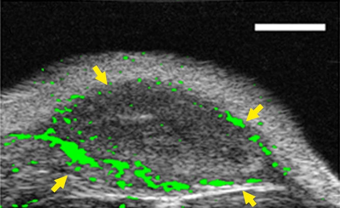
Figure 4a:

(a–c) Transverse color-coded US images of subcutaneous human ovarian cancer xenograft (scale bar = 1 mm) imaged longitudinally after intravenous administration of MBEndoglin in the same mouse at three tumor stages (a, small; b, medium; c, large). Note that targeted contrast-enhanced US imaging signal (shown as green areas overlaid on B-mode images) was highest in small tumors and decreased as tumors grew larger. (d) Representative immunoblots of ovarian cancer lysates from small, medium, and large tumors show decreasing expression levels as tumors grow larger (upper row). α Tubulin was used as a loading control (lower row).
