Abstract
Previous studies showed that the insertion of the intramuscular tendons of the deltoid muscle formed three discrete lines. The purpose of the present study was to establish a new dividing method of the deltoid muscle into various anatomical segments based on the distribution of the intramuscular tendons with their insertions (anatomical study). We further hoped to clarify the relationship between the anatomical segments and their activity pattern assessed by positron emission tomography with [18F]-2-fluoro-deoxyglucose (FDG–PET; PET study). Sixty cadaveric shoulders were investigated in the anatomical study. Three tendinous insertions of the deltoid muscle to the humerus were identified. Then, the intramuscular tendons were traced from their humeral insertions to the proximal muscular origins. The extent of each anatomical segment of the muscle including its origin and insertion was determined through careful dissection. Six healthy volunteers were examined using FDG–PET for the PET study. PET images were obtained after exercise of elevation in the scapular plane. On the PET images, margins of each anatomical segment of the deltoid muscle were determined using magnetic resonance images. Then, the standardized uptake value in each segment was calculated to quantify its activity. The anatomical study demonstrated that the deltoid muscle was divided into seven segments based on the distribution of its intramuscular tendons. The PET study revealed that the intake of FDG was not uniform in the deltoid muscle. The area with high FDG intake corresponded well to the individual muscular segments separated by the intramuscular tendons. We conclude that the deltoid muscle has seven anatomical segments, which seem to represent the functional units of this muscle.
Keywords: anatomy, deltoid, function, intramuscular tendon, positron emission tomography
Introduction
The deltoid muscle has been classically divided into three anatomical portions: the anterior; the middle; and the posterior portions. The anterior deltoid takes its origin from the lateral one-third of the clavicle as well as the anterior acromion; the middle deltoid originates from the lateral margin of the acromion; and the posterior deltoid from the scapular spine (Williams & Warwick, 1980). It has been believed that the activation pattern of this muscle during shoulder motion is different among these three portions (Reinold et al. 2007). Even in the same anatomical portion, its function may vary as the direction of muscle fibers changes gradually with changing the sites of their origins and insertions.
It has been reported that the intramuscular tendons play important roles for the force transmission to bone (Lieber & Fridén, 2001; Huijing, 2003; Finni, 2006). In the deltoid muscle, the insertion of the intramuscular tendons forms three discrete lines (Klepps et al. 2004). Klepps et al. also described that the classical three parts of the deltoid muscle based on its origin did not correspond to the three insertions at the humeral shaft. More recently, Leijnse et al. (2008) proposed a generic model of deltoid muscle consisting of multiple segments through their detailed anatomical observations. All these reports suggest that the deltoid muscle has more complex anatomical architecture than has been believed. Thus, the distribution of these intramuscular tendons should be taken into consideration when we assess the function of this muscle.
Recent studies revealed that the function of skeletal muscles could be evaluated successfully using positron emission tomography with [18F]-2-fluoro-deoxyglucose (FDG–PET; Fujimoto et al. 1996; Ohnuma et al. 2006; Tashiro et al. 1999). FDG–PET is a nuclear medicine tool for non-invasive quantification of both regional blood flow and tissue glucose metabolism in vivo. There are two major advantages to applying FDG–PET on skeletal muscles. First, the activity of the whole muscle can be quantified non-invasively. Second, FDG–PET can visualize the working pattern of each small segment in a large muscle during any type of exercise by synchronizing with computed tomography or magnetic resonance imaging (MRI). These advantages enable us to assess the function of each segment in a large muscle, such as the deltoid.
Based on these facts, the aim of the present study was to establish a new dividing method of the deltoid muscle based on the distribution of the intramuscular tendons as well as their insertions. We further hoped to clarify the relationship between these anatomical segments and their activity patterns assessed by FDG–PET.
Materials and methods
Anatomical study
Sixty shoulder girdles were obtained from 30 embalmed cadavers. There were 15 males and 15 females, with an average age of 82 years (range, 68–91 years). No specimens had prior history of shoulder surgery.
After removing skin, we first investigated the surface anatomy of the deltoid muscle and observed their muscle fibers superficially (Fig. 1). Then, the origin as well as insertion of this muscle was carefully dissected to clarify their detailed anatomy. As Rispoli et al. (2009) reported, the tendinous insertion of the deltoid muscle consists of three distinct lines, which form an M-shape (Fig. 2). These tendinous insertions were named as anterior, middle and posterior insertions, respectively. At this level, the muscle belly of the deltoid was first divided into three parts. Then, the intramuscular end tendons were identified, which were traced proximally to their origins. The muscle fibers were divided into ‘segments’ by their attaching intramuscular tendons. At the origin of the muscle fibers, the anatomical landmarks that indicated the proximal attachment of each segment were identified. The bony facets where each segment originated were determined and their width was measured by a digital caliper. Finally, the extent of each segment of the deltoid muscle was compared with that of the conventional three portions.
Fig. 1.
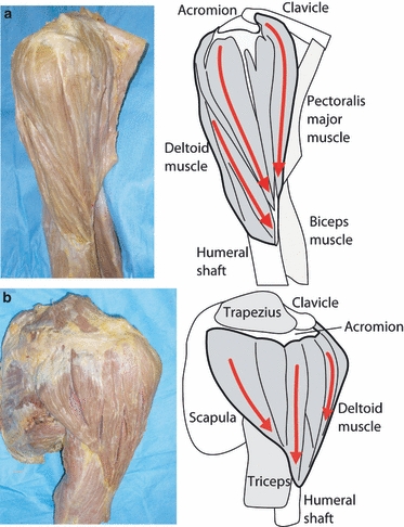
Surface anatomy of the deltoid muscle. (a) The anterolateral view of the deltoid muscle. The muscle fibers run in the anteroinferior direction (red arrows), which converge the anterior side of the deltoid tubercula. (b) The posterior view of the deltoid muscle.
Fig. 2.
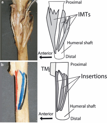
Anatomy of the insertion of the deltoid into the humeral shaft. (a) Three intramuscular tendons insert individually into the humeral shaft (IMTs, intramuscular tendons). (b) The insertions of the intramuscular tendons form three discrete lines (TMj, teres major muscle).
PET study
Six healthy volunteers without any histories of shoulder pain or trauma were examined using FDG–PET. There were four males and two females, with an average age of 74 years (range; 67–85 years). These subjects underwent FDG–PET with the protocol, which we previously established (Omi et al. 2010). All subjects refrained from eating and drinking for at least 3 h before the examination. The FDG was dissolved in approximately 2 mL saline, which was then injected intravenously via the cubital vein. The mean dose and standard deviation of injected FDG were 86.5 and 8.0 MBq, respectively. Exercise of scaption (elevation in the scapular plane) for 10 min was performed before and after injection of FDG. The exercise consisted of 200 repetitions of scaption between 0 and 90 ° of elevation with 250-g weights around the wrists. PET images were collected 40 min after the injection with a whole-body positron camera (SET-2400W; Shimazu, Kyoto, Japan). To quantify the muscle activities in each muscle segment, it was necessary to determine its exact location on the PET images. For this purpose, an MRI scan was performed for image fusion (Signa Horizon LX 1.5T Ver.9.1; GE Healthcare, Milwaukee, WI, USA). The measurement conditions were as follows: repetition time/echo time was 3000/85 ms; number of excitations was 1; the field of view was 46 cm; number of matrix was 512 × 512; slice thickness was 3 mm; and a slice gap was 1.5 mm. A T2-weighted transverse MR image with fat suppression was used to determine the outer margin of each segment in the deltoid muscle on the PET image at the same level. The volumes of interest (VOI) in each portion of the muscle defined on MR images were superimposed onto the registered PET images using a software, Dr View/LINUX (AJS, Tokyo, Japan) for evaluation of radioactivity in each muscle. After fusion of PET and MR images, the standardized uptake values (SUVs) in each segment of the deltoid muscle were calculated to quantify their activity with the following equation:
Based on the definition of SUV, this equation can be modified as follows:
 |
Statistical analysis was performed using StatMate III statistical software (version 3.16; Atms, Tokyo, Japan). Statistical significance of difference in activity level (SUV) between six segments was examined for SUVs using one-way anova. A P-value < 0.05 was considered as statistically significant.
Results
Anatomical study
Segments of the deltoid muscle
The presence of three insertions (anterior, middle and posterior) was confirmed in all shoulders. Among them, a thick intramuscular tendon (anterior end tendon), which attached to the anterior insertion, was identified (Fig. 2A). The direction of this tendon was almost parallel to the humeral shaft. Intramuscular tendons also inserted to the middle and posterior insertions, respectively (middle and posterior end tendons). The direction of these two tendons was superoposterior against the humeral shaft.
At the level of the proximal deltoid muscle, the anterior and posterior end tendons branched into three intramuscular tendons, respectively (Fig. 3a). A total of seven intramuscular tendons were identified at this level (anterior tendon: 3; middle tendon: 1; posterior tendon: 3). These intramuscular tendons were named A1, A2 and A3 tendons (anterior intramuscular tendons); M1 tendon (middle intramuscular tendon); and P1, P2 and P3 tendons (posterior intramuscular tendons), respectively. Consequently, the deltoid muscle was divided into seven segments, including A1, A2, A3, M1, P1, P2 and P3 segments based on the attachment to these seven intramuscular tendons (Fig. 3c).
Fig. 3.
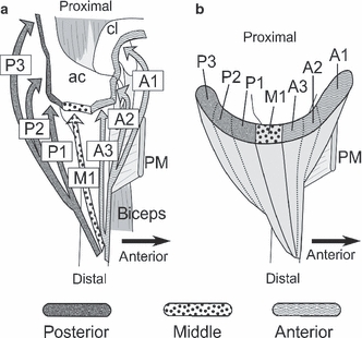
(a) Directions of the intramuscular tendons. Arrows indicate the direction of each intramuscular tendon (A1, A2, A3, M1, P1, P2 and P3, intramuscular tendons; ac, acromion; cl, clavicle; PM, pectoralis major muscle). (b) Seven segments at the proximal part of the deltoid muscle on the transverse plane.
Proximal origins and their landmarks
The anterior part of the deltoid muscle widely spread over the clavicle, and the anterior surface and the anterior-third of the lateral acromion. The middle part was relatively narrow, which attached to the mid-third of the lateral aspect of the acromion. The posterior part attached to the posterior-third of the lateral acromion as well as the scapular spine.
There were several anatomical landmarks for dividing each segment at the proximal origin of the deltoid muscle. The border between A1 and A2 segments located approximately 5 mm medial from the acromioclavicular joint in all specimens (Fig. 4a). The bony landmark between A2 and A3 segments was the anterolateral corner of the acromion. From the lateral aspect of the acromion, three segments (A3, M1 and P1) originated (Fig. 4a). There were two small bony tubercula on the lateral border of the acromion, which separated these segments (A3, M1 and P1) (Fig. 4b). These two bony tubercula were seen in 56 specimens (93.3%). The lateral border of the acromion was divided into three facets by these bony tubercula, which were named as the anterior, middle and posterior facets (Fig. 3b). A3, M1 and P1 segments originated from the anterior, middle and posterior facets, respectively. The width of each facet was 19.5 ± 4.1 mm (mean ± standard deviation), 14.2 ± 4.0 mm and 17.9 ± 5.0 mm, respectively. P1 and P2 segments were divided by the posterior angle of the acromion, where the posterior tendon originated. On the other hand, no bony landmark was identified between P2 and P3 segments at their origin.
Fig. 4.
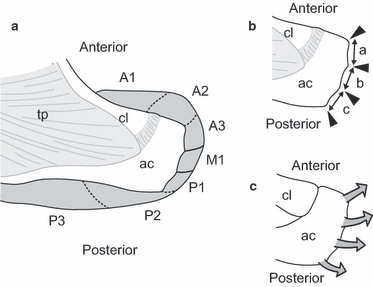
The landmarks at the origin of the deltoid muscle. (a) Origin of each segment. The border between A1 and A2 locates approximately 5 mm medial from the acromioclavicular joint. There are no particular structures between P2 and P3 at the proximal part of the deltoid (ac, acromion; cl, clavicle; tp, trapezius). (b) There are two bony tubercula at the lateral border of the acromion. These bony tubercula separate the lateral border of the acromion into three facets: a, anterior facet; b, middle facet; c, posterior facet. (c) Four intramuscular tendons originate from the anterior angle, two bony tubercula and posterior angle of the acromion, respectively.
The relationship between the classical three portions and the segments
The classical clavicular portion corresponded well to the A1 segment in the new dividing method (Fig. 4a). The acromial portion consisted of A2, A3, M1 and P1 segments, and the spinal portion was compatible to P2 and P3 segments. In other words, classical clavicular, acromial and spinal portions consisted of one, four and two segments, respectively.
Anatomical variations
In the present series, some anatomical variations were observed. In four shoulders (6.7%), the M1 segment had two distinct intramuscular tendons. The middle insertion formed a single line or double lines in these shoulders. As a result, these shoulders had four acromial facets and five proximal tendons at the lateral aspect of the acromion.
PET study
Because there was no particular landmark between P2 and P3 segments on MR images, we could not differentiate these two segments on PET images (Fig. 5). Thus, PET analysis was done among the six segments, for example A1, A2, A3, M1, P1 and P2 + P3. The PET images revealed that the intake of FDG was not uniform in the deltoid muscle. The area with high intake represented a dotted pattern in the upper level of the deltoid, which corresponded well to the anatomically defined muscular segments (Fig. 6a). At the middle and lower level of the deltoid, the area with high intake was not separated. The SUV of each segment was shown in Fig. 6b. A3 and M1 showed relatively higher values of SUV than those of other segments, including A1, A2, P1 and P2 + P3 (Fig. 6b). Statistically, the SUVs in both A3 and M1 segments were significantly higher than that in P2 + P3 (P< 0.05).
Fig. 5.
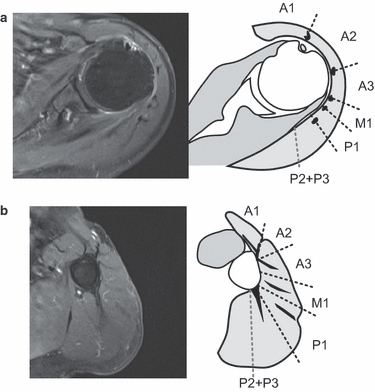
Dividing method of the deltoid muscle on MR images. The intramuscular tendons are clearly depicted in a T2-weighted transverse MR image with fat suppression. The deltoid muscle is divided based on the distribution of intramuscular tendons. The straight lines show the border between each muscle segment. The proximal intramuscular tendons locate at the borders of each segment. For the distal insertions, A3, M1 and P1 intramuscular tendons exist at their center. On the other hand, A1, A2 and P3 have their distal intramuscular tendons at the margin of the segments. (a) At proximal deltoid level, (b) At distal deltoid level.
Fig. 6.
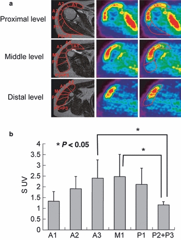
(a) Axial MRI and PET images of the proximal, middle and distal levels of the deltoid. At proximal level, the segments divided on MRI well correspond to the dotted FDG intake pattern in the PET images. (b) SUVs of each segment. The standardized uptake values (SUVs) of A3 and M1 are relatively higher than that of other segments. Especially, both A3 and M1 activities were significantly greater than that of P2 + P3 (P< 0.05).
Discussion
In the clinical practice, the classical distinction of the deltoid muscle into three portions based on the origin of these portions is widely used. Although this method seemed to be very convenient to understand the anatomy of this muscle, one should keep in mind that these portions may not reflect the function of this muscle.
In 1911, Fick described in his textbook that the deltoid muscle had seven functional segments (Fick, 1911). Since then, several studies have been carried out concerning the intramuscular architectures of the deltoid muscle. Brown and Wickham divided the deltoid muscle into seven segments based on its superficial anatomy as well as the activation levels assessed by electromyogram (EMG; Wickham & Brown, 1998; Brown & Wickham, 2006; Brown et al. 2007). Unfortunately, however, it seemed that neither of them took the morphology of the intramuscular tendons into consideration. Recently, Leijnse suggested the dissection model of the deltoid muscle should be based on the morphology of the deltoid origin and end tendons (Leijnse et al. 2008). In the present study, we confirmed that the deltoid muscle could be divided into seven segments with their intramuscular tendons. It was also interesting to note that the classical acromial portion had four segments (A2, A3, M1 and P1), which attached individually to the different bony facets. The presence of bony tubercules on the acromion could be useful landmarks for surgeons to identify each segment.
To assess the muscle activity, EMG has been widely used as a standard technique (Kronberg et al. 1990; McMahon et al. 1995; Reddy et al. 2000; Kelly et al. 2005; Reinold et al. 2007; Yasojima et al. 2008; Cordasco et al. 2009). However, there was a major disadvantage to applying this method to the deltoid muscle. A fine needle electrode used for EMG could only reflect the muscle activities of its small portion. Based on these facts, we assessed the function of the deltoid muscle using FDG–PET in the present study. In the PET images, the FDG intake represented a dotted pattern consistent with the separated segments at the upper level of the deltoid. These results indicated that the deltoid muscle worked as segments during the arm elevation, which corresponded well to the anatomically defined seven muscular segments with the intramuscular tendons. Therefore, we assumed that these anatomical seven segments reflect the functional units of the deltoid muscle. Because the SUV values after exercise in all segments were greater than the SUV values at rest, we assumed that they worked synergistically during the arm elevation in the scapular plane. The SUV value of deltoid muscle at rest was approximately 0.7 in our previous study (Omi et al. 2010). On the other hand, A3 and M1 represented higher SUVs than other segments. Moreover, the difference in SUV between these two segments and P2 + P3 was statistically significant. These results might suggest that the mid-part of the acromial portion including A3 and M1 played a great role in elevating the arm in the scapular plane.
There were several limitations in the present study. First, we did not investigate the relationship between the nerve endings and the muscle segments in the present study. Second, we failed to differentiate P2 and P3 segments on MR images because there were no particular structures between these segments. Consequently, the muscle activities in these two segments could not be precisely differentiated. Third, only the scaption exercise was performed in the present study. Future studies using FDG–PET with exercise in various directions would be necessary to clarify the detailed function of the deltoid muscle.
Conclusion
The deltoid muscle could be divided into seven segments separated by their intramuscular tendons. The active muscular regions in the deltoid muscle were separated on the PET images by their intramuscular tendons. Based on these results, we assumed that the anatomical seven segments corresponded well to the functional units of the deltoid muscle.
Acknowledgments
The authors would like to thank Professor Mari Dezawa, MD, PhD, and Dr Jian-lin Zuo, MD, for their support.
Author contributions
Yoshimasa Sakoma was a principal investigator who investigated all of the cadaver specimens with Yoshiaki Itoigawa. Nobuhisa Shinozaki examined the PET and MRI. Nobuyuki Yamamoto and Hirotaka Sano (corresponding author) analyzed the data of PET experiments as well as the anatomical measurements. Eiji Itoi and Toshifumi Ozaki were senior investigators, and supervised this project.
References
- Brown JM, Wickham J. Neuromotor coordination of multisegmental muscle during a change in movement direction. J Musculoskelet Res. 2006;10:63–74. [Google Scholar]
- Brown JM, Wickham J, McAndrew DJ, et al. Muscles within muscles: coordination of 19 muscle segments within three shoulder muscles during isometric motor tasks. J Electromyogr Kinesiol. 2007;17:57–73. doi: 10.1016/j.jelekin.2005.10.007. [DOI] [PubMed] [Google Scholar]
- Cordasco FA, Chen NC, Backus SI, et al. Subacromial injection improves deltoid firing in subjects with large rotator cuff tears. HSS J. 2010;6:30–36. doi: 10.1007/s11420-009-9127-6. [DOI] [PMC free article] [PubMed] [Google Scholar]
- Fick R. Handbuch der Anatomie und Mekanik der Gelenke. Jena: Gustav Fischer; 1911. [Google Scholar]
- Finni T. Structural and functional features of human muscle-tendon unit. Scand J Med Sci Sports. 2006;16:147–158. doi: 10.1111/j.1600-0838.2005.00494.x. [DOI] [PubMed] [Google Scholar]
- Fujimoto T, Itoh M, Kumano H, et al. Whole-body metabolic map with positron emission tomography of a man after running. Lancet. 1996;348:266. doi: 10.1016/s0140-6736(05)65572-9. [DOI] [PubMed] [Google Scholar]
- Huijing PA. Muscular force transmission necessitates a multilevel integrative approach to the analysis of function of skeletal muscle. Exerc Sport Sci Rev. 2003;31:167–175. doi: 10.1097/00003677-200310000-00003. [DOI] [PubMed] [Google Scholar]
- Kelly BT, Williams RJ, Cordasco FA, et al. Differential patterns of muscle activation in patients with symptomatic and asymptomatic rotator cuff tears. J Shoulder Elbow Surg. 2005;14:165–171. doi: 10.1016/j.jse.2004.06.010. [DOI] [PubMed] [Google Scholar]
- Klepps S, Auerbach J, Calhon O, et al. A cadaveric study on the anatomy of the deltoid insertion and its relationship to the deltopectoral approach to the proximal humerus. J Shoulder Elbow Surg. 2004;13:322–327. doi: 10.1016/j.jse.2003.12.014. [DOI] [PubMed] [Google Scholar]
- Kronberg M, Nemeth G, Brostrom LA. Muscle activity and coordination in the normal shoulder. An electromyographic study. Clin Orthop Relat Res. 1990;257:76–85. [PubMed] [Google Scholar]
- Leijnse JN, Han SH, Kwon YH. Morphology of deltoid origin and end tendons – a generic model. J Anat. 2008;213:733–742. doi: 10.1111/j.1469-7580.2008.01000.x. [DOI] [PMC free article] [PubMed] [Google Scholar]
- Lieber RL, Fridén J. Clinical significance of skeletal muscle architecture. Clin Orthop Relat Res. 2001;383:140–151. doi: 10.1097/00003086-200102000-00016. [DOI] [PubMed] [Google Scholar]
- McMahon PJ, Debski RE, Thompson WO, et al. Shoulder muscles forces and tendon excursions during glenohumeral abduction in the scapular plane. J Shoulder Elbow Surg. 1995;4:199–208. doi: 10.1016/s1058-2746(05)80052-7. [DOI] [PubMed] [Google Scholar]
- Ohnuma M, Sugita T, Kokubun S, et al. Muscle activity during a dash shown by 18F-fluorodeoxyglucose positron emission tomography. J Orthop Sci. 2006;11:42–45. doi: 10.1007/s00776-005-0972-y. [DOI] [PubMed] [Google Scholar]
- Omi R, Sano H, Ohnuma M, et al. Function of the shoulder muscles during shoulder elevation: an assessment using positron emission tomography. J Anat. 2010;216:643–649. doi: 10.1111/j.1469-7580.2010.01212.x. [DOI] [PMC free article] [PubMed] [Google Scholar]
- Reddy AS, Mohr KJ, Pink MM, et al. Electromyographic analysis of the deltoid and rotator cuff muscles in persons with subacromial impingement. J Shoulder Elbow Surg. 2000;9:519–523. doi: 10.1067/mse.2000.109410. [DOI] [PubMed] [Google Scholar]
- Reinold MM, Macrina LC, Wilk KE, et al. Electromyographic analysis of the supraspinatus and deltoid muscles during 3 common rehabilitation exercises. J Athl Train. 2007;42:464–469. [PMC free article] [PubMed] [Google Scholar]
- Rispoli DM, Athwal GS, Sperling JW, et al. The anatomy of the deltoid insertion. J Shoulder Elbow Surg. 2009;18:386–390. doi: 10.1016/j.jse.2008.10.012. [DOI] [PubMed] [Google Scholar]
- Tashiro M, Fujimoto T, Itoh M, et al. 18F-FDG PET imaging of muscle activity in runners. J Nucl Med. 1999;40:70–76. [PubMed] [Google Scholar]
- Wickham J, Brown JM. Muscles within muscles: the neuromotor control of intra-muscular segments. Eur J Appl Physiol. 1998;78:219–225. doi: 10.1007/s004210050410. [DOI] [PubMed] [Google Scholar]
- Williams P, Warwick L. Gray's Anatomy. Edinburgh: Churchill Livingstone; 1980. [Google Scholar]
- Yasojima T, Kizuka T, Noguchi H, et al. Differences in EMG activity in scapular plane abduction under variable arm positions and loading conditions. Med Sci Sports Exerc. 2008;40:716–721. doi: 10.1249/MSS.0b013e31816073fb. [DOI] [PubMed] [Google Scholar]


