Abstract
Cerebrovascular complications have been reported to occur in patients with neurocysticercosis (NCC). We report a patient who presented with relapsed subarachnoid hemorrhage possibly linked to NCC. In addition, we performed a literature review of all of the reported cases of aneurysmal and non-aneurysmal hemorrhagic cerebrovascular events associated with NCC. We identified 11 such cases. The majority of the individuals were young males (mean: 38 years; 70% males). Four cases (36%) had aneurysms. Four (36%) others had negative cerebral angiograms and therefore classified as non-aneurysmal, while the remaining three (28%) did not report sufficient information to classify them. All cases with aneurysmal hemorrhage underwent successful surgical repair of the aneurysms. Seven patients received albendazole (including three who had had surgery). Three patients died; all three presented in the pre-albendazole era. In summary, neurocysticercosis should be considered in the differential diagnosis of hemorrhagic cerebrovascular events in young patients without classical vascular risk factors who have lived or visited NCC-endemic areas.
Introduction
Neurocysticercosis (NCC) caused by the larval stage of Taenia solium is the most common helminth infection of the human central nervous system worldwide.1 Neurocysticercosis has become an important emerging infection in the United States. This has largely been driven by the influx of immigrants from highly endemic regions into the United States and widespread access to neuroimaging.2
Neurocysticercosis is a disease with protean manifestations, which depends upon the number of parasites, their location, and the degree of host inflammatory response. Most common clinical manifestations include late onset epilepsy or symptoms of intracranial hypertension.3 Cerebrovascular complications have been reported to occur in 4–12% of patients with NCC.4 In the majority of these cases, the diagnoses were ischemic cerebrovascular events associated with vasculitis and/or thrombosis from surrounding cysts in both small- and large-diameter vessels. Subarachnoid hemorrhages have been noted in subarachnoid neurocysticercosis and have been associated with cerebral aneurysms in some, but not all, cases.4–8
Despite recent advances, treatment of NCC remains suboptimal at this time. Treatment with antihelmintics, corticosteroids, antiepileptic drugs, and surgical interventions constitute some alternatives in the management of the disease.9 Here we report a patient who presented with a relapsed non-aneurysmal subarachnoid hemorrhage possibly associated with subarachnoid cysticercosis. In addition, we systematically reviewed the literature to summarize all of the reported cases of hemorrhagic cerebrovascular events associated with NCC.
Material and Methods
We searched the English literature in December 2009 with PubMed using the search terms [Stroke AND neurocysticercosis], [Subarachnoid hemorrhage AND neurocysticercosis], and [cerebral hemorrhage AND neurocysticercosis]. We also searched references from previous literature. Abstracts were reviewed, and papers that presented original cases of hemorrhagic cerebrovascular events associated with neurocysticercosis were reviewed in detail.
Results
Case report.
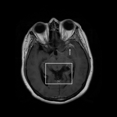
A 39-year-old Hispanic female was admitted with a 1-day history of severe bilateral frontal headache not associated with visual disturbances, nausea, vomiting, or neurological deficits. She was afebrile and her physical exam, including a detailed neurologic examination, was within normal limits. She had moved to Houston, Texas ∼15 years earlier from a small rural desert town on the Pacific coast of Mexico, where she used to be a farmer. Two years earlier, she had been diagnosed with a subarachnoid hemorrhage located around the left Sylvian fissure. Multiple magnetic resonance imaging (MRI) studies and cerebral angiograms done at that time did not show any evidence of aneurysms or vascular malformations. Up until her current hospitalization, she had remained asymptomatic. However, on admission an MRI of her brain revealed a new left subarachnoid hemorrhage involving the left suprasellar cistern, interpeduncular cistern, left ambient cistern, and again the left Sylvian fissure. Additionally, the images showed dilatation of the lateral and third ventricles, and the aqueduct of Sylvius, with obstruction caused by cysts associated with leptomeningeal enhacement of the supracerebellar cistern (Figure 1). A lumbar puncture revealed an opening pressure of 15 mm H2O, 18 red blood cells/mL, 20 white blood cells/mL (78% lymphocytes and 5% eosinophils), glucose of 88 mg/dL, and protein of 28 mg/dL. Bacterial, fungal, and mycobacterial cultures of the cerebral spinal fluid were all negative. She was also found to have peripheral eosinophilia (absolute eosinophil count of 900 cells/mL). A repeat cerebral angiogram was again negative for aneurysms. Enzyme-linked immunotransfer blot for cysticercosis in serum was positive. As a result, the patient had a ventriculoperitoneal shunt placed because of the obstructive hydrocephalus. Subsequently, she was given dexamethasone and albendazole for a total of 4 weeks. She fully recovered without any neurological deficits.
Figure 1.

T1-weighted axial magnetic resonance image of the brain revealing an acute subarachnoid hemorrhage around the Sylvian fissure (arrow) and racemose cyst within the supracerebellar cistern (square), causing a non-communicating hydrocephalus with dilatation of the third and lateral ventricles, and the aqueduct of Sylvius.
Review of the literature.
We identified 10 other cases with aneurysmal or non-aneurysmal hemorrhagic cerebrovascular events associated with neurocysticercosis in the literature (Table 1). The mean age was 38 years (median: 33). Most cases were males (70%, 7 of 10 cases; data was not available for one case). Four cases (36%) were found to have an aneurysmal hemorrhagic event. Four (36%) had negative cerebral angiograms and therefore had a non-aneurysmal hemorrhagic event. Three (28%) did not report sufficient information to categorize them as aneurysmal or non-aneurysmal. Location of lesions was varied but perhaps the most common presentation involved subarachnoid hemorrhage in the Sylvian fissure (28%, 3 of 11 cases). All three patients who had examination of cerebrospinal fluid (CSF) showed evidence of anti-cysticercal antibodies in the CSF. All cases with aneurysmal hemorrhagic events underwent successful surgical repair of the aneurysms. Seven patients received albendazole (including three who had had surgery) usually along with corticosteroids. Three patients died; all presented in the pre-albendazole era.
Table 1.
Aneurysmal and non-aneurysmal CNS hemorrhage caused by neurocysticercosis (NCC)*
| Report | Age/ sex | Clinical presentation | CSF | Imaging studies | Aneurysm location/ morphology | Surgery | Medication | Outcome |
|---|---|---|---|---|---|---|---|---|
| 1. Zee and others,10 1980 | 23/M | Headache, nausea, vomiting, AMS, and CN impairment | NA | Large right temporal hematoma | Right distal MCA /mycotic | Clipping of the proximal artery | NA | NA |
| 2. Iwanowski and others,11 1987 | 56/F | NA | NA | Focal hemorrhage in basal ganglia. Solitary cyst in temporal horn | NA | NA | NA | Died |
| 3. Iwanowski and others,11 1987 | 54/M | Hemiparesis | NA | Subarachnoid hemorrhage with solitary cyst on quadrigeminal plate | NA | NA | NA | Died |
| 4. Alarcon and others,4 1992 | NA | Motor deficits | NA | Hemorrhage into a frontal lobe cyst† | NA | None | NA | Died |
| 5. Soto-Hernandez and others,5 1996 | 32/M | Headaches, nausea, vomiting, AMS, and meningeal signs | 6,486 cells (77% PMN and 5% E), glucose 10 mg/dL, protein 1,560 mg/dL. ELISA for NCC (+) | Subarachnoid hemorrhage, hydrocephalus, subarachnoid cysts at the suprasellar, and pontine cisterns and anterior aspect of right temporal lobe | Right branch of AICA/fusiform | Wrapping | Albendazole for 10 days and steroids | Improved |
| 6. Sawhney and others,12 1998 | 10/M | Headache, nausea, vomiting, and partial seizures | NA | Left parietal hematoma and multiple cysts | Normal angiogram | NA | Albendazole, steroids, (duration unknown) | No improvement |
| 7. Huang and others,6 2000 | 32/M | Headache, nausea, and dysphasia | NA | Subarachnoid hemorrhage and multiple cysts in the left Sylvian fissure | Left M2 MCA Branch/pseudofusiform | Clipping | Albendazole, (duration unknown) | Improved |
| 8. Tellez-Zenteno and others,7 2003 | 32/F | Headache, dysarthria, left hemiparesis, and psychomotor agitation | 3 cells, glucose 55 mg/dL, protein 33 mg/dL, ELISA for NCC (+) | Right lenticulo-capsular hemorrhage around a large cystic lesion and cysts in right parietal lobe | Normal angiogram | None | Albendazole for 15 days and steroids | Improved |
| 9. Tellez-Zenteno and others,7 2003 | 34/M | Headache, nausea, vomiting, diplopia, gait disorder, psychomotor agitation, CN impairment | 107 cells (97% L), glucose 19 mg/dL, protein 695 mg/dL, ELISA for NCC (+) | Hemorrhagic lesion and a cyst in the ambiens cistern | Normal angiogram | None | Albendazole for 8 days and steroids | Improved |
| 10. Kim and others,8 2005 | 69/M | AMS | NA | Subarachnoid hemorrhage in the right Sylvian fissure, large right temporal lobe hematoma, and a cyst within the fissure | Right MCA, distal branch of ATA/fusiform | Trapping | Albendazole for 2 months | Improved |
| 11. Current case, 2010 | 39/F | Headaches | 20 cells (78% L, 5% E), glucose 88 mg/dL, protein 28 mg/dL | Subarachnoid hemorrhage around the left Sylvian fissure and multiple cysts within the basilar cistern | Normal angiogram | None | Albendazole for 4 weeks and steroids | Improved |
AICA = anteriorinferior cerebellar artery; AMS = altered mental status; ATA = anterior temporal artery; CN = cranial nerve; CNS = central nervous system; CSF = cerebrospinal fluid; E = eosinophils; ELISA = enzyme-linked immunosorbent assay; L = lymphocytes; MCA = middle cerebral artery; NA = information not available; PMN = polymorphonuclear cells.
Autopsy findings.
Discussion
Although cerebral cysticercosis (particularly subarachnoid cysticercosis) has been linked to the occurrence of cerebrovascular events, most studies have focused on ischemic events. In our review we found 11 cases with aneurysmal or non-aneurysmal hemorrhagic cerebrovascular events associated with cysticercosis.
The majority of cases occurred in young individuals presumptively without classic vascular risk factors. The fact that neurocysticercosis involving the subarachnoid space was the most common finding observed underscores the progressive nature and poor evolution, if not properly managed, of this type of neurocysticercosis.
The pathogenesis of subarachnoid hemorrhage in neurocysticercosis is not clear. The presence of clumps of subarachnoid cysts within the cisterns of the cerebrospinal fluid may cause an inflammatory exudate, which could result in thickening of the leptomeninges. Blood vessels trapped within this dense exudate might undergo invasion of the vessel wall by inflammatory cells, leading to endarteritis and endothelial hyperplasia (cysticercotic angiitis).5,7 In a previous study, 15 (53%) of 28 patients with subarachnoid cysticercosis who underwent cerebral angiography had evidence of cerebral arteritis in middle size arteries (middle cerebral artery and posterior cerebral artery).13 Almost certainly this severe inflammatory process could lead not only to ischemic cerebrovascular events but also hemorrhagic events. The mechanism of blood vessel rupture might result directly from the marked inflammatory process of a small penetrating artery,7 from erosion of a degenerating cyst in the wall of a small vessel or through the formation of an inflammatory aneurysm.5,6,8,10 Inflammatory aneurysms are usually located at distal intracranial arteries, not at bifurcations like congenital aneurysms, and are more commonly fusiform in shape.8 Direct clipping of inflammatory aneurysms is also more difficult than with congenital aneurysms.5,6 The wall of inflammatory aneurysms and parent vessels are extremely friable, and the possibility of intraoperative rupture is higher. In addition, inflammatory aneurysms are fusiform so clipping of the aneurysm neck while preserving the parent artery is technically challenging. As a result, they are generally secured by wrapping,5 clipping of the proximal artery,10 or trapping.8
Among those patients who had examination of the CSF, all of them showed positive anti-cysticercal antibodies. This could be caused by a large parasite (antigenic) burden leading to an intense inflammatory response in the CSF. Recently, it has been demonstrated that detection of cysticercal antigens by monoclonal antibody-based enzyme-linked immunosorbent assay (ELISA) in the CSF suggests active subarachnoid cysticercosis.14 Hence, we hypothesize that application of this technique in cases of hemorrhagic cerebrovascular events associated with neurocysticercosis may be warranted.
After albendazole became available in the mid-1990s, all cases of hemorrhagic stroke associated with cysticercosis were treated with a course of this antihelmintic, usually along with corticosteroids and surgical therapy, if an aneurysm was present. All of these patients experienced improvement of their symptoms. Whether albendazole, as part of the administered therapeutic regimen, contributed to the improvement of the condition remains speculative at this time. In addition, there seems to be consensus on the need of surgical intervention in cases of aneurysmal hemorrhagic events associated with neurocysticercosis; however, its role, if any, in cases of non-aneurysmal hemorrhagic events needs to be investigated.
In summary, neurocysticercosis should be considered in the differential diagnosis of aneurysmal and non-aneurysmal hemorrhagic cerebrovascular events in young patients without classical vascular risk factors who have lived in or traveled to areas where NCC is endemic. Further studies of the pathophysiology, diagnosis, therapy, and prognosis of hemorrhagic cerebrovascular complications associated with neurocysticercosis are clearly needed.
Footnotes
Authors' addresses: George M. Viola, The University of Texas MD Anderson Cancer Center, Department of Medicine, Division of Infectious Diseases, Infection Control and Employee Health, Houston, TX, E-mail: GMViola@mdanderson.org. A. Clinton White Jr., Director, Infectious Disease Division, Department of Internal Medicine, University of Texas Medical Branch, Galveston, TX, E-mail: acwhite@utmb.edu. Jose A. Serpa, Department of Medicine, Section of Infectious Diseases, Baylor College of Medicine, Houston, TX, E-mail: jaserpaa@bcm.edu.
References
- 1.Garcia HH, Del Brutto OH, Nash TE, White AC, Jr, Tsang VC, Gilman RH. New concepts in the diagnosis and management of neurocysticercosis (Taenia solium) Am J Trop Med Hyg. 2005;72:3–9. [PubMed] [Google Scholar]
- 2.Serpa JA, Yancey LS, White AC., Jr Advances in the diagnosis and management of neurocysticercosis. Expert Rev Anti Infect Ther. 2006;4:1051–1061. doi: 10.1586/14787210.4.6.1051. [DOI] [PubMed] [Google Scholar]
- 3.Garcia HH, Del Brutto OH. Neurocysticercosis: updated concepts about an old disease. Lancet Neurol. 2005;4:653–661. doi: 10.1016/S1474-4422(05)70194-0. [DOI] [PubMed] [Google Scholar]
- 4.Alarcon F, Hidalgo F, Moncayo J, Vinan I, Duenas G. Cerebral cysticercosis and stroke. Stroke. 1992;23:224–228. doi: 10.1161/01.str.23.2.224. [DOI] [PubMed] [Google Scholar]
- 5.Soto-Hernandez JL, Gomez-Llata Andrade S, Rojas-Echeverri LA, Texeira F, Romero V. Subarachnoid hemorrhage secondary to a ruptured inflammatory aneurysm: a possible manifestation of neurocysticercosis: case report. Neurosurgery. 1996;38:197–199. doi: 10.1097/00006123-199601000-00045. discussion 9–200. [DOI] [PubMed] [Google Scholar]
- 6.Huang PP, Choudhri HF, Jallo G, Miller DC. Inflammatory aneurysm and neurocysticercosis: further evidence for a causal relationship? Case report. Neurosurgery. 2000;47:466–467. doi: 10.1097/00006123-200008000-00042. discussion 7–8. [DOI] [PubMed] [Google Scholar]
- 7.Tellez-Zenteno JF, Negrete-Pulido O, Cantu C, Marquez C, Vega-Boada F, Garcia Ramos G. Hemorrhagic stroke associated to neurocysticercosis. Neurologia. 2003;18:272–275. [PubMed] [Google Scholar]
- 8.Kim IY, Kim TS, Lee JH, Lee MC, Lee JK, Jung S. Inflammatory aneurysm due to neurocysticercosis. J Clin Neurosci. 2005;12:585–588. doi: 10.1016/j.jocn.2004.07.018. [DOI] [PubMed] [Google Scholar]
- 9.Nash TE, Singh G, White AC, Rajshekhar V, Loeb JA, Proano JV, Takayanagui OM, Gonzalez AE, Butman JA, DeGiorgio C, Del Brutto OH, Delgado-Escueta A, Evans CA, Gilman RH, Martinez SM, Medina MT, Pretell EJ, Teale J, Garcia HH. Treatment of neurocysticercosis: current status and future research needs. Neurology. 2006;67:1120–1127. doi: 10.1212/01.wnl.0000238514.51747.3a. [DOI] [PMC free article] [PubMed] [Google Scholar]
- 10.Zee CS, Segall HD, Miller C, Tsai FY, Teal JS, Hieshima G, Ahmadi J, Halls J. Unusual neuroradiological features of intracranial cysticercosis. Radiology. 1980;137:397–407. doi: 10.1148/radiology.137.2.6968919. [DOI] [PubMed] [Google Scholar]
- 11.Iwanowski L, Wislawski J. Neuropathologic analysis of 8 undiagnosed cases of cerebral cysticercosis. Neurol Neurochir Pol. 1987;21:390–395. [PubMed] [Google Scholar]
- 12.Sawhney IM, Singh G, Lekhra OP, Mathuriya SN, Parihar PS, Prabhakar S. Uncommon presentations of neurocysticercosis. J Neurol Sci. 1998;154:94–100. doi: 10.1016/s0022-510x(97)00206-2. [DOI] [PubMed] [Google Scholar]
- 13.Barinagarrementeria F, Cantu C. Frequency of cerebral arteritis in subarachnoid cysticercosis: an angiographic study. Stroke. 1998;29:123–125. doi: 10.1161/01.str.29.1.123. [DOI] [PubMed] [Google Scholar]
- 14.Rodriguez S, Dorny P, Tsang VC, Pretell EJ, Brandt J, Lescano AG, Gonzalez AE, Gilman RH, Garcia HH. Detection of Taenia solium antigens and anti-T. solium antibodies in paired serum and cerebrospinal fluid samples from patients with intraparenchymal or extraparenchymal neurocysticercosis. J Infect Dis. 2009;199:1345–1352. doi: 10.1086/597757. [DOI] [PMC free article] [PubMed] [Google Scholar]


