Abstract
A method for creating a tissue model using a female rabbit for laparoscopic simulation exercises is described. The specimen is called a Rabbit Tissue Model (RTM). Dissection techniques are described for transforming the rabbit carcass into a small, compact unit that can be used for multiple training sessions. Preservation is accomplished by using saline and refrigeration. Only the animal trunk is used, with the rest of the animal carcass being discarded. Practice exercises are provided for using the preserved organs. Basic surgical skills, such as dissection, suturing, and knot tying, can be practiced on this model. In addition, the RTM can be used with any pelvic trainer that permits placement of larger practice specimens within its confines.
Keywords: Laparoscopy, Simulation, Laparoscopic Trainer
INTRODUCTION
Laparoscopic training involves many levels that the surgeon must master. Practice in both the simulation laboratory and the animal operating room should, ideally, precede the human operating room. Traditionally, surgeons have practiced on “pelvic trainers” of different varieties. These devices permit a safe haven for learning instrument handling maneuvers and mastery of basic surgical skills, such as dissecting, suturing, and knot tying. The trainers are used both by novice surgeons just starting out and experienced ones wanting to refine a particular skill. Many materials can be used for practicing on these trainers. In the past, cuts of chicken, organ meats, or molded plastic or sponge models have been used for this purpose.
Practice on natural tissue is preferable to practice on artificial models. The resemblance to human tissue is closer and should better prepare surgeons for the operating room. Rabbits are an easily obtained laboratory animal and often used in surgical research. The technique described below permits salvage of the rabbit carcasses after sacrifice and allows their use as training material for laparoscopic pelvic trainers.
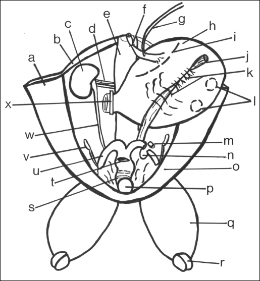
Figure 1 shows an illustration of the Rabbit Tissue Model (RTM) and the anatomy used during practice sessions. The stomach can be filled and used to practice dissecting techniques with both the laparoscopic scissors and cautery. Interrupted, running, or purse-string suturing can be practiced on the different organs.
Figure 1.
Drawing of a rabbit carcass model.
- (a) Inner right abdominal wall flap
- (b) Posterior abdominal muscles
- (c) Right kidney
- (d) Right renal vessels
- (e) Spine and retroperitoneal structures
- (f) Tie anchoring esophagus to spine
- (g) Catheter through esophagus into stomach
- (h) Stomach distended with saline
- (i) Site for an electrocautery incision
- (j) Sigmoid remnant embedded in stomach
- (k) Interrupted sutures
- (l) Site for purse-string sutures
- (m) Segment of left uterine horn removed prior to anastomosis
- (n) Opening in broad ligament
- (o) Inner left abdominal wall flap
- (p) Urinary bladder
- (q) Left thigh with intact femoral vessels
- (r) Site of disarticulation of leg
- (s) Circumferential incision in body of uterus for end-to-end anastomosis
- (t) Incision site in body of uterus for running anastomosis
- (u) Right horn of uterus
- (v) Right ovary
- (w) Right ureter
- (x) Clamp across pylorus
METHODS
Mature female rabbits are preferred because of their large bicornate uterus. The larger the animal, the better the specimen. After sacrificing the animals in a humane fashion, they are immediately skinned, and a laparotomy is performed by a midline longitudinal incision extending from the xiphoid to the pubis. Bilateral subcostal incisions then follow to create 2 triangular abdominal wall muscle flaps. Attention is then directed to the lower abdominal cavity. The urinary bladder is identified and emptied by exerting pressure on the dome.
A segment of rectosigmoid measuring approximately 6 inches is cleaned free of stool and preserved. Care is taken to tie off the vascular pedicle to the bowel without injuring the renal vessels. The portal vein is identified and tied off separately. The remaining large and small bowel is then excised to the level of the pylorus, followed by removal of the liver. The opening into the stomach through the pylorus is then gently stretched and the contents within the lumen extracted and the cavity irrigated copiously with normal saline.
The esophagus is then identified and gently dissected free. An entry is made into the diaphragm and dissection of the esophagus carried out to the upper-most portion of the thorax, at which point the organ is transected. A tie is then placed at the end of the esophagus and the stump sutured to the top of the remaining spinal musculature to hold it in place.
Attention is then directed to the lower extremities. The legs are removed at the knee joint leaving the thighs attached to the pelvis. The upper half of the animal carcass is then removed below the costal margins and the spine transected an inch above the level of the diaphragmatic insertion. Ideally, this is the point at which the animal carcass is also bled, if prior entry into the vasculature has not already accomplished this. However, on farm animals traditional slaughtering and bleeding techniques at the neck are done before proceeding with the above dissections.
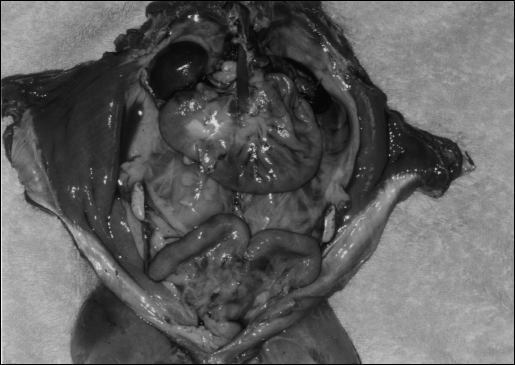
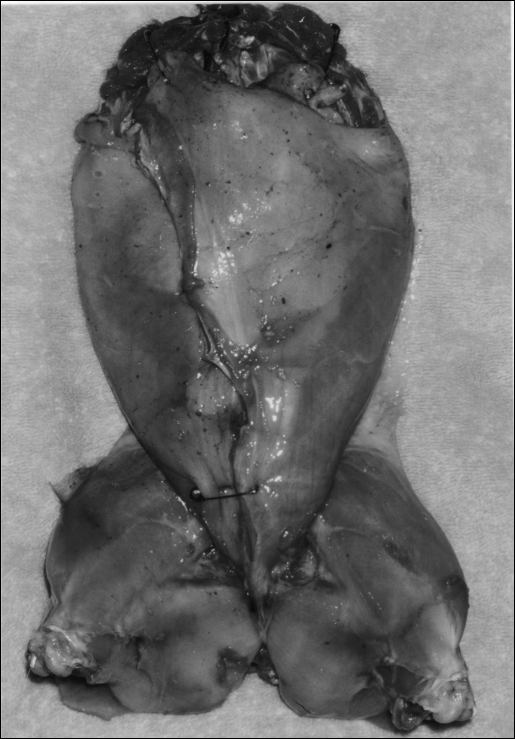
The preserved internal organs of the RTM are demonstrated in Figure 2. Clearly seen are the triangular muscle flaps, kidneys, esophagus, stomach, ovaries, and the bicornate uterus. The specimen is then rinsed a final time with saline and the abdominal flaps approximated with large safety pins as illustrated in Figure 3. Prior to freezing, the specimen is placed in an airtight plastic bag to preserve the tissue architecture and prevent drying and hardening. When needed, the specimen can be defrosted and kept moist with frequent applications of saltwater during the exercises. This model can be used repeatedly and frozen between use. When finished, simply discard as any other meat product.
Figure 2.
Rabbit Tissue Model with open abdominal flaps and visualization of the remaining internal organs, after disembowelment.
Figure 3.
Rabbit Tissue Model completed and ready for storage. Safety pigs used to close triangular abdominal wall flaps.
DISCUSSION
Many different lessons can be practiced using the RTM. The kidneys, renal vessels, ureters, the bicornate uterus, and ovaries can be used to practice laparoscopic dissecting techniques. Simple, running, interrupted suturing, and stapling practice can be performed on the inner and outer muscle flaps of the RTM. The esophagus and the bicornate uterus can be used to practice catheterization of tubular structures. The stomach can be filled with saline through a catheter in the esophagus and the pylorus closed off with an atraumatic clamp to hold the liquid in place. While the stomach is distended, an electrocautery incision can be placed and 2 muscular flaps raised. To leave the underlying mucosa intact during this exercise, careful dissection is required. The sigmoid remnant can be used to represent a ureter. Approximating the raised stomach muscular flaps over this remnant can simulate the reimplantation of a ureter. Purse-string suturing can also be practiced on the distended stomach. End-to-end anastomoses can be performed on both the horns and body of the rabbit uterus, as well as on the esophagus.
CONCLUSIONS
Surgeons at different levels of competency in laparoscopic surgery can benefit from the RTM. To obtain maximum flexibility during the exercises, a pelvic trainer large enough to accommodate the carcass is needed. The Laparoscopic Ring Simulation Trainer is the author's trainer of choice. It easily accommodates specimens of different sizes and provides a greater number of options in instrument positioning above the selected specimens.1
Although mature female rabbits provide ample practice material, the harvesting techniques described above can also be used, with minor alterations, to create tissue models of larger animals like dogs or pigs. When using a pig, the above preparation can be modified to leave behind the liver and gallbladder. This allows the practice of a laparoscopic simulation cholecystectomy. In the case of the pig, the entire bowel must be removed because the soft stool does not allow a sigmoid remnant to be preserved. The large pig kidneys provide a great resemblance to human kidneys. The pig bicornate uterus can be used as a bowel substitute. This permits surgeons to perform laparoscopic manual or stapler anastomoses in preparation for similar bowel anastomosis in humans.
The RTM provides ample practice material for laparoscopic simulation exercises. This is an inexpensive model to produce and the animals are not alive during the practice sessions. Laboratory animals designated for sacrifice, after termination of research protocols, can be recycled for this purpose. In the absence of laboratory-designated animals, farm animals can be obtained and used in a similar fashion.
Footnotes
Disclosure: The above manuscript entitled “Laparoscopic Ring Simulation Trainer” is an original document written by Dr. Marelyn Medina and is self-funded. Dr. Medina is president of Boriquen Creative Systems, that produces the laparoscopic simulation trainer called MedinaTrainer for sale.
References:
- 1. Medina M. The laparoscopic ring simulation trainer, JSLS 2002;6(1):69–75 [PMC free article] [PubMed] [Google Scholar]


