Abstract
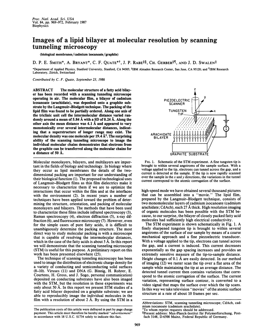
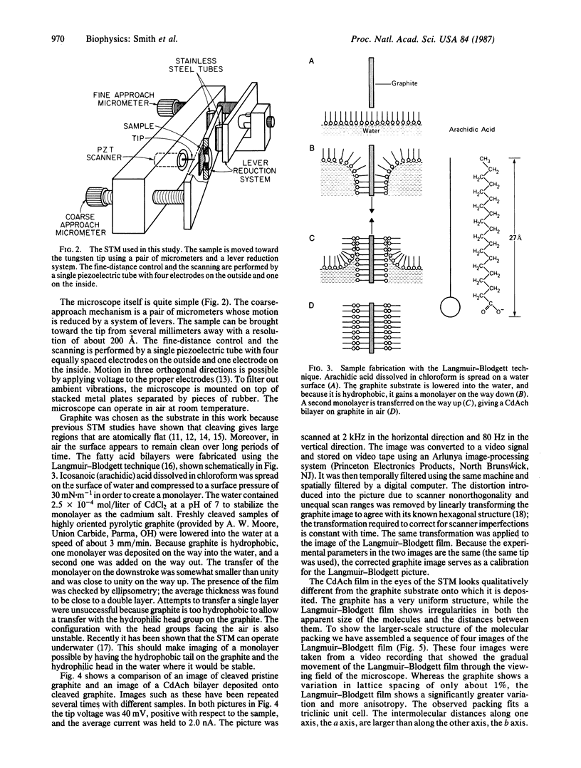
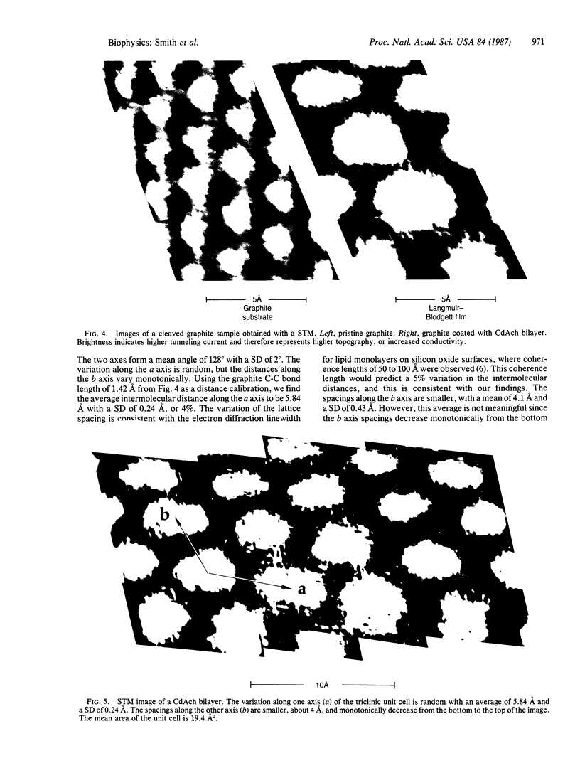
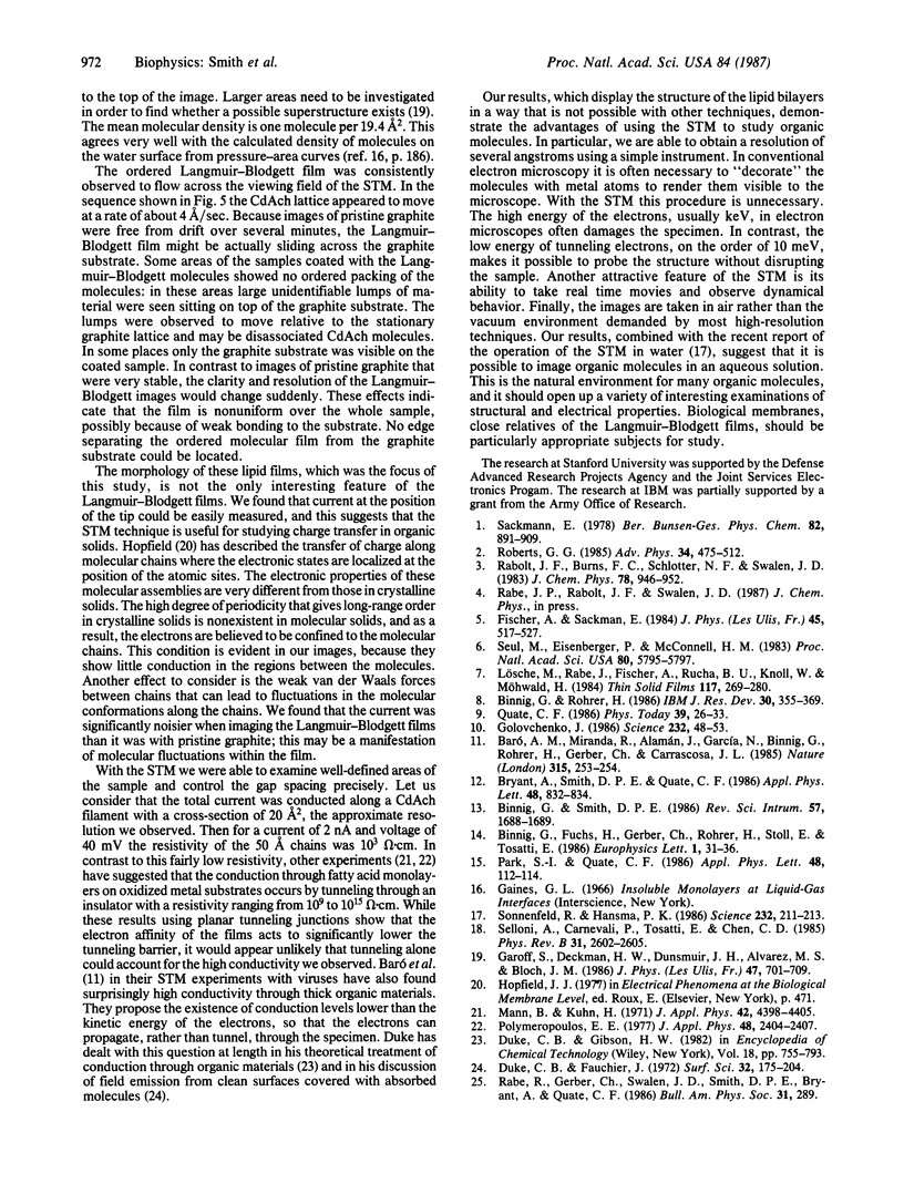
The molecular structure of a fatty acid bilayer has been recorded with a scanning tunneling microscope operating in air. The molecular film, a bilayer of cadmium icosanoate (arachidate), was deposited onto a graphite substrate by the Langmuir-Blodgett technique. The packing of the lipid film was found to be partially ordered. Along one axis of the triclinic unit cell the intermolecular distance varied randomly around a mean of 5.84 A with a SD of 0.24 A. Along the other axis the mean distance was 4.1 A and appeared to vary monotonically over several intermolecular distances, indicating that a superstructure of longer range may exist. The molecular density was one molecular per 19.4 A2. The surprising ability of the scanning tunneling microscope to image the individual molecular chains demonstrates that electrons from the graphite can be transferred along the molecular chains for a distance of 50 A.
Full text
PDF


Images in this article
Selected References
These references are in PubMed. This may not be the complete list of references from this article.
- Baró A. M., Miranda R., Alamán J., García N., Binnig G., Rohrer H., Gerber C., Carrascosa J. L. Determination of surface topography of biological specimens at high resolution by scanning tunnelling microscopy. Nature. 1985 May 16;315(6016):253–254. doi: 10.1038/315253a0. [DOI] [PubMed] [Google Scholar]
- Golovchenko J. A. The tunneling microscope: a new look at the atomic world. Science. 1986 Apr 4;232(4746):48–53. doi: 10.1126/science.232.4746.48. [DOI] [PubMed] [Google Scholar]
- Hornak J. P., Szumowski J., Rubens D., Janus J., Bryant R. G. Breast MR imaging with loop-gap resonators. Radiology. 1986 Dec;161(3):832–834. doi: 10.1148/radiology.161.3.3786741. [DOI] [PubMed] [Google Scholar]
- Selloni A, Carnevali P, Tosatti E, Chen CD. Voltage-dependent scanning-tunneling microscopy of a crystal surface: Graphite. Phys Rev B Condens Matter. 1985 Feb 15;31(4):2602–2605. doi: 10.1103/physrevb.31.2602. [DOI] [PubMed] [Google Scholar]
- Seul M., Eisenberger P., McConnell H. M. X-ray diffraction by phospholipid monolayers on single-crystal silicon substrates. Proc Natl Acad Sci U S A. 1983 Sep;80(18):5795–5797. doi: 10.1073/pnas.80.18.5795. [DOI] [PMC free article] [PubMed] [Google Scholar]
- Sonnenfeld R., Hansma P. K. Atomic-resolution microscopy in water. Science. 1986 Apr 11;232(4747):211–213. doi: 10.1126/science.232.4747.211. [DOI] [PubMed] [Google Scholar]


