Abstract
We propose the design of a teaching system named Interpretation and Diagnosis of Mammograms (INDIAM) for training students in the interpretation of mammograms and diagnosis of breast cancer. The proposed system integrates an illustrated tutorial on radiology of the breast, that is, mammography, which uses education techniques to guide the user (doctors, students, or researchers) through various concepts related to the diagnosis of breast cancer. The user can obtain informative text about specific subjects, access a library of bibliographic references, and retrieve cases from a mammographic database that are similar to a query case on hand. The information of each case stored in the mammographic database includes the radiological findings, the clinical history, the lifestyle of the patient, and complementary exams. The breast cancer tutorial is linked to a module that simulates the analysis and diagnosis of a mammogram. The tutorial incorporates tools for helping the user to evaluate his or her knowledge about a specific subject by using the education system or by simulating a diagnosis with appropriate feedback in case of error. The system also makes available digital image processing tools that allow the user to draw the contour of a lesion, the contour of the breast, or identify a cluster of calcifications in a given mammogram. The contours provided by the user are submitted to the system for evaluation. The teaching system is integrated with AMDI—An Indexed Atlas of Digital Mammograms—that includes case studies, e-learning, and research systems. All the resources are accessible via the Web.
Key words: Education, medical, computer-assisted instruction, computers in medicine, digital mammography
Introduction
Medical education is a challenge because it requires a vast range of material; intellectual, visual, and tactile skills; and the incorporation of large amounts of factual information. Traditionally, medical teaching is based on texts, lectures, and bedside teaching, with self-guided individual learning based on books.1 The traditional medical teaching and individual learning methods can be complemented with electronic learning systems delivered via the Internet, leading to the so-called e-learning paradigm. With the advancement of Web technology, e-learning has been a topic of interest in recent years and has become an important trend in medical education.
Alves et al.1 described their experience in designing a multi-agent system for e-learning in the medical area. The proposed system is based on the client/server network architecture, where the server holds the data warehouse, medical data, and the software agent that delivers medical content; on the client side, the interface is based via Internet technology. Shaw et al.2 and Johnson et al.3 presented the Adele system, which includes the pedagogical agent Adele, for assisting students as they assess and diagnose medical and dental patients in a clinical setting. In a typical use of Adele, the student is presented with a computer simulation of a clinical problem. The integrated system is downloaded and run on the client’s side. Shyu et al.4 proposed a system to establish a virtual medical school as the platform of an e-learning center, which provides a problem-based e-learning environment, and utilizes the hospital information system to capture and store clinical cases; medical students and residents can access the clinical cases online.
An e-learning platform consists of various complex activities, such as an authoring system that helps in creating and exchanging content, a learning management system that stores and manages content, and a run-time system that interacts with the user. These activities can be perceived and modeled as processes and executed as workflows. Vossen and Westerkamp5 present extensive bibliographical references and a prototypical system using workflow analysis technique to mange activities of learning.
The activities that compose an e-learning environment can be implemented as Web Services to support flexible module composition, interoperability, and reuse. A Web Service is a stand-alone software component that has a unique Uniform Resource Identifier (URI) that operates on the Web and offers possibilities to access services in distributed environments.5 The basic premises is that Web Services have a provider and users, and can be combined to build new services with more comprehensive functionality. Various works have been presented in the literature utilizing Web Services in the e-learning environment.5–10
In particular, applications related to the interpretation of mammograms and the diagnosis of breast cancer could derive significant benefits from the use of an appropriate ontology. Few works address the development of an ontology to represent the knowledge in the diagnosis of breast cancer. The most general ontology, termed Breast Cancer Imaging Ontology,11 developed within the project Medical Imaging with Advanced Knowledge Technologies, includes information regarding ultrasound, mammography, and magnetic resonance images, besides histopathology and clinical history of the patient. Podsiadly-Marczykowska et al. proposed MammoOnt,12 an ontology that represents information related to mammography. Rose et al.13 proposed an ontology limited to the terms available in the Digital Database for Screening Mammography (DDSM),14 with the aim of developing a Web Service that is able to retrieve mammographic images in a well-supported format. Although some ontologies have been developed, they are not available for reuse such as Breast Cancer Imaging Ontology,11 or they are not adequately general so as to be reused for the proposed work, such as the ontology proposed by Rose et al.13.
An ontology is a formal and explicit specification of an abstraction, i.e., a simplified vision of the world or domain that we wish to represent for a specific purpose. An ontology shares a domain of knowledge, defining a common vocabulary.15 An ontology can be used by software agents to establish a common agreement regarding the concepts and relationships of a specific domain of knowledge.16 An ontology defines the primary concepts in an application area, as well as the relationships among those concepts. It is independent of particular algorithms, such as database schema. Unlike a database, an ontology can express complex relationships among the concepts represented.17
An ontology can be used to formalize the representation of a knowledge domain, to describe a common and defined vocabulary for the data of a given application, to provide unified access to information through ontology-based querying for both human and computational processing, to improve management and integration of application data, and to facilitate data mining.
An ontology can be represented as a graph, where the nodes are connected by directed arcs (arrows). The nodes are concepts (implemented as classes) in the ontology, and the directed arcs are the relationships (implemented as properties) between the concepts.
In this paper, we propose the design of an e-learning system oriented to a specific problem to assist medical students in the interpretation of mammograms and diagnosis of breast cancer that we call INDIAM: INterpretation and DIAgnosis of Mammograms. The activities that compose the proposed e-learning system are modeled as Web Services, which can be reused by other medical e-learning environments. The information required to model the application knowledge (interpretation of mammograms and diagnosis of breast cancer) is represented by an ontology that is associated with an illustrated tutorial and a mammographic database. The resources available in INDIAM guide the medical student step-by-step in analyzing a mammogram correctly, simulate diagnosis of breast cancer with appropriate feedback, answer questions of the student according to the knowledge modeled by the ontology, and navigate through the illustrated hypermedia tutorial. The proposed e-learning system makes available a graphical interface via the Web with digital image processing tools that permit the student to draw the contours of lesions, the contour of the breast, and boundaries of clusters of microcalcifications during activities to simulate interpretation of diagnosis.
The paper is organized as follows. “An Overview of the Proposed e-Learning System: INDIAM” presents the general architecture of the INDIAM e-learning system.18,19 “The Application Knowledge Base” provides a description of each component of the application knowledge base, composed of a mammographic database and a breast cancer ontology.18 “The Breast Cancer Tutorial” presents a description of an illustrated tutorial on breast cancer.18 “The Web Services” contains the details of the Web services made available by INDIAM and their functionalities.18,19 “Evaluation of INDIAM” presents a methodology being used to evaluate the proposed e-learning system. “Integration of INDIAM into AMDI—An Indexed Atlas of Digital Mammograms” gives a succinct description of AMDI, an Indexed Atlas of Digital Mammograms,20–23 including a mammogram registration module, a research system module, and the e-learning system INDIAM. “Discussion and Conclusion” presents the concluding remarks.
An Overview of the Proposed e-Learning System: INDIAM
INDIAM is designed to assist a medical student or resident in the interpretation of mammograms and diagnosis of breast cancer, and is being developed using Web Services. Figure 1 illustrates a general overview of the system. INDIAM makes available a tutorial that uses education techniques to guide the users (doctors, students, or researchers) through concepts related to the diagnosis of breast cancer. It also makes available a Web Service to simulate the analysis and diagnosis of breast cancer (Diagnosis Simulation) using cases retrieved from a mammographic database, a Web Service to train the student in the interpretation of mammograms (Diagnosis Tutor), a Web Service to answer questions (Aquest) posed by the user, and a service to help the student to search the Web (Search) for information related to breast cancer diagnosis. The e-learning system integrates an ontology that provides controlled and consistent vocabularies to describe concepts and relationships, thereby enabling knowledge sharing.24 The system makes available a user-friendly graphical interface that is configured according to the service being provided.
Fig 1.
An overview of the architecture of INDIAM.
The Application Knowledge Base
The knowledge of the application is composed of a mammographic database and on ontology that represent the knowledge of the radiologist in breast cancer diagnosis.
The Mammographic Database
The mammographic database was modeled using PostgreSQL with image-handling extension (PostgreSQL-IE), an extended relational database management system (XRDBMS), that includes facilities to organize visual and conventional data and answer queries based on image content.23,25,26 It includes cases with all of the available mammographic views, radiological findings, diagnosis proven by biopsy, the patient’s clinical history, and information regarding the life style of the patient. Each exam of each case includes four views (two views of each breast: cranio-caudal or CC and medio-lateral oblique or MLO). To address the teaching aspects, the database links each mammogram with the contour of the breast, the boundary of the pectoral muscle (MLO views only), the contours of masses (if present), the regions of clusters of calcifications and the number of calcifications (if present), and the locations and details of any other features of interest. The contours of masses and regions of clusters of calcifications may be drawn interactively by an expert radiologist when including a new case in the database. The mammographic database also supports in inclusion of several mammographic exams of the same patient performed at different instants of time. This information can be used for temporal analysis of the breast to assess the natural modifications that occur during the life of a woman or to analyze interval cancer.
The Ontology
In this work, we propose the development of an ontology termed BreastCancerOnto that represents information regarding radiological findings in mammograms based on the Breast Imaging Reporting and Data System (BI-RADS) classification.27 The proposed ontology was designed by an expert breast radiologist, provides a complete system of X-ray mammography, and is freely available via Web.18 The ontology is part of the knowledge base of INDIAM,18,19 a system that makes it possible to search information via the Web considering the semantics of the search term and answer questions from the user, such as:
Which types of radiological findings are associated with malignant lesions?
Which categories of BI-RADS are associated with radiological findings and special cases with characteristics of malignancy?
Which procedure must be followed in the case of malignant diganosis?
What are the radiological findings associated with BI-RADS category 5?
How can a lesion associated with an oval form be classified?
Because the information in BreastCancerOnto is exhaustive, a vast range of questions may be asked: the “answer–question” or Aquest Web Service is designed to provide flexible and user-friendly interface for this purposed, as described in “Aquest: The Answer–Question Web Service.”
The proposed ontology has been implemented in Ontology Web Language28 using the Protégé Framework29 and the inference engine termed Jena.30 In its current version, BreastCancerOnto possesses 21 classes, 92 proprieties with 86 of them having an inverse property, and 161 instances. Figure 2 illustrates a section of Protégé showing the 21 classes available in BreastCancerOnto. Table 1 illustrates a set of properties and instances of the class “shape.”
Fig 2.
A section Protégé with the 21 classes available in BreastCancerOnto.
Table 1.
Properties and Instances of the Class “Shape”
| Properties | Instances |
|---|---|
| Is_associated_with_Shape-Birads | Round |
| Is_associated_with_Shape-Density | Oval |
| Is_associated_with_Shape-Margin | Lobulated |
| Inversed_of_is_associated_with_Neoplay-Shape | Irregular |
| Inversed_of_is_associated_with_FibroglandularDensity-Shape | |
| Inversed_of_is_associated_with_Lesions-shape | |
| Inversed_of_is_associated_with_State-Shape | |
| Inversed_of_is_associated_with_Texture-Shape | |
| Inversed_of_is_associated_with_Behavior-Shape | |
| Has_mammogram | |
| Has_title | |
| Has_paragraph | |
| Has_synonyms | |
| Has_hints | |
| Has_document | |
| Has_picture |
The Breast Cancer Tutorial
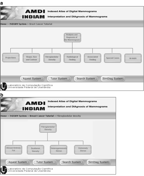
The INDIAM e-learning system provides a tutorial on breast radiology using hypertext and hypermedia technologies, which guides the users (doctors, students, or researchers) through the concepts related to the diagnosis of breast cancer. The tutorial is associated with BreastCancerOnto, includes informative text about specific subjects, provides access to a bibliography, and makes available tools to retrieve similar cases from the mammographic database. The breast cancer tutorial accesses and can be accessed by the Web Services described in “The Web Services.” The tutorial uses education techniques to provide to the user an easy strategy to navigate through the content of the tutorial. Figure 3a shows the initial graphical user interface for the tutorial. By choosing the option fibroglandular density, a new window is presented to the user with all categories according to the BI-RADS classification: almost entirely fat, scattered density, heterogeneously dense, and extremely dense; see Figure 3b. Figure 4 illustrates the content for the option heterogeneously dense. The links available in the left-top part of the window make it possible to obtain additional information on the subject being studied. The link termed References searches the Web using search Web Service, as described in “The Web Services.” The search takes into account the semantics of the term heterogeneously dense in the context related to breast cancer diagnosis. Figure 5 shows the results of the search. The link Relationships presents all the terms represented in the ontology related to heterogeneously dense, as shown in Figure 6. The links Documents and Database Cases bring from the ontology the documents previously stored that are related to the term and bring from the mammographic database new example cases, respectively.
Fig 3.
a The initial graphical user interface of the Breast Cancer Tutorial. b The interface after choosing the option fibroglandular density.
Fig 4.
The tutorial available for the topic heterogeneously dense.
Fig 5.
An illustration of the links obtained by the search using the term heterogeneously dense.
Fig 6.
The interface that displays the relationships between the term heterogeneously dense and the terms represented in BreastCancerOnto.
The Web Services
Web Services offer possibilities to access services in a distributed environment via the Web. When associated with an ontology, Web Services can incorporate semantics into the related activities. The proposed e-learning system is composed of four principal Web Services: the Search Web Services that makes available a tutorial on the diagnosis of breast cancer associated with the ontology, the Aquest Web Service that answers questions from the user based on BreastCancerOnto, the Diagnosis-Tutor Web Service to guide the beginner student through the steps of analysis of a mammogram; and the Diagnosis-Simulation Web Service that makes available a graphical user interface to stimulate the diagnosis of a given mammogram retrieved from the mammographic database.
Search Web Service
The Search Web Service makes use of Google and/or Yahoo engines to help the student to search information in the Web related to breast cancer diagnosis. The search engine takes into account the semantics represented in BreastCancerOnto, making the search more precise (with reduction of false positives). Figure 7 illustrates the graphical user interface available in the system. Figure 5 illustrates the result of a search with the term heterogeneously dense.
Fig 7.
The graphical user interface of the Search Web Service.
Aquest: The Answer–Question Web Service
This Web Service allows the user to pose questions to INDIAM using an oriented strategy to frame the questions. The Aquest Web Service integrates the Breast Cancer Tutorial, the Search Web Service, and BreastCancerOnto. A general overview of the architecture of Aquest is shown in Figure 8.
Fig 8.
A general overview of the architecture of Aquest.
Although BreastCancerOnto is able to answer any type of questions related to radiology of the breast, the Aquest Web Service limits the structure of the questions to four grammatical classes, with the last two of them being optional: (1) a question type, (2) a noun that represents a class of ontology, (3) a verb that represents a property, and (4) an adjective that represents an instance of the ontology.
Questions can be connected by the logical operators AND or OR. The question types are limited to what/which/how and allow the student to obtain information from the e-learning system by crossing classes of BreastCancerOnto with the associated properties. The Aquest Web Service uses the API Jena30 as the inference engine.
The student is not required to know the names of the classes, properties, or instances modeled by the ontology. Aquest Web Service provides a graphical user interface that guides the user through the terms represented in the ontology, as illustrated in Figure 9. A more flexible way to frame a question to the e-learning system is by using a natural language; however, this approach is beyond the scope of the present work.
Fig 9.
Aquest Web service interface.
The answers obtained by the Aquest Web Service are represented as links to the Breast Cancer Tutorial. This facility makes it possible to obtain additional information for each answer given by the system, as described in “The Breast Cancer Tutorial.”
Diagnosis-Tutor Web Service
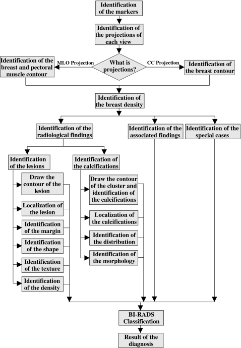
The Diagnosis-Tutor Web Service guides the user through the mammogram-interpretation flowchart (Fig. 10), designed by an expert radiologist. In a general way, the Diagnosis-Tutor Web Service has the following objectives: (1) provide an ergonomic environment for the presentation of the content, (2) guide the students through the content and assist them to reach the correct diagnosis, (3) provide a flexible and friendly environment to answer questions, and (4) present questions to guide the student during the learning process. The Diagnosis-Tutor Web Service communicates with the ontology, mammographic database, and the tutorial to send hints and appropriate feedback to the user. This service uses the Aquest Web Service, described in “Aquest: The Answer–Question Web Service,” to provide the answer–question user interaction when required by the user.
Fig 10.
The steps in analyzing a mammogram represented by a flowchart designed by an expert radiologist.
The Diagnosis Tutor presents to the student a mammographic case retrieved from the mammographic database (displaying the associated four views) to be analyzed and limits the actions of the user according to the steps proposed in the mammogram-interpretation flowchart. During the learning process, the student is required to fill out, in sequence, all the fields of each step of the diagnosis represented in the flowchart. If he or she makes a mistake, the Diagnosis Tutor provides feedback with the correct answer and gives the student the option to return to the previous step or to continue the analysis. Options for framing questions using Aquest or for searching in the Web using the Search Web Service are always available.
Figure 11 shows a stage in the analysis of a mammogram where the student has to draw the contour of the lesion in a given view. Digital image processing tools including zoom in, zoom out, and enhancement are available in the system to help the student in identifying the boundary of the lesion. At the point illustrated in Figure 11, the student has already identified the projection, the breast contour, and the identification of the fibroglandular density.
Fig 11.
Step of the Diagnosis Tutor to draw the contour of the lesion in a given view.
Diagnosis-Simulation Web Service
The Diagnosis-Simulation Web Service simulates an environment for the interpretation of mammograms and breast cancer diagnosis. The service displays the four views of a mammographic case retrieved from the mammographic database. The student analyzes each pair of views and asks for a specific report form to submit his or her diagnosis. The system makes available four report forms: to draw the contour of the breast and the pectoral muscle, to identify global radiological findings, radiological findings associated with lesions, and radiological findings associated with microcalcifications. After filling out the forms, the reports are submitted and are checked by the system; the system provides appropriate feedback in the case of errors. In addition to the analysis of the report, this service makes available the same digital image processing tools used in the Diagnosis-Tutor Web Service with which the student can draw the contour of the breast, delineate and remove the pectoral muscle (for MLO views), identify clusters of microcalcifications and indicate the position of each once, and draw the contours of lesions. The digital image processing tools was developed by the laboratory of scientific computation of Federal University of Uberlândia, and has zoom and enhancement techniques. The management system of INDIAM evaluates the degree of similarity between the answer of the user and the data stored in the mammographic database, helping the user if necessary. Figure 12 illustrates the graphical user interface for the Diagnosis-Simulation Web Service, with the lesion report form.
Fig 12.
Graphical user interface of the Diagnosis Simulation Web Service.
Evaluation of INDIAM
To evaluate the INDIAM system, we propose a methodology that involves students and experts in radiology of the breast. The methodology is composed of a set of forms and scripts with the aim of guiding the student through the analysis of the system. Each module that integrates INDIAM is evaluated separately. Each student receives the same script previously stored in a text file. The script contains instructions to be followed for each module:
Breast Cancer Tutorial—The script asks the student to surf through the map of contents, searching for the term related with the nomenclature of the BI-RADS.
Answer–Question System—The script requires the student to pose five queries to the Answer–Question System using the connective operators OR and AND. After that, he or she is required to pose five new queries using the connective operators.
Search System—The script guides the student to search the Web with a search term related to X-ray mammography.
Diagnosis-Tutor System—The script guides the student to select randomly a case in the mammographic database and follow the workflow, as proposed by the system.
Diagnosis-Simulation System—The script guides the student to select randomly a case in the mammographic database and carry out the diagnosis. In this module, the user is required to draw the boundary of the pectoral muscle and the contour of the breast, and to identify the density of the parenchyma. Any mass present should be classified according to BI-RADS.
At the end of the tests, each student is asked to fill out forms with her or his evaluation of each module. The forms include
A qualitative evaluation of the system, with analysis of the graphical user interface, and the occurrence of failure during the use of the system, and an evaluation of the available facilities to aid in the learning process.
A confidence evaluation, with analysis of the degree of confidence in the system during the steps of interpretation and diagnosis of mammograms. In this case, the answers given by the system are analyzed by an expert in breast radiology.
A quantitative evaluation, with analysis of the completeness of the information stored in the knowledge base of the system. A general evaluation of the system is also provided by the user.
The proposed methodology is being evaluated by ten students in medicine and two experts in radiology at the Universidade Federal de Uberlândia, Brazil.
Integration of INDIAM into AMDI—An Indexed Atlas of Digital Mammograms
AMDI20,22,23 is a system that integrates modules to permit the addition of new cases into the mammographic database by authorized radiologists and to assist research and education activities in breast cancer through a flexible and easy-to-use interface via the Web. Figure 13 shows a schematic representation of the flow of information in AMDI.
Fig 13.
An overall view of AMDI.
The research system called SISPRIM26,31,32—Sistema de Pesquisa para Recuperação de Imagens Mamograficas, that is, Research System for Retrieval Mammographic Images—integrated with AMDI, allows physicians and oncologists to study possible statistical correlation between the incidence of breast cancer and the life style of the patient. For this purpose, some of the important items of data supported by AMDI relate to information regarding the patient, including basic data about the clinical history of the patient as well as lifestyle, such as food habits, exercise, diet, and the use of antidepressive medication, tobacco, or alcohol. The SISPRIM research system also allows content-based image retrieval to assist the radiologist in breast cancer diagnosis. The physician can realize queries such as “Return all images that are similar to the given reference image, including the point that the patient uses an antidepressant” or “Return all images that are similar to the given reference image and have a malignant diagnosis.”
AMDI provides a tool that allows the user to download cases from the database, so as to make the information available to authorized medical and research communities interested in breast cancer diagnosis.
Discussion and Conclusion
In this work, we have presented INDIAM, an e-learning system to assist a medical student in the interpretation of mammograms and diagnosis of breast cancer. The proposed system is available via the Web and integrates an application knowledge base, a tutorial in X-ray mammography, and four Web Services.
The application knowledge base of INDIAM includes a mammographic database with complete clinical information and details regarding the lifestyle of the patient; global and local radiological findings, and an ontology termed BreastCancerOnto that represents the knowledge of the radiologist for the analysis of a mammographic case.
The activities that compose INDIAM were designed using Web Services that take benefit of sharing the Web Services with other e-learning environments, facilitating the upgrading of the proposed e-learning system, and the reuse of the Web Services designed by other providers. INDIAM is composed of four Web Services: Diagnosis Tutor is a Web Service that guides the student through the stages necessary to reach a diagnosis according to the steps designed by an expert radiologist. Diagnosis Simulation is a Web Service that evaluates the diagnosis done by a student and returns appropriate feedback in the case of errors. Both services use digital image processing tools that allow the user to draw contours of breast, lesions, and clusters of microcalcifications. The remaining two components are Search and Aquest Web Services; the latter provides facilities for constructing questions. Further work is being conducted to develop an interface using a natural language.
The proposed system also makes available an illustrated tutorial on breast cancer radiology designed using techniques of distance education.
INDIAM is integrated with AMDI—an Indexed Atlas of Digital Mammograms—an atlas available via the Web that also includes a research system to retrieve images based on content and a tool for inserting new mammographic cases by registered radiologists.
To evaluate the INDIAM system, we have proposed a methodology that involves students in medicine and radiologists specialized in X-ray mammography. The evaluation is in progress.
References
- 1.Alves V, Neves J, Nelas L, Marreiros F: Web-based medical teaching using a multi-agent system. Proceedings of AI2005, the Twenty-Fifth SGAI International Conference on Innovative Techniques and Applications of Artificial Intelligence, Applications and Innovations in Intelligent Systems XIII, Cambridge, UK, 25:14, 2005
- 2.Shaw E, Johnson WL, Ganeshan R: Pedagogical agents on the Web. Proceedings of the Third Annual Conference on Autonomous Agents, Seattle, WA, 1999, pp 283–290
- 3.Johnson WL, Shaw E, Marshall A, Labore C: Evolution of the user interaction: the case of Agent Adele. Proceedings of the 8th International Conference on Intelligent User Interfaces, Miami, Florida, 2003, pp 93–100
- 4.Shyu F-M, Liang Y-F, Hsu W-TA, Luh J-J, Chen H-S. A problem-based e-learning prototype system for clinical medical education. MedInfo. Amsterdam, The Netherlands: IOS; 2004. pp. 983–987. [PubMed] [Google Scholar]
- 5.Vossen G, Westerkamp P: E-learning as a Web service. Proceedings of the 7th International Database Engineering and Applications Symposium, Asunción, Paraguay, pp 242–249, 2003
- 6.Vossen G, Westerkamp P. Maintenance and exchange of learning objects in a Web services based e-learning system. Electronic Journal of e-Learning. 2004;2(2):293–304. [Google Scholar]
- 7.Moreale E, Vargas-Vera M. Semantic services in e-learning: an argumentation case study. International Forum of Educational Technology & Society. 2004;7:112–128. [Google Scholar]
- 8.Dickinson I, Wooldridge M: Towards practical reasoning agents for the semantic web. International Conference on Autonomaus Agents. New York, NY: ACM, 827–834, 2003
- 9.Vargas-Vera M, Motta E, Domingue J: AQUA: an ontology-driven question answering system. Proceedings AAAI Spring Symposium, Stanford, CA, 2003, pp 53–57
- 10.Moldt D, Ortmann J: A conceptual and practical framework for web-based processes in multi-agent systems. Proceedings of the Third International Joint Conference on Autonomaus Agents and Multiagent Systems, AAMAS, New York, NY, 2004, pp 1464–1465
- 11.Dasmahapatra S, Hu B, Lewis P, Shadbolt N: Ontology-based medical image annotation with description logics. In: 15th IEEE International Conference on Tools with Artificial Intelligence, Sacramento, CA, 2003, pp 77–82
- 12.Podsiadly-Marczykowska T, Goszczynka H, Guzik A: Towards an OWL mammographic ontology. In: The 3rd European Medical and Biological Engineering Conference, 4 pages on CDROM, Prague, Czech Republic, 2005
- 13.Rose C, Turi D, Williams A, Wolstencroft K, Taylor C: Web services for the DDSM and digital mammography research. In: Lecture Notes in Computer Science. Springer, Berlin, Germany, 2003, pp 376–385
- 14.Digital Database for Screening Mammography. http://marathon.csee.usf.edu/Mammography/Database.html. Accessed June, 2007
- 15.Gruber TR, Olsen G, Runkel JT. The configuration design ontologies and the VT elevator domain theory. Int J Human-Comput Stud. 1996;44:569–598. doi: 10.1006/ijhc.1996.0024. [DOI] [Google Scholar]
- 16.Schumacher M. Security engineering with patterns origins, theoretical model, and new applications. Lecture Notes in Computer Science—LNCS 2754. New York, NY: Springer; 2003. pp. 29–44. [Google Scholar]
- 17.Nusen MA. Medical informatics: searching for underlying components. Methods Inf Med. 2002;41:12–19. [PubMed] [Google Scholar]
- 18.Guliato D, Bôaventura RS, Rangayyan RM, Simedo MS, Macedo AAM: A teaching system for assistence in the interpretation of mammograms and the diagnosis of breast cancer. In: Proceedings of the International Special Topics Conference on Information Technology in Biomedicine—IEEE ITAB2006, volume October, Ioannina, Greece, 2006, 6 pages on CDROM
- 19.Bôaventura RS, Guliato D, Simedo MS, Maia MA, Macedo TAA, Rangayyan RM: INDIAM—um sistema de ensino para auxiliar estudantes na interpretação de mamogramas e diagnóstico de câncer de mama via Web. In WIM 2007—VII Workshop de Informática Médica, page 10 pages on CDROM, Porto de Galinhas, PE, Brazil, June 2007
- 20.Guliato D, Caetano M, Rangayyan RM, Marques PMA, Janones FR, de Deus Vinícus, Lima SC: AMDI: An indexed atlas of digital mammograms available via the Web. In: 7th International Workshop on Digital Mammography, Durham, NC, 2004, 4 pages on CDROM
- 21.Guliato D, Caetano M, Janones FR, de Deus V, Lima SC, Rangayyan RM, Bôaventura RS, Marques PMA: AMDI: An indexed atlas of digital mammograms availablevia the Web. III Latin American Congress on Biological Engineering, IFMBE Proceedings, 5, 2004, 4 pages on CDROM
- 22.Guliato D, de Melo EV, Bôaventura RS, Janones FR, de Deus V, Ranggayan RM: AMDI: An atlas to integrate case studies e-learning, and research systems via the Web. Proceedings of the IASTED International Conference on telehealth. Banff, AB, Canada, 2005, pp 69–74
- 23.Guliato D, Bôaventura RS, Melo EV, Rangayyan RM. AMDI: an indexed atlas of digital mammograms that integrates case studies, e-learning, and research systems via the web. In: Suri JS, Rangayyan RM, editors. Recent Advances in Breast Imaging, Mammography, and Computer-aided Diagnosis of Breast Cancer, Chapter 15. Bellingham, WA: SPIE; 2006. pp. 529–555. [Google Scholar]
- 24.Gruber TR. Toward principles for the design of ontologies used for knowledge sharing. Int J Human-Comput Stud. 1995;43:907–928. doi: 10.1006/ijhc.1995.1081. [DOI] [Google Scholar]
- 25.Stonebraker M, Rowe LA, Hirohama M. The implementation of Postgres. Knowledge and Data Engineering. 1990;2(1):125–142. doi: 10.1109/69.50912. [DOI] [Google Scholar]
- 26.Guliato D, Rangayyan RM, Melo EV, Soares RC: A system for content-based image retrieval and analysis of mammograms using PostgresSQL with image-handling extension. In: Proceedings of the Fifth IASTED International Conference on Biomedical Engineering, pp 402–408, Innsbruck, Austria, February 2007.
- 27.American College of Radiology. Breast Imaging Reporting and Data System BI-RADS. American College of Radiology, Reston, VA, 4th edition, 2004
- 28.OWL. Web ontology language overview—w3c. http://www.w3.org/TR/owl-features/. Accessed on June 2006
- 29.Protégé. Documentation. http://protégé.stanford.edu/doc/users.html. Accessed in: June 2006
- 30.Jena. A semantic web framework for java. http://jena.sourceforge.net/documentation.html. Accessed in June 2006
- 31.Melo EV, Guliato D, Rangayyan RM, Soares RS: SISPRIM—sistema de pesquisa com suporte para recuperação de imagens por conteúdo. In: WIM2006—VI Workshop de Informatica Medica, Vila Velha, ES, Brazil, Junho 2006.
- 32.Guliato D, Melo EV, Rangayyan RM, Carvalho JD, Soares RC. PostgreSQL-IE: An Image-handling Extension for PostgreSQL. Journal of Digital Imaging. 2008;January 23:18214614. doi: 10.1007/s10278-007-9097-5. [DOI] [PMC free article] [PubMed] [Google Scholar]


