Abstract
A temporal subtraction image, which is obtained by subtraction of a previous image from a current one, can be used for enhancing interval changes (such as formation of new lesions and changes in existing abnormalities) on medical images by removing most of the normal structures. However, subtraction artifacts are commonly included in temporal subtraction images obtained from thoracic computed tomography and thus tend to reduce its effectiveness in the detection of pulmonary nodules. In this study, we developed a new method for substantially removing the artifacts on temporal subtraction images of lungs obtained from multiple-detector computed tomography (MDCT) by using a voxel-matching technique. Our new method was examined on 20 clinical cases with MDCT images. With this technique, the voxel value in a warped (or nonwarped) previous image is replaced by a voxel value within a kernel, such as a small cube centered at a given location, which would be closest (identical or nearly equal) to the voxel value in the corresponding location in the current image. With the voxel-matching technique, the correspondence not only between the structures but also between the voxel values in the current and the previous images is determined. To evaluate the usefulness of the voxel-matching technique for removal of subtraction artifacts, the magnitude of artifacts remaining in the temporal subtraction images was examined by use of the full width at half maximum and the sum of a histogram of voxel values, which may indicate the average contrast and the total amount, respectively, of subtraction artifacts. With our new method, subtraction artifacts due to normal structures such as blood vessels were substantially removed on temporal subtraction images. This computerized method can enhance lung nodules on chest MDCT images without disturbing misregistration artifacts.
Key words: Temporal subtraction, nonlinear warping, computer-aided diagnosis, chest CT, image registration
Introduction
Detection of subtle lesions on computed tomography (CT) images is a difficult task for radiologists because subtle lesions such as small lung nodules tend to be low in contrast, and a large number of CT images must be interpreted in a limited time. A temporal subtraction image, which is obtained by subtraction of a previous image from a current one, can be used for enhancing interval changes (such as formation of new lesions and changes in existing abnormalities) on medical images by removal of most normal structures. Therefore, for detection of lesions in chest radiographs, the temporal subtraction method has been applied successfully to clinical cases, leading to an improvement of radiologists’ diagnostic accuracy and a reduction of their reading time.1,2
With the subtraction method, it is important to employ an image-warping technique for accurately deforming the previous image to match the current image. If the warping is incorrect, normal structures may produce artifacts in the subtraction image, and the image quality can be degraded. Many warping techniques for two-dimensional (2-D) images in chest radiography have been developed in the field of computer-aided diagnosis.1,3–7 In the temporal subtraction obtained with multiple-detector computed tomography (MDCT) volume images that is used in thoracic examinations, it is necessary to employ a more complex three-dimensional (3-D) registration and warping of lung regions between the current and previous images.
Itai et al. proposed a computerized method for providing temporal subtraction images with MDCT volume images.8–11 Although the quality of the subtraction images was relatively good in general, misregistration appeared as artifacts in these images. They have also noted the same type of artifacts in another approach to temporal subtraction images obtained with MDCT.12
In this study, we developed a new 3-D voxel-matching method for reducing the artifacts caused by normal structures, including blood vessels, in order to obtain accurate subtraction of two thoracic MDCT images.
Materials and Methods
Image Database
We used two types of MDCT images obtained with multislice CT scanners (Light Speed QXi, GE, Milwaukee, WI, USA; and Aquilion, Toshiba, Japan), with four row detectors in the Light Speed QXi scanner and 16 row detectors in the Aquilion scanner, to develop a new method for the temporal subtraction technique applied to volume data. The matrix size for each slice image was 512 × 512, and the voxel size ranged from 0.488 to 0.712 mm (mean, 0.646 mm) on the x- and y-axes and 5.00 or 1.00 mm on the z-axis. In order to increase the resolution in the z direction, we employed interpolated images between slice images so that the resolution in three directions was isotropic. The volume data used in this study consisted of ten normal cases and ten abnormal cases with lung nodules. All cases included both previous and current CT images.
3-D Nonlinear Image Warping by Use of 3-D Voxel Matching and 3-D Image Subtraction
One of the important problems in temporal subtraction images obtained from chest images and thoracic CT is that subtraction images commonly include artifacts which are called subtraction artifacts here. The subtraction artifacts are created by slight differences in the size, shape, and/or location of anatomic structures such as blood vessels, nodules, chest walls, ribs, and other lung and cardiac structures, which are included in both the current and previous images. This slight difference can be as small as 1 pixel in 2-D images and 1 voxel in 3-D images, which may cause disturbing subtraction artifacts in temporal subtraction images that may be difficult to distinguish from new abnormalities or changes in existing lesions. Thus, it is very important to remove these subtraction artifacts from temporal subtraction images. It should be noted that these subtraction artifacts would disappear when the anatomic structures included in both current and previous images are identical, which, in general, is not the case. For example, a temporal subtraction image obtained from two successive CT scans can have noticeable subtraction artifacts on very small lung structures due to pulsation.
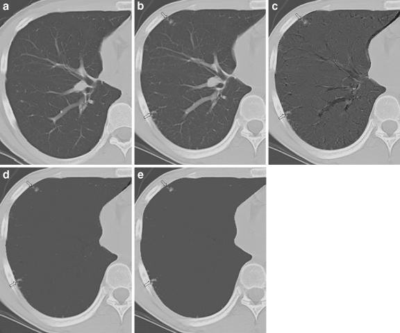
A new voxel-matching technique is an efficient method for substantially removing these subtraction artifacts, as described below. An image-warping technique such as that described in the “Appendix” is first applied to the current and the previous image, as shown in Figure 1a,b, respectively, in order to obtain shift vectors5 which represent the extent of deformation (or warping) of the previous image relative to the current image. Based on these shift vectors on the current image, the previous image can be warped to produce a temporal subtraction image. However, the temporal subtraction image obtained by the subtraction of the warped previous image from the current image would usually contain considerable subtraction artifacts, as shown in Figure 1c, even if the general appearance of the warped previous image would be very similar to that of the current image.
Fig 1.
Comparison of a previous image, b current image, and temporal subtraction images obtained c without and d with the voxel-matching technique by use of a 3 × 3 × 3 kernel and e a 5 × 5 × 5 kernel.
With the voxel-matching technique, for a given location in the current image, we initially identify the corresponding location in the warped previous image. We then search a voxel in the previous image (or in the warped previous image) within a small search volume, which may be called a kernel, such as a cube of 3 × 3 × 3 centered at the corresponding location, in order to identify the matched voxel which has a voxel value identical or nearly equal to the voxel value at the given location in the current image. This search of the matched voxel is repeated for all of the voxels in the current image. The warped previous image is then replaced by the matched-voxel warped previous image in which the voxel values are generally identical to or nearly equal to the voxel values in the current image except for the voxel values in new lesions or changes in existing abnormalities. Therefore, an improved temporal subtraction image with significantly reduced artifacts can be obtained by subtraction of the matched-voxel warped previous image from the current image, as shown in Figure 1d,e. With the voxel-matching technique, it is possible to remove subtraction artifacts due to very slight differences in the size, shape, and location of normal anatomic structures, thus producing very smooth temporal subtraction images except for new abnormalities, as clearly illustrated in Figure 1d,e. In addition, the majority of the noise in CT images can be removed by use of the voxel-matching technique, as shown by the smooth background in the temporal subtraction images in Figure 1d,e.
However, it should be noted that, when the size of a new lesion is extremely small such as 1 or 2 voxels, this tiny lesion would be removed. The temporal subtraction technique, however, has been applied generally to the detection of relatively large abnormalities; for example, the size of small lung nodules may be in the range of 2 mm or larger. If the size of a nodule is 1 mm or less, such a nodule might be eliminated by this technique. The size of a search volume can be made larger than 3 × 3 × 3, such as 5 × 5 × 5, which may remove some additional large artifacts, as shown in Figure 1e, but may remove some small lesions and also tend to reduce the size of large lesions. In addition, a search volume can be changed to take various forms, including a sphere and irregular shapes.
The voxel-matching method is one of the optimization registration tools for removal of the subtraction artifacts in a temporal subtraction technique, and it can be applied to 2-D images such as chest radiographs by use of a pixel-matching method instead of the voxel-matching in 3-D images. It is important to note that, with the voxel-matching or the pixel-matching technique, the voxel values or pixel values in the previous image must be approximately equal to those in the current image. This condition is usually satisfied in CT images obtained with the same scanner. However, chest radiographs generally have different pixel values because of the variation in exposure conditions, and thus it is necessary to adjust the pixel values for one of the chest images such as a previous image, so that the majority of the pixel values in the current image are comparable to those in the previous image. It should be noted that the voxel-matching and the pixel-matching technique can be applied not only to temporal subtraction images but also to other subtraction medical images such as digital subtraction angiography images, CT angiography images, and bilateral subtraction and contralateral subtraction images.
In general, the temporal subtraction technique can display temporal changes, but normal structures still appear as subtraction artifacts such as blood vessels on subtraction images (see Fig. 1c). On the other hand, the voxel-matching method can reduce the subtraction artifacts (see Fig. 1d). With voxel-matching, each voxel in the current image is registered or matched to the one in the previous image, which is the most similar to the current voxel, to reduce any slight difference in the temporal subtraction image.
In order to apply the new voxel-matching method for a substantial reduction in subtraction artifacts on the subtraction images, it is important initially to provide a proper registration of anatomic structures, which can be obtained successfully by use of global and local matching techniques.4 With the voxel-matching method, the correspondence not only between the structures but also between the voxel values in the current and the previous images is determined.
Results
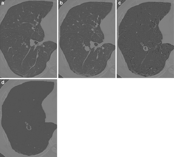
We applied this temporal subtraction method to images of 20 chest cases obtained with MDCT. The difference in time between the previous and current examination was in the range from 3 to 6 months. Figure 2 illustrates the results of the use of our temporal subtraction method. Figure 2a,b are the previous and current images, respectively, where the size of a nodule in Figure 2b is larger than that in Figure 2a. Figure 2c,d show the subtraction images without and with use of the voxel-matching technique. It is shown in Figure 2d that subtraction artifacts are substantially removed with a clean and smooth background, illustrating clearly several small nodules and one large ring-shaped nodule that indicate a temporal change in the size of the nodule. It may be noted also that the shape and size of the ring in Figure 2d is slightly different from that in Figure 2c; this is due to the use of the voxel-matching technique.
Fig 2.
Comparison of a previous image, b current image, and temporal subtraction images obtained c without and d with the voxel-matching technique by use of a 3 × 3 × 3 kernel.
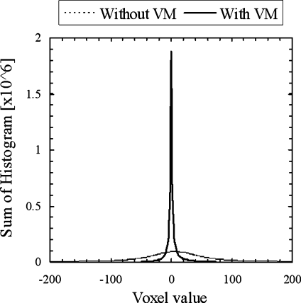
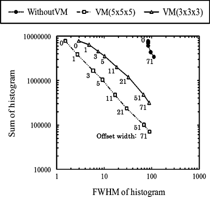
In order to evaluate the usefulness of the voxel-matching technique for removal of subtraction artifacts, we compared two histograms, without and with voxel matching, as shown in Figure 3, which were obtained from all voxels in the lungs in the 3-D temporal subtraction images. These histograms include voxels due to subtraction artifacts and a small fraction of nodules corresponding to interval changes. It is apparent in Figure 3 that the distribution of voxel values with the voxel-matching technique is much narrower than that without the technique, thus indicating that the majority of subtraction artifacts were removed by use of the voxel-matching technique. It should be noted that the voxel values included in this narrow histogram for the temporal subtraction image with the voxel-matching technique are due to image noise in CT and also to extremely low-background structures such as small blood vessels and parenchymal patterns in the lungs, which are usually not visualized in temporal subtraction images. In order to remove this low-level background, we employed an offset window, which is placed over a small range of voxel values, such as an offset window width of 5 to range from −2 to +2; the voxel values from −2 to 2 are then removed. Then, the subtraction image is shown by removing those voxel values in the offset width, the effect of which was examined at the offset width of 0, 1, 3, 5, 11, 21, 51, and 71, as illustrated in Figure 4.
Fig 3.
Comparison of histograms of voxel values in lungs for temporal subtraction images obtained without and with the voxel-matching technique.
Fig 4.
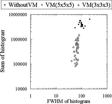
Relationship between the sum and the FWHM of histograms of voxel values in lungs for temporal subtraction images, obtained without and with the voxel-matching technique by use of 3 × 3 × 3 and 5 × 5 × 5 kernels, when the offset width is varied from zero to 71.
The magnitude of artifacts remaining in subtraction images was examined by the use of the full width at half maximum (FWHM) and the sum of a histogram of voxel values, which may indicate the average contrast and the total amount, respectively, of subtraction artifacts. Figure 4 shows the relationship between the sum and the FWHM of these histograms obtained without and with the voxel-matching technique by use of two kernels of 3 × 3 × 3 and 5 × 5 × 5, when the offset width was changed from 0 to 71. At very small offset widths, the sums of the histograms for the three conditions above were comparable, whereas the FWHM of histograms with the voxel-matching technique was extremely low, which implies that the contrast of the subtraction artifacts obtained with the voxel-matching technique would be extremely low and would not be visualized. As the offset width increases, the sum of histogram decreases, although the FWHM increases. At a large offset width of 71, the FWHMs of the three conditions become comparable, but the sum of the histograms is reduced substantially by use of the voxel-matching technique, which clearly indicates the usefulness of the voxel-matching technique in providing a substantial reduction of subtraction artifacts. Figure 5 shows the sum and the FWHM for temporal subtraction images obtained from 20 clinical cases without and with the voxel-matching technique at an offset width of 51. It is apparent that the subtraction artifacts in these cases were largely reduced by use of the voxel-matching technique with a kernel size of 3 × 3 × 3 or 5 × 5 × 5.
Fig 5.
Illustration of the sum and the FWHM of histograms of voxel values for temporal subtraction images on 20 clinical cases without and with the voxel-matching technique.
Conclusion
In this study, we have developed a new method for removing subtraction artifacts in temporal subtraction images applied to thoracic CT images. With our new method, subtraction artifacts due to normal structures such as blood vessels were substantially removed on temporal subtraction images. This computerized method can enhance lung nodules on chest MDCT images without disturbing misregistration artifacts. Preliminary results indicated that our temporal subtraction method may be useful for radiologists in the detection of interval changes on MDCT images.
Appendix: A Method of Temporal Subtraction on 3D Thoracic Images
A. Overall scheme of the temporal subtraction method
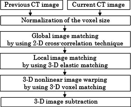
For a temporal subtraction method, registration between the current and the previous image is the most important task because the image quality on the subtraction image would be degraded due to some artifacts caused by incorrect registration. In our temporal subtraction method, the registration is achieved by global image matching, local image matching, and 3-D nonlinear image-warping techniques as illustrated in Figure 6. First, the voxel size of the previous and current images is normalized by using a linear interpolation technique. Then, we employ a global matching technique to correct for the global displacement caused by variation in patient positioning. For more accurate registration, a local matching technique based on 3-D elastic matching is applied to obtain a shift vector for each voxel, which represent the extent of warping of the previous image relative to the current image. The previous image is then warped by use of shift vectors nonlinearly. In addition, the voxels of the warped previous image are matched to those of the current image by using the voxel-matching technique developed in this study. Finally, the matched-voxel warped previous image is subtracted from the current image, thus providing a temporal subtraction image.
Fig 6.
Illustration of the overall scheme of a 3-D temporal subtraction method.
B. Global matching by use of a 2-D template matching technique
A global shift vector, which can correct for global temporal displacement caused by patient positioning, is determined on each current slice image. In this matching process, 2-D template matching based on a 2-D cross-correlation method is employed.8 First, blurred images are obtained from the previous and the current images by use of a Gaussian filter (kernel size 15 × 15), and we reduce the matrix size from 512 × 512 to 128 × 128 in the x–y plane for reducing the computation time and for matching only of large structures. In each current slice image, a rectangular region including the lung area is selected as a template image. We move the template image on the previous slice image in order to determine the global shift vector which is obtained from the template location with the maximum of the 2-D cross-correlation value, which indicates the similarity between the current and the previous slice images.
C. Local matching by use of a 3-D elastic matching technique
In order to achieve a high accuracy in matching the current and the previous image, we employ the local matching technique to determine local shift vectors. First, a number of the template and the search area volumes of interest (VOIs) are automatically located within the lung regions in the current and previous images, respectively. The matrix sizes of the template and the search area VOIs are 32 × 32 × 16 and 64 × 64 × 32, respectively. The distances between the adjacent VOIs are 16 pixels in the x–y plane and 8 pixels in the z direction. Then a 3-D cross-correlation value for each VOI pair is calculated with translation (producing shift vector) of the template VOI in the search area VOI. The local shift vector for each VOI pair is determined when the 3-D cross-correlation value becomes the maximum.9
The shift vector which is used for image warping is obtained by a combination of the global shift vector and the local shift vector determined previously. However, the orientation and the amplitude of the shift vector tend to suddenly change in comparison with those of the adjacent shift vectors due to noise in MDCT images. To overcome this problem, Itai et al.10 employ a 3-D elastic matching method for smoothing shift vectors.
With a 2-D elastic matching method, it is possible to obtain the shift vector, which preserves a high cross-correlation value and high consistency over the other shift vectors, as Li et al.13 have mentioned. We employed a 3-D elastic matching technique to deal with the shift vector in 3-D space. In the elastic matching method, the smoothed shift vector can be obtained by minimizing of a cost function that is a weighted sum of an internal and an external energy. The internal energy is given by the squared sum of the first- and second-order derivative values of the shift vectors. The smoother the shift vectors, the smaller the internal energy. On the other hand, the external energy is equal to the negative value of the 3-D cross-correlation value which is obtained with the VOI pair. A shift vector with a large correlation value can provide a small external energy. Therefore, with this elastic matching method, the smoothed shift vector can be obtained by taking into account not only the similarity between the current and the previous images but also the consistency of the shift vectors. With the smoothed shift vectors obtained, the shift vectors in all voxels in the previous image are determined by use of a tri-interpolation method.
Footnotes
USPHS Grants CA62625, CA98119.
References
- 1.Kakeda S, Kamada K, Hatakeyama Y, et al. Effect of temporal subtraction technique on interpretation time and diagnostic accuracy of chest radiography. Am J Roentgenol. 2006;187:1253–1259. doi: 10.2214/AJR.05.1270. [DOI] [PubMed] [Google Scholar]
- 2.Sakai S, Yabuuchi H, Matsuo Y, et al. Integration of temporal subtraction and nodule detection system for digital chest radiographs into picture archiving and communication system (PACS): four-year experience. J Digit Imaging. 2007;21:91–98. doi: 10.1007/s10278-007-9014-y. [DOI] [PMC free article] [PubMed] [Google Scholar]
- 3.Kano A, Doi K, MacMahon H, et al. Digital image subtraction of temporally sequential chest images for detection of interval change. Med Phys. 1994;21:445–461. doi: 10.1118/1.597308. [DOI] [PubMed] [Google Scholar]
- 4.Ishida T, Ashizawa K, Engelmann R, et al. Application of temporal subtraction for detection of interval changes on chest radiographs: Improvement of subtraction images using automated initial image matching. J Digit Imaging. 1999;12:77–86. doi: 10.1007/BF03168846. [DOI] [PMC free article] [PubMed] [Google Scholar]
- 5.Ishida T, Katsuragawa S, Nakamura K, et al. Iterative image warping technique for temporal subtraction of sequential chest radiographs to detect interval change. Med Phys. 1999;26:1320–1329. doi: 10.1118/1.598627. [DOI] [PubMed] [Google Scholar]
- 6.Katsuragawa S, Tagashira H, Li Q, et al. Comparison of temporal subtraction images obtained with manual and automated methods of digital chest radiography. J Digit Imaging. 1999;12:166–172. doi: 10.1007/BF03168852. [DOI] [PMC free article] [PubMed] [Google Scholar]
- 7.Difazio MC, MacMahon H, Xu XW, et al. Digital chest radiography: effect of temporal subtraction images on detection accuracy. Radiology. 1997;202:447–452. doi: 10.1148/radiology.202.2.9015072. [DOI] [PubMed] [Google Scholar]
- 8.Ishida T, Katsuragawa S, Abe H, et al: Development of 3D CT temporal subtraction based on nonlinear 3D image warping technique. In: Proc. of the 91st Radiological Society of North America: 111, 2005
- 9.Ishida T, Katsuragawa S, Kawashita I, et al. Temporal subtraction on 3D CT images by using nonlinear image warping technique. Int J Comput Assist Radiol Surg. 2006;1:468. [Google Scholar]
- 10.Itai Y, Kim H, Ishikawa S, et al.: 3D elastic matching for temporal subtraction employing thorax MDCT image. In: Proc of the World Congress on Med Phys and Biomedical Engineering 2181–2191, 2006
- 11.Itai Y, Kim H, Ishiwaka S, et al: Development of temporal subtraction multislice CT images by using a 3D local matching with a genetic algorithm. In: Proc The 92nd Radiological Society of North America 779, 2006
- 12.Takao H, Doi I, Watanabe T, et al. Temporal subtraction of thin-section thoracic computed tomography based on a 3-dimensional nonlinear geometric warping technique. J Comput Assist Tomogr. 2006;30:283–286. doi: 10.1097/00004728-200603000-00023. [DOI] [PubMed] [Google Scholar]
- 13.Li Q, Katsuragawa S, Doi K. Improved contralateral subtraction images by use of elastic matching technique. Med Phys. 2000;27:1934–1943. doi: 10.1118/1.1287112. [DOI] [PubMed] [Google Scholar]


