Abstract
Purpose
The study introduces reference data for a computer-aided analysis. The semiautomated computer-aided diagnostic system provides the estimation of joint space width at the distal interphalangeal joints, considering gender-specific and age-related changes.
Patients and methods
869 subjects (351 female/518 male) with hand x-rays were included and underwent measurements of joint space distances at the distal interphalangeal articulation (JSD-DIP) of the second to the fifth finger using computer-aided joint space analysis (CAJSA).
Results
Data showed a notable age-related decrease of CAJSA parameters, and an accentuated age-related joint space narrowing in women. Males showed a significantly wider JSD-DIP (+ 16.7%) compared to the female cohort for all age groups. Both men and women revealed an accentuated decrease of JSD-DIP (total) in the age group from 10 to 15 years (for men −10.5% and for women −17.6%). After the age of 21 years a continuous decline of the JSD-DIP (total) is observed.
Conclusion
Our data present gender-specific and age-related normative reference data for computer-aided joint space analysis, which provide a valid and reliable differentiation between disease-related joint space narrowing and age-related joint space narrowing, particularly in patients with osteoarthritis of the fingers.
Key words: Computer-aided diagnosis, distal interphalangeal joint, joint space distance, normative reference value, osteoarthritis, rheumatoid arthritis
INTRODUCTION
The hand is a common site of peripheral osteoarthritis, which is underestimated as a cause of disability and, in addition, its effect on quality of life may be considerable.1 The most common sites for finger osteoarthritis are the distal interphalangeal joints (DIP), proximal interphalangeal joints (PIP), and the base of the thumb.2–5 Osteoarthritis of the hand has an incidence rate of approximately 100/100,000 person per year.6 Cross sectional studies have estimated the prevalence of radiographic hand osteoarthritis in those patients over 65 years as ranging from 64 to 78% in men and 71 to 99% in women.7–10
Conventional radiography is a widely available and cost-effective method, and remains the standard of diagnosis for the detection and quantification of joint alteration in the course of finger osteoarthritis.11 The assessment of disease is performed by visualization of changes in joint space width as an indirect sign of cartilage loss. The disadvantage of conventional imaging is the limited sensitivity in detecting an early narrowing of the joint space.12 The conventional assessment of joint space width is influenced by the subjective scoring depending on the experience of the clinician, and is characterized by a remarkable interobserver variation of the measurements.13
Computer-aided diagnosis (CAD) has been consequently refined and improved, and is increasingly accepted in the field of radiological diagnostics.14–16 Although early measurements of joint space widths were carried out only on large joints, especially at the hip and knee,17–20 the new computer-aided joint space analysis (CAJSA, Version 1.3.6; Sectra; Sweden) is a recently developed approach, which conducts semiautomated measurements of joint space distances at the distal interphalangeal articulation of the second to the fifth finger. In recent studies, CAJSA have been able to detect and quantify disease-related joint-space narrowing of the metacarpal–phalangeal joint (MCP) caused by the course and severity of rheumatoid arthritis, which is accelerated in early RA.21,22
The aim of this study is to introduce reference data for computer-aided joint space analysis (CAJSA), a semiautomated computer-aided diagnostic system for the measurements of joint space distances, and to quantify gender-specific and age-related differences regarding the distal interphalangeal articulation.
PATIENTS AND METHODS
Patients
This prospective study enrolled 869 subjects (351 female, 518 male). All subjects underwent measurements of the joint space distance of the distal interphalangeal articulation (JSD-DIP in centimeters) using CAJSA technology.
Mean age was 35.5 years with a standard deviation of 19.2 years and an age range of 6.1 to 95.3 years (females: mean age 37.9 ± 20.8 years, males: 33.2 ± 17.5 years). Digital radiographs of the hand were taken from each subject between 2001 and 2005 who were admitted to the University clinic because of fracture exclusion. Each hand radiograph was read by two musculoskeletal radiologists for evidence of osteoarthritis using the Kellgren–Lawrence grading system with a standard atlas23: grade 0 = normal joint; grade 1 = small osteophyte of doubtful significance; grade 2 = definite osteophyte; grade 3 = osteophyte and joint space narrowing; grade 4 = severe joint space narrowing. In cases of ambiguity, a third musculoskeletal radiologist reviewed the radiographs.
Exclusion criteria were determined by an extensive questionnaire, and included: visible metallic material (ie, splints and material after osteosynthesis; n = 899), signs of fracture (n = 4,867), amputation (n = 38), endocrinological diseases known to affect bone metabolism (eg, hyper/hypoparathyroidism, Cushing disease; n = 245), rheumatic diseases (eg, rheumatoid arthritis; n = 1,237), renal disorders (n = 1,084), genetic diseases (n = 23), oncological diseases (n = 1,255), medication with bone-influencing drugs (eg, steroids, vitamin D, or calcium intake; n = 182), Kellgren–Lawrence grade higher than 1 (n = 534) and incorrect hand positioning (n = 103).
Methods
Measurement of Joint Space Width (By Computer-aided Joint Space Analysis)
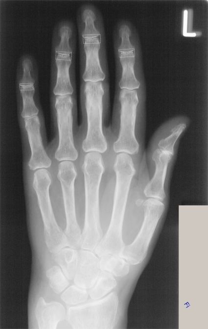
The computer-aided joint space analysis (Version 1.3.6; Sectra; Sweden) was used to determine JSD-DIP based on radiographs of the hand (Fig. 1). All digital plain radiographs of the hand were acquired by a Siemens Multix device (Siemens, Erlangen, Germany) under the following standardized conditions: filter 1.0, film focus distance 1 m, aluminum 80, tube voltage 42 kV, exposure level 4 mAs, AGFA Scopix Laser 2 B 400 (Agfa, Cologne, Germany).
Fig. 1.
Semiautomatic measurement of the distal interphalangeal joint space (II–V) by the computer-aided joint space analysis (CAJSA, Version 1.3.6; Sectra; Sweden). The region of interest (ROI) is semiautomatic positioning by the operator. In the figure, the ROIs are in projection of the distal interphalangeal joint II to V. The software causes a filtering of the edge of the ROI and the distance between the bones is defined as the average distance between the two involved edges.
The system performs a continual self-checking to maintain quality of the digital imaging, stopping the process when imaging becomes inferior (ie, consecutively incorrect depictions of anatomical structures).
The technique analyzes a finger joint by detection of the joint edges within a rectangular region of interest as defined by the user. Each region of interest was drawing around the joint by the same musculoskeletal radiologist. The positioning of the region of interest to specify a particular joint is the only operator-dependent interaction during the entire measurement process. The software causes a filtering of the edge of the ROI and automatically detects the tips of the two specified bones. A 1.5-cm-long edge path across each bone is further determined and the distance between the two edges is measured as a function of the horizontal position. The mean average and standard deviation of the distance over a moving interval of 0.8 cm is calculated. The distance between the bones is defined to be over the edge interval for which the standard deviation is minimal. In our investigations, the measurement of the joint spaces was methodically established for the distal interphalangeal joint II–V and distances were given in centimeters.
Short-term Precision of Joint Space Measurement (By Computer-aided Joint Space Analysis)
The intraradiograph reproducibility (measurements of 10 images of the same hand with repositioning) for the CAJSA parameters showed the following coefficients of variation, where the notation II through V denotes the second through fifth fingers, respectively:
 |
 |
Ethics
All examinations were performed in accordance with the rules and regulations of the local human research and ethics committee. As a special note, the authors emphasize that all radiographs used for CAJSA-calculations were performed as part of routine clinical care (eg, exclusion of fractures caused by relevant trauma); no additional radiographs were obtained only for study purposes.
Data Analysis
The objective of the statistical analysis was to establish normative reference values of the joint space distance for the distal interphalangeal joint of the second to fifth digit. Comparison of CAJSA parameters versus age were done via regression analysis. The significance of sex-dependent changes was calculated with the Mann–Witney U test. The significance of differences between the age group under 10 years and above 86 years were also calculated with the Mann–Witney U test. Statistical analysis was performed using SPSS version 10.13® (SPSS, Chicago, Illinois, USA) for Windows.
RESULTS
CAJSA Parameters Depend on Age-related Changes
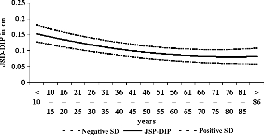
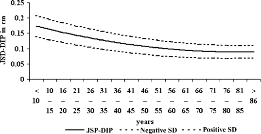
For all individuals, the CAJSA system reliably recognized the distal interphalangeal articulations. This study revealed a close relationship between age and all established parameters. Therefore all correlations between age and parameters of CAJSA were significant negative for women (−0.54<r< −0.63, p<0.001) as well as for men (−0.46<r< −0.57, p<0.001) whereas JSD-DIP for the female subjects (Fig. 2) continuously showed a closer association to age compared with the JSD-DIP in men (Fig. 3). The highest correlation was observed for JSD-DIP III versus age (r= −0.63, p<0.001) in the female group.
Fig. 2.
Age-related normative values of Joint Space Distance of the Distal-Interphalangeal joint (JSD-DIP total) and standard deviations (SD) determined on women (n = 351).
Fig. 3.
Age-related normative values of Joint Space Distance of the Distal-Interphalangeal joint (JSD-DIP total) and standard deviations (SD) determined on men (n = 518).
In men JSD-DIP (total) significantly decreased (−57.9%; p<0.001) from 0.19 ± 0.03 cm (age< 10 years) to 0.08 ± 0.02 cm (age>86 years) in a continuous manner (Tables 2 and 3). In women, JSD-DIP (total) also presented a significant and continuous reduction (−52.9%; p<0.001) from 0.17 ± 0.03 cm (age< 10 years) to 0.08 ± 0.03 cm (age>86 years; Tables 1 and 3). A similar decline was observed for JSD-DIP III in males (−57.9%; p<0.001). In females, JSD-DIP (II to IV) revealed a significant reduction of −50.0% (p<0.001) between ages <10 and >86 years. A maximal decrease for JSD-DIP V in men (−61.1%; p<0.001) from 0.18 ± 0.02 cm (age< 10 years) to 0.07 ± 0.01 cm (age>86 years) and in women (−56.3%; p< 0.001) from 0.16 ± 0.04 cm (age<10 years) to 0.07 ± 0.03 cm (age>86 years) was documented. For JSD-DIP II and IV, similar results with −47.1% (p<0.001) versus −60.0% (p<0.001) were obtained for men.
Table 2.
Mean and Standard Deviation for Joint Space Distance of the Distal Interphalangeal Joint (Men, n = 518)
| Age in Years | N | JSD, in Centimeters | ||||
|---|---|---|---|---|---|---|
| DIPa II Mean (SD) | DIPa III Mean (SD) | DIPa IV Mean (SD) | DIPa V Mean (SD) | DIPa Total Mean (SD) | ||
| <10 | 15 | 0.17 (0.05) | 0.19 (0.02) | 0.20 (0.04) | 0.18 (0.02) | 0.19 (0.03) |
| 10–15 | 26 | 0.17 (0.03) | 0.18 (0.04) | 0.17 (0.04) | 0.15 (0.03) | 0.17 (0.04) |
| 16–20 | 96 | 0.14 (0.02) | 0.15 (0.03) | 0.14 (0.03) | 0.12 (0.02) | 0.14 (0.03) |
| 21–25 | 82 | 0.13 (0.03) | 0.13 (0.03) | 0.13 (0.03) | 0.11 (0.02) | 0.13 (0.03) |
| 26–30 | 60 | 0.13 (0.02) | 0.13 (0.03) | 0.12 (0.03) | 0.11 (0.02) | 0.12 (0.03) |
| 31–35 | 46 | 0.13 (0.03) | 0.13 (0.03) | 0.12 (0.03) | 0.11 (0.02) | 0.12 (0.03) |
| 36–40 | 38 | 0.13 (0.03) | 0.13 (0.03) | 0.12 (0.03) | 0.10 (0.02) | 0.12 (0.03) |
| 41–45 | 36 | 0.12 (0.02) | 0.12 (0.03) | 0.12 (0.03) | 0.10 (0.02) | 0.12 (0.03) |
| 46–50 | 28 | 0.12 (0.03) | 0.12 (0.03) | 0.11 (0.03) | 0.10 (0.03) | 0.11 (0.03) |
| 51–55 | 20 | 0.12 (0.02) | 0.12 (0.02) | 0.11 (0.03) | 0.10 (0.01) | 0.11 (0.02) |
| 56–60 | 14 | 0.11 (0.02) | 0.11 (0.02) | 0.09 (0.03) | 0.08 (0.02) | 0.10 (0.02) |
| 61–65 | 15 | 0.11 (0.03) | 0.10 (0.02) | 0.09 (0.02) | 0.08 (0.02) | 0.10 (0.02) |
| 66–70 | 12 | 0.10 (0.02) | 0.10 (0.02) | 0.09 (0.02) | 0.08 (0.02) | 0.09 (0.02) |
| 71–75 | 8 | 0.10 (0.03) | 0.10 (0.03) | 0.09 (0.02) | 0.08 (0.04) | 0.09 (0.03) |
| 76–80 | 11 | 0.10 (0.02) | 0.10 (0.02) | 0.09 (0.02) | 0.08 (0.01) | 0.09 (0.02) |
| 81–85 | 3 | 0.09 (0.2) | 0.10 (0.02) | 0.09 (0.02) | 0.08 (0.03) | 0.09 (0.02) |
| >86 | 8 | 0.09 (0.03) | 0.08 (0.01) | 0.08 (0.01) | 0.07 (0.01) | 0.08 (0.02) |
| Total | 518 | 0.13 (0.03) | 0.12 (0.03) | 0.12 (0.03) | 0.10 (0.02) | 0.12 (0.03) |
aDIP = Distal Interphalangeal joint
SD = Standard deviation
JSD = Joint Space Distance
Table 3.
Changes of Joint Space Distances Between the Age Groups <10 and >86 Years
| Reduction Between Groups <10 and >86 Years, in Percent | ||
|---|---|---|
| Men (n = 518) | Women (n = 351) | |
| JSD-DIPa II | −47.1% (p < 0.001) | −50.0% (p < 0.001) |
| JSD-DIPa III | −57.9% (p < 0.001) | −50.0% (p < 0.001) |
| JSD-DIPa IV | −60.0% (p < 0.001) | −50.0% (p < 0.001) |
| JSD-DIPa V | −61.1% (p < 0.001) | −56.3% (p < 0.001) |
| JSD-DIPa total | −57.9% (p < 0.001) | −52.9% (p < 0.001) |
aJSD-DIP = Joint Space Distance of the Distal Interphalangeal joint, in centimeters
Table 1.
Mean and Standard Deviation for Joint Space Distance of the Distal Interphalangeal Joint (Women, n = 351)
| Age in Years | N | JSD, in Centimeters | ||||
|---|---|---|---|---|---|---|
| DIPa II Mean (SD) | DIPa III Mean (SD) | DIPa IV Mean (SD) | DIPa V Mean (SD) | DIPa Total Mean (SD) | ||
| <10 | 12 | 0.16 (0.03) | 0.18 (0.03) | 0.18 (0.03) | 0.16 (0.04) | 0.17 (0.03) |
| 10–15 | 16 | 0.14 (0.02) | 0.15 (0.03) | 0.14 (0.02) | 0.12 (0.02) | 0.14 (0.02) |
| 16–20 | 42 | 0.13 (0.02) | 0.13 (0.03) | 0.12 (0.02) | 0.10 (0.02) | 0.12 (0.02) |
| 21–25 | 45 | 0.12 (0.02) | 0.12 (0.02) | 0.12 (0.01) | 0.10 (0.01) | 0.12 (0.02) |
| 26–30 | 35 | 0.12 (0.03) | 0.12 (0.03) | 0.11 (0.03) | 0.10 (0.02) | 0.11 (0.03) |
| 31–35 | 32 | 0.12 (0.02) | 0.12 (0.02) | 0.11 (0.02) | 0.10 (0.02) | 0.11 (0.02) |
| 36–40 | 22 | 0.12 (0.02) | 0.11 (0.02) | 0.10 (0.02) | 0.10 (0.01) | 0.11 (0.02) |
| 41–45 | 22 | 0.11 (0.03) | 0.11 (0.02) | 0.10 (0.02) | 0.09 (0.01) | 0.10 (0.02) |
| 46–50 | 24 | 0.11 (0.02) | 0.11 (0.02) | 0.10 (0.02) | 0.09 (0.01) | 0.10 (0.02) |
| 51–55 | 24 | 0.10 (0.02) | 0.09 (0.02) | 0.09 (0.01) | 0.08 (0.01) | 0.09 (0.02) |
| 56–60 | 16 | 0.10 (0.02) | 0.09 (0.02) | 0.08 (0.02) | 0.08 (0.02) | 0.09 (0.02) |
| 61–65 | 11 | 0.10 (0.02) | 0.09 (0.02) | 0.08 (0.01) | 0.08 (0.02) | 0.09 (0.02) |
| 66–70 | 14 | 0.09 (0.02) | 0.09 (0.02) | 0.08 (0.02) | 0.07 (0.02) | 0.08 (0.02) |
| 71–75 | 7 | 0.08 (0.02) | 0.09 (0.02) | 0.08 (0.01) | 0.07 (0.02) | 0.08 (0.02) |
| 76–80 | 9 | 0.08 (0.03) | 0.09 (0.03) | 0.08 (0.02) | 0.07 (0.01) | 0.08 (0.02) |
| 81–85 | 12 | 0.08 (0.03) | 0.09 (0.02) | 0.08 (0.02) | 0.07 (0.01) | 0.08 (0.02) |
| >86 | 8 | 0.08 (0.03) | 0.09 (0.02) | 0.08 (0.02) | 0.07 (0.03) | 0.08 (0.03) |
| Total | 351 | 0.11 (0.02) | 0.11 (0.02) | 0.10 (0.02) | 0.09 (0.02) | 0.10 (0.02) |
aDIP = Distal Interphalangeal joint
SD = Standard deviation
JSD = Joint Space Distance
An accentuated decrease of JSD-DIP (total) in the age group from 10 to 15 years (for men −10.5% with p<0.05, and for women −17.6% with p<0.01) and in the age group from 16 to 20 years (for men −17.6% with p<0.01 and for women −14.3% with p<0.01) was found for both genders. The annual joint space narrowing up to the age of 20 years (annual joint space narrowing between the age of 10 up to 20 years, given as percentage per year) was −1.9% (p<0.05) for men and −2.1% (p<0.05) for women. Beginning with the age of 21 years (annual joint space narrowing between the ages of 21 up to 90 years, given as percentage per year) males revealed a decrease of −0.4% (p<0.05) and females showed a reduction of −0.5% (p<0.05).
Gender-related Differences of CAJSA Parameters
The results showed that women had a significantly smaller total JSD-DIP (mean −16.7%; p<0.01; Table 4), compared to men. The female JSD-DIP II, JSD-DIP III, and JSD-DIP V are significantly decreased with −15.4% (p<0.01), −8.3% (p<0.05), and −10.0% (p<0.05) in comparison to males. The pronounced difference was observed for JSD-DIP IV with a change of −16.7% (p<0.01).
Table 4.
Comparison of Normative Values (JSD-DIP) Between Men and Women
| Men | Women | Relative Difference Between Men and Women (%) | Significance (Mann–Witney U Test) | |
|---|---|---|---|---|
| Mean (SD) | Mean (SD) | |||
| JSD-DIPa II (in cm) | 0.13 (0.03) | 0.11 (0.02) | 15.4 | p < 0.01 |
| JSD-DIPa III (in cm) | 0.12 (0.03) | 0.11 (0.02) | 8.3 | p < 0.05 |
| JSD-DIPa IV (in cm) | 0.12 (0.03) | 0.10 (0.02) | 16.7 | p < 0.01 |
| JSD-DIPa V (in cm) | 0.10 (0.02) | 0.09 (0.02) | 10.0 | p < 0.05 |
| JSD-DIPa total (in cm ) | 0.12 (0.03) | 0.10 (0.02) | 16.7 | p < 0.01 |
aJSD-DIP = Joint Space Distance of the Distal Interphalangeal joint
DISCUSSION
The aim of this study is to establish normative gender-specific and age-related reference values for computer-aided joint space analysis (CAJSA), to establish CAJSA into the clinical routine. Our data evaluated age-related and gender-specific changes of the distal interphalangeal joint space width in a normal population.
Short-term Precision of Different Articulations
CAJSA technology now provides measurements of the JSD-MCP, which may be published for patients with rheumatoid arthritis. Böttcher et. al.24 has shown an excellent short-term precision regarding the metacarpal–phalangeal articulation between 0.59% (the metacarpal–phalangeal joint II) to 0.99% (the metacarpal–phalangeal joint IV). Measurements of JSD at the proximal and distal interphalangeal joints are more complex, because these joints have a bicompartmental configuration which results in a varied width of each compartment following minor rotations of the hand. A minor rotation during x-ray imaging results in impaired reproducibility for the proximal interphalangeal articulation and distal interphalangeal articulation. Böttcher et al documented a limited intraradiograph reproducibility of JSD of the proximal interphalangeal joint using the CAJSA version 1.3.5.24 After improvement of contour-finding procedures based on the new CAJSA software version (1.3.6), the actual results revealed a moderate short-term precision ranging from 1.32% (JSD-DIP IV) to 1.59% (JSD-DIP III).
Influence of Gender and Age on JSD-DIP
Present theories of the pathogenesis of osteoarthritis suggest that both systemic and local factors affect the likelihood of osteoarthritis onset in the small hand joints.25,26 Local factors such as higher grip strength and hand trauma have been associated with the development of hand osteoarthritis.27,28 Obesity is also reported to be a significant risk factor for the development of knee osteoarthritis,29,30 but its association with hand osteoarthritis is less clear. Some studies have reported no association between high body mass index and hand osteoarthritis in the elderly subjects, whereas other studies have found a positive association, particularly with carpometacarpal osteoarthritis.31–33 It is thought that systemic factors such as age, sex, race, and genetics predispose for hand osteoarthritis.34 Age and sex have well described effects on the prevalence of hand osteoarthritis.35 Jones et al36 presented a high prevalence of hand osteoarthritis with manifestation at the distal interphalangeal articulation in women (70%), as compared to men (57%). Furthermore, age and sex have well described effects on the severity of hand osteoarthritis,35 but there is less information as to whether the age gradient is different between the sexes.37 Our results reveal a remarkable impact of the occurrence of finger osteoarthritis in women. The JSD-DIP (total) for women is considerably smaller (−16.7%) than in the male cohort.
JSD Measurement in Clinical Practice
Hand radiography has been claimed to be the best method for clinical determination of hand osteoarthritis.7–9,38,39
However, precise verification of hand osteoarthritis remains problematic,39 because no absolute clinical, radiological, or pathological standard for the diagnosis exists.40 Using the normative values estimated by CAJSA, a radiological standard of JSD for affected distal interphalangeal joints is now available. Consecutive cutoff values based on these normative data can improve the detection of initial cartilage destruction in early osteoarthritis; the narrowing of JSD-DIP may now be measured and evaluated in detail.
The reference values established by this technology allow the detection and quantification of cartilage dissolution in the early stages of joint-affecting disorders (ie, osteoarthritis and rheumatoid arthritis) based on a direct comparison between normative values and the data of the patient. Further studies should focus on the applicability to other articulations and determine the influence on therapeutic strategies in patients with osteoarthritis.
CONCLUSION
Computer-aided techniques have shown remarkable potential in the measurement of several radio-geometrical features; in particular CAJSA has provided a cost-effective approach for the quantification of joint space distances using digital radiographs of the hand. The present study provides normative reference values for both women and men, revealing significant gender-specific differences in joint space width for the two sexes in all age groups. Additionally, this new CAD method allows an excellent overview of the narrowing of JSD-DIP during aging. Therefore, multicenter studies with larger gender-specific age groups will be required to confirm the age-associated joint space narrowing. These normative values are expected to enhance the process of identification of patients suffering from osteoarthritis. Further prospective studies are required to determine the predictive value of CAJSA to detect patients with early onset of joint osteoarthritis and to introduce the CAJSA technique as a cost-effective, widely available, and precise method in the clinical routine.
Acknowledgments
The authors thank Monika Arens (managing director, Arewus GmbH) and Anders Rosholm, PhD, for the use of the Computer-aided joint space analysis equipment and Rüdiger Vollandt, PhD, for the statistical advice. Finally, the authors would also like to thank Dieter Felsenberg, MD (Berlin, Germany), and Claus C. Glueer, PhD (Kiel, Germany) for their comments regarding this study.
Abbreviations
- CAD
Computer-aided diagnosis
- CAJSA
Computer-aided joint space analysis
- CV
Coefficient of variation
- JSD
Joint space distance
- JSD-MCP
Joint space distance of the metacarpal–phalangeal joint
- JSD-DIP
Joint space distance of the distal–interphalangeal joint
- SD
Standard deviation
- JSD-PIP
Joint space distance of the proximal-interphalangeal joint
References
- 1.Hart DJ, Spector TD: Definition and epidemiology of ostearthritis of the hand: a review. Osteoarthr Cartil (suppl A):2–7, 2000 [DOI] [PubMed]
- 2.Acheson RM, Chan YK, Clementt AR. New Haven Survey of joint diseases XII: distribution and symptoms of osteoarthritis in the hands with reference to handedness. Ann Rheum Dis. 1970;29:275–286. doi: 10.1136/ard.29.3.275. [DOI] [PMC free article] [PubMed] [Google Scholar]
- 3.Plato CC, Norris AH. Osteoarthritis of the hand age-specific joint digit prevalence rates. Am J Epidemiol. 1979;19:169–180. doi: 10.1093/oxfordjournals.aje.a112672. [DOI] [PubMed] [Google Scholar]
- 4.Butler WJ, Hawthorne VM, Mikkelsen WM, et al. Prevalence of radiographically defined osteoarthritis in the finger and wrist joints of adult residents of Tecumseh, Michican, 1962–1965. J Clin Epidemiol. 1988;41:467–473. doi: 10.1016/0895-4356(88)90048-0. [DOI] [PubMed] [Google Scholar]
- 5.Egger P, Cooper C, Hart DJ, Doyle DV, Coggon D, Spector TD. Pattern of joint involvement in osteoarthritis of the hand. The Chingford study. J Rheumatol. 1995;22:1509–1513. [PubMed] [Google Scholar]
- 6.Oliveria SA, Felson DT, Reed JI, Cirillo PA, Walker AM. Incidence of symptomatic hand, hip and knee osteoarthritis among patients in a health maintenance organization. Arthritis Rheum. 1995;38:1134–1141. doi: 10.1002/art.1780380817. [DOI] [PubMed] [Google Scholar]
- 7.Mikkelsen WM, Duff IF. Age–sex prevalence of radiographic abnormalities of the joints of the hand, wrist and cervical spine of adult resident of Tecumseh. Michigan community Health Study area 1962–1965. J Chronic Dis. 1970;23:151–159. doi: 10.1016/0021-9681(70)90092-5. [DOI] [PubMed] [Google Scholar]
- 8.Swanson AB, Swanson GG. Osteoarthritis in the hand. Clin Rheum Dis. 1985;11:393–419. [PubMed] [Google Scholar]
- 9.Saase JLCM, Romunde LKJ, Cats A, Vandenbroucke JP, Valkenburg HA. Epidemiology of osteoarthritis, Zoetermeer survey. Comparison of radiological osteoarthritis in a Dutch population with that in ten other populations. Ann Rheum Dis. 1989;48:21–80. doi: 10.1136/ard.48.4.271. [DOI] [PMC free article] [PubMed] [Google Scholar]
- 10.Cauley JA, Kwoh K, Egeland G, et al. Serum sex hormones and severity of osteoarthritis of the hand. J Rheumatol. 1993;20:1170–1175. [PubMed] [Google Scholar]
- 11.Dahaghin S, Bierma-Zeinstra SMA, Ginai AZ, et al. Prevalence and pattern of radiographic osteoarthritis and association with pain and hand disability (the Rotterdam study) Ann Rheum Dis. 2005;64:682–687. doi: 10.1136/ard.2004.023564. [DOI] [PMC free article] [PubMed] [Google Scholar]
- 12.Sharp JT, Heijde D, Angwin J, et al. Measurement of joint space width and erosion size. J Rheumatol. 2005;32:2456–2461. [PubMed] [Google Scholar]
- 13.Duryea J, Jiang Y, Zakharevich M, Genant HK. Neutral network based algorithm to quantify joint space width in joints of the hand for arthritis assessment. Med Phys. 2000;27:1185–1194. doi: 10.1118/1.598983. [DOI] [PubMed] [Google Scholar]
- 14.Pietka E, Gertych A, Pospiech-Kurkowska S, Cao F, Huang HK, Gilzanz V. Computer-assissted bone age assessment: graphical user interface for image processing and comparison. J Digit Imaging. 2004;17:175–188. doi: 10.1007/s10278-004-1006-6. [DOI] [PMC free article] [PubMed] [Google Scholar]
- 15.Malich A, Fischer DR, Facius M, et al. Effect of breast density on computer aided detection. J Digit Imaging. 2005;19:1871–1889. doi: 10.1007/s10278-004-1047-x. [DOI] [PMC free article] [PubMed] [Google Scholar]
- 16.Alvarez RE. Lung field segmenting in dual-energy subtraction chest X-ray images. J Digit Imaging. 2004;17:45–56. doi: 10.1007/s10278-003-1701-8. [DOI] [PMC free article] [PubMed] [Google Scholar]
- 17.Dacre JE, Huskisson EC. The automatic assessment of knee radiographs in ostheoarthritis using digital image analysis. Br J Rheumatol. 1989;28:506–510. doi: 10.1093/rheumatology/28.6.506. [DOI] [PubMed] [Google Scholar]
- 18.Buckland-Wright JC, MacParlane DG, Lynch JA, Jasani MK. Quantitative microfocal radiography detects changes in OA knee joint space width in patients in placebo controlled trial of NSAID therapy. J Rheumatol. 1995;22:937–943. [PubMed] [Google Scholar]
- 19.Duryea J, Li J, Peterfy CG, Gordon C, Genant HK. Trainable rule-based algorithm for the measurement of joint space width in digital radiographic images of the knee. Med Phys. 2000;27:580–591. doi: 10.1118/1.598897. [DOI] [PubMed] [Google Scholar]
- 20.Conrozier T, Jousseaume CA, Mathieu P, et al. Quantitative measurement of joint space narrowing progression in hip osteoarthritis: a longitudinal retrospective study of patients treated by total hip arthoplasty. Br J Rheumatol. 1998;37:961–968. doi: 10.1093/rheumatology/37.9.961. [DOI] [PubMed] [Google Scholar]
- 21.Böttcher J, Pfeil A, Rosholm A, et al. Computerized quantification of joint space narrowing and periarticular demineralization in patients with rheumatoid arthritis based on Digital X-ray Radiogrammetry. Invest Radiol. 2006;41:36–44. doi: 10.1097/01.rli.0000191594.76235.a0. [DOI] [PubMed] [Google Scholar]
- 22.Böttcher J, Pfeil A, Rosholm A, et al. Computerized digital imaging techniques provided by digital radiogrammetry as new diagnostic tool in rheumatoid arthritis. J Digit Imaging. 2006;19:279–288. doi: 10.1007/s10278-006-0263-y. [DOI] [PMC free article] [PubMed] [Google Scholar]
- 23.Kellgren J, Lawrence J. Radiological assessment of osteoarthrosis. Ann Rheum Dis. 1957;16:494–502. doi: 10.1136/ard.16.4.494. [DOI] [PMC free article] [PubMed] [Google Scholar]
- 24.Böttcher J, Pfeil A, Rosholm A, et al. Digital X-Ray Radiogrammetry combined with semi-automated analysis of joint space distances as a new diagnostic approach in rheumatoid arthritis—A cross-sectional and longitudinal study. Arthritis Rheum. 2005;52:3850–3859. doi: 10.1002/art.21606. [DOI] [PubMed] [Google Scholar]
- 25.Felson DT, Zhang Y. An update on the epidemiology of knee and hip osteoarthritis with a view to prevention. Arthritis Rheum. 1998;41:1343–1355. doi: 10.1002/1529-0131(199808)41:8<1343::AID-ART3>3.0.CO;2-9. [DOI] [PubMed] [Google Scholar]
- 26.Arokoski JPA, Jurvelin J, Väätäinen U, Helminen HJ. Normal and pathological adaptation of articular cartilage to joint loading: review. Scand J Med Sci Sports. 2000;10:186–198. doi: 10.1034/j.1600-0838.2000.010004186.x. [DOI] [PubMed] [Google Scholar]
- 27.Sowers M, Lachance L, Hochberg M, Jamadar D. Radiographically defined osteoarthritis of the hand and knee in young and middle-aged African American and Caucasian women. Osteoarthritis Cartilage. 2000;8:69–77. doi: 10.1053/joca.1999.0273. [DOI] [PubMed] [Google Scholar]
- 28.Chaisson CE, Zhang Y, Sharma L, Felson DT. Higher grip strength increases the risk of incident radiographic osteoarthritis in proximal hand joints. Osteoarthritis Cartilage. 2000;8(Supplement A):S29–S32. doi: 10.1053/joca.2000.0333. [DOI] [PubMed] [Google Scholar]
- 29.Lau EC, Cooper C, Lam D, Chan VN, Tsang KK, Sham A. Factors associated with osteoarthritis of the hip and knee in Hong Kong Chinese: obesity, joint injury and occupational activities. Am J Epidemiol. 2000;152:855–862. doi: 10.1093/aje/152.9.855. [DOI] [PubMed] [Google Scholar]
- 30.Cooper C, Snow S, McAlindon TE, et al. Risk factors for the incidence and progression of radiographic knee osteoarthritis. Arthritis Rheum. 2000;43:995–1000. doi: 10.1002/1529-0131(200005)43:5<995::AID-ANR6>3.0.CO;2-1. [DOI] [PubMed] [Google Scholar]
- 31.Hochberg MC, Lethbridge-Cejku M, Plato CC, Wigley FM, Tobin JD. Factors associated with osteoarthritis of the hand in males: data from the Baltimore Longitudinal Study of Aging. Am J Epidemiol. 1991;134:1121–1127. doi: 10.1093/oxfordjournals.aje.a116015. [DOI] [PubMed] [Google Scholar]
- 32.Hart DJ, Spector TD. The relationship of obesity, fat distribution and osteoarthritis in women in the general population. The Chingford study. J Rheumatol. 1993;20:331–335. [PubMed] [Google Scholar]
- 33.Sturmer T, Gunther KP, Breener H. Obesity, overweight and patterns of osteoarthritis: the Ulm Osteoarthritis Study. J Clin Epidemiol. 2000;53:307–313. doi: 10.1016/S0895-4356(99)00162-6. [DOI] [PubMed] [Google Scholar]
- 34.Haara MM, Manninen P, Kröger H, et al. Osteoarthritis of finger joints in Finns aged 30 or over: prevalence, determinants, and association with mortality. Ann Rheum Dis. 2003;62:151–158. doi: 10.1136/ard.62.2.151. [DOI] [PMC free article] [PubMed] [Google Scholar]
- 35.Lawrence JS, Bremner JM, Biers F. Osteoarthritis. Prevalence in the population and relationship between symptoms and x-ray changes. Ann Rheum Dis. 1966;25:1–24. [PMC free article] [PubMed] [Google Scholar]
- 36.Jones G., Colley HM., Stankovich J.M. A Cross sectional study of the association between sex, smoking, and other lifestyle factors and osteoarthritis of the hand. J Rheumatol. 2002;29:1719–1724. [PubMed] [Google Scholar]
- 37.Oliveria SA, Felson DT, Reed JI, Cirillo PA, Walker AM. Incidence of symptomatic hand, hip, and knee osteoarthritis among patients in a health maintenance organization. Arthritis Rheum. 1995;38:1134–1141. doi: 10.1002/art.1780380817. [DOI] [PubMed] [Google Scholar]
- 38.Hochberg MC, Lane NE, Pressman AR, et al. The association of radiographic changes of osteoarthritis of the hand and hip in elderly women. J Rheumatol. 1995;22:2291–2294. [PubMed] [Google Scholar]
- 39.Hart D, Spector T, Egger P, Coggon D, Cooper C. Defining osteoarthritis of the hand for epidemiological studies: the Chingford study. Ann Rheum Dis. 1994;53:220–223. doi: 10.1136/ard.53.4.220. [DOI] [PMC free article] [PubMed] [Google Scholar]
- 40.Spector TD, Cooper C. Radiological assessment of osteoarthritis: whither Kellgren and Lawrence? Osteoarthritis Cartilage. 1993;1:203–206. doi: 10.1016/S1063-4584(05)80325-5. [DOI] [PubMed] [Google Scholar]


