Abstract
DicomWorks is freeware software for reading and working on medical images [digital imaging and communication in medicine (DICOM)]. It was jointly developed by two research laboratories, with the feedback of more than 35,000 registered users throughout the world who provided information to guide its development. We detail their occupations (50% radiologists, 20% engineers, 9% medical physicists, 7% cardiologists, 6% neurologists, and 8% others), geographic origins, and main interests in the software. The viewer’s interface is similar to that of a picture archiving and communication system viewing station. It provides basic but efficient tools for opening DICOM images and reviewing and exporting them to teaching files or digital presentations. E-mail, FTP, or DICOM protocols are supported for transmitting images through a local network or the Internet. Thanks to its wide compatibility, a localized (15 languages) and user-friendly interface, and its opened architecture, DicomWorks helps quick development of non proprietary, low-cost image review or teleradiology solutions in developed and emerging countries.
Key words: Computers, digital imaging and communication in medicine (DICOM), DICOM viewer, teleradiology
BACKGROUND
When the first picture archiving and communication systems (PACS) became widely available in the mid 1990s, radiologists had to face a reality: their daily job was going to be dramatically transformed as new tools were mandatory to absorb the growing number of images that imaging modalities produced [computed tomography (CT), magnetic resonance imaging (MRI), and ultrasound (US)]. Technology imposed itself in the daily work of many, and words like digital imaging and communication in medicine (DICOM), Joint Picture Exchange Group images (JPEG), picture archiving and communication system (PACS), radiology information system (RIS), and health level 7 standard progressively became familiar to radiologists.
The first PACS systems focused on productivity, efficiency of primary interpretation, and integration with the RIS, but left out some very important parts of the specialty: personal archiving, teaching files, and presentations. The lack of these important features was very frustrating because it was difficult to export PACS images for presentations and personal use. This led us to build a simple tool that could convert complex DICOM images to simple JPEG images and import them into Microsoft PowerPoint (Microsoft, Redmond, WA, United States). Today, in spite of true efforts from PACS vendors, these problems persist because the installation of concurrent third-party applications on diagnosis work stations is usually forbidden to avoid software incompatibilities, CPU usage, or network bandwidth interference that can alter the station’s reliability and performance.
Now that the vast majority of radiology institutions or departments have moved to digital, several challenges are revealed:
Inside radiology departments, there is a growing need for additional, secondary viewing stations because primary interpretation stations provided with modalities or the PACS are not always available in sufficient number. Additionally, there is a need for portability: many radiologists wish to review images in their office or on their laptop and prepare presentations at home. In all these cases, additional low-cost DICOM stations are desirable, but rarely included in PACS projects or department equipment plans, as their profitability would be too poor.
Outside radiology departments, clinicians have many difficulties reviewing their studies on digital removable media, both because their knowledge of digital imaging is limited (DICOM, windowing, etc...) and because their equipment is often inadequate. Aside from modern PACS-equipped institutions, reading a study on a DICOMDIR CD-ROM with a preinstalled “auto executable” software may not be easy due to hardware (slow processor, inadequate graphic card) or system incompatibilities (a few work only with Windows 95) and lack of features.
There is a growing demand for teleradiology applications in developed and emerging countries.
METHODS
Application Design
Development was initiated using Microsoft Visual Basic version 5.0 (Microsoft) because this environment allowed rapid application development and fast interface modifications. This environment was insufficient for critical memory manipulations, such as image-processing algorithms. A more powerful component specifically dedicated to image processing was developed using Microsoft Visual C++ (Microsoft). It was included in the preexisting Visual Basic project using the ActiveX technology. No reference to license-based or commercial DICOM libraries was made, but the DICOM standard was strictly followed, thanks to the public availability of the standard.1 An important work of testing was done to make this 32-bit application work on all Microsoft Windows systems (from the first version of Windows 95 to the latest version of Windows XP) with no additional software or driver, as well as on low-end personal computers (the minimal hardware requirement is a Pentium II class processor with 64 megabytes of RAM, and a 640 × 480 × 16-bit display). No reference to hardware-dependant components (like Microsoft DirectX technology) was made to avoid further incompatibilities. The following descriptions apply to version 1.3.5.
Client Installation
The program2 is available in 15 languages and can be downloaded from the Internet and installed at no charge. Some advanced functions (such as archiving and compression of images) are locked until the user registers (for free) on the web site and provides simple information about the interests that he/she has in the program. The standard package includes a Microsoft PowerPoint export plug-in, which adds the feature of a direct export of the DICOM images to PowerPoint slides. Additional packages can be downloaded for free to add DICOM Store Service Class Provider and User (STORE SCP and STORE SCU) functionalities.
RESULTS
Since its first version in September, 2000, more than 150,000 downloads were counted, and since 2003, more than 35,000 of these users registered the software to unlock special features (e-mail, presentation software, and AVI exports). They provided personal information (name, country, occupation, interests, and other programs used in the same field) to help the development team to know them better. Table 1 details their main occupations, interests, and geographical origins.
Table 1.
Distribution of DicomWorks Registered Users Between January, 2003, and May, 2006, Depending on their Occupation, Interest in the Software, Geographic Region, and Country Development Index
| Teleradiology Users | All Users | |
|---|---|---|
| Number of registered users | n = 442 | n = 35,839 |
| By occupation | ||
| Radiologists | 50% | |
| Engineers (PACS administrators, PACS engineers, IT technicians) | 20% | |
| Medical physicists | 9% | |
| Cardiologists | 7% | |
| Neurology (neurologists, neurosurgeons) | 6% | |
| Others | 8% | |
| By interest | ||
| Viewing images | 8,996 (25%) | |
| Export to presentation software | 1,478 (4%) | |
| DICOM tags edition/anonymization | 943 (3%) | |
| Export to AVI (cine-loop) | 756 (2%) | |
| Teleradiology | 442 (1%) | |
| Others (testing, not specified) | 23,223 (65%) | |
| By geographic region | ||
| European Union | 29% (n = 126) | 42% (including Germany 8.6%; France 5.7%; UK 5.2%) |
| North America | 42% (n = 184) | 36% (including USA 33.%; Canada 2.7%) |
| South America | 10% (n = 45) | 6.8% (including Brazil 2.4%; Mexico 1.3%) |
| Asia | 4% (n = 17) | 5.1% (including Japan 2.1%; China 1.4%) |
| India | 5% (n = 24) | 3.1% (including India 2.9%) |
| Australia | 6% (n = 27) | 2.6% (including Australia 2.2%) |
| Middle East | 1% (n = 3) | 2.3% (including Turkey 0.8%; Israël 0.5%) |
| Africa | 2% (n = 7) | 1.5% (including South Africa 0.5%; Egypt 0.4%) |
| Russia | <1% (n = 1) | 0.7% (including Russian Federation 0.7%; Ukraine 0.2%) |
| By development indexa | ||
| Top 30 countries (HDI range from 0.963 down to 0.878) | 71% (n = 316) | 78.3% (including United States 33%; Germany 8.6%; France 5.7%) |
| High development (n = 57) HDI>=0.8 | 84% (n = 371) | 85.7% (n = 30,727) (including United States 33%; Germany 8.6%; France 5.7%) |
| Moderate development (n = 88) 0.5<HDI<=0.799 | 16% (n = 71) | 13.3% (n = 4,766) (India 2.9%; Brazil 2.4%; China 1.4%) |
| Low development (n = 32) (HDI < 0.5) | 0% | 0.1% (n = 39) (Senegal n = 6; Nigeria n = 4; Tanzania n = 4) |
HDI = Human Development Index
aBased on the 2005 UN Human Development Index10
Including DicomWorks in the Images Workflow
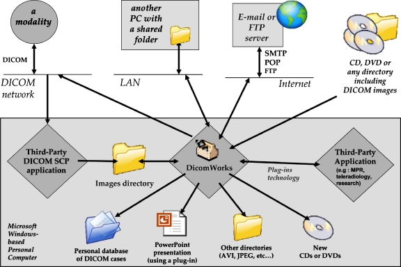
DicomWorks 1.3.5 can open DICOM images on the local disk or local area network (LAN). This includes the ability to open unsorted or sorted directories of DICOM images (a sorted list of patients and studies becomes available) or DICOMDIR indexed removable media (CD-ROM or DVD-ROM), which are faster to scan. All formats of DICOM images are supported: uncompressed or compressed (RLE, lossy and lossless JPEG), monochrome or color, single or multiframe. One helpful feature allows the user to set a directory as a favorite (similar to what you can do with an Internet browser) and search it with a simple double-click on its name. When a directory is set as a favorite, patients, studies, series, or images can simply be dragged and dropped on and from it. This allows easy manipulation of large sets of DICOM images, and the creation of databases of real (unaltered) DICOM images, especially helpful for research. DICOM STORE SCP and SCU are freely available with DicomWorks, respectively, with a small application based on the OFFIS DICOM toolkit3 and as an external plug in, based on the CTN code.4 If query/retrieve is necessary (FIND SCU, MOVE SCU, FIND SCP, and MOVE SCP DICOM services), a separate, free, third-party DICOM server is required: the ConQuest DICOM server5 directory structure is recognized. Both applications can run together on the same machine. A DICOM creation module allows the creation of DICOM images from JPEG and BMP files, but also directly from a video card or TWAIN-compatible scanner capture. Additionally, DICOM images can be imported and exported to third-party applications (such as 3-dimensional processing, for example), using a plug-ins technology that allows third-party developers to include their application in DicomWorks (Fig. 1).
Fig 1.
The main import sources and export destinations of DicomWorks. The application can import images from a local hard disk, network hard disk, removable media (DICOMDIR support), e-mail accounts, FTP, or a DICOM network with the help of a third-party DICOM SCP application. Images can be modified and exported to numerous destinations (local hard disk, e-mail, FTP, DICOM archive, or directly in a Microsoft PowerPoint presentation). The plug-ins technology allows conversion and exportation to any other file format or destination.
Reviewing a Study
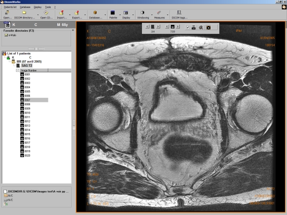
All sources of images are displayed identically as a hierarchical list of patients, studies, series, and images that can be expanded by double-clicking on their icon (Fig. 2). Any type of DICOM image can be displayed (CT, MR, US, x-ray angio, mammography, nuclear medicine, secondary capture...). A fast and intuitive “cine-loop” review of images can be performed by clicking on an image and using the mouse wheel or keyboard arrows. Simple buttons allow the browsing of series and images, marking of images in a separate palette, and switching the display of application overlays. Image browsing can be performed either exclusively with the keyboard, exclusively with the mouse, or with both. It is possible to split the screen to compare up to four series with optional synchronization. This allows the comparison of two images of the same organ at the same slice location with different modalities or protocols (CT and MR, for example).
Fig 2.
Screenshot of the main screen of version 1.3.5 with a study opened (axial T2 weighted sequence of a prostate MRI examination). On the left part of the screen, a direct access to favorite directories of DICOM images is possible (blue folder), whereas the current directory is sorted in patients, studies, series, and images. All levels are expandable (such as here at the image level). On the right part of the screen, a maximum of the screen surface is dedicated to the image. Patient and image data are overlaid. A simple toolbar allows easy navigation through the series and images. Window leveling, pan, and zoom are performed using the mouse, and the mouse wheel allows scrolling in the series. Important functions (measures, screen splitting, etc...) are accessible from the toolbar.
Education and Research Functions
Below are listed the most relevant functions dedicated to research or education:
DICOM tags edition/anonymization: The application includes a powerful tool for anonymizing studies, removing or changing patient and study details. It is also possible to find duplicates and remove and insert new DICOM tags in any file.
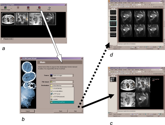
Exporting to Microsoft PowerPoint (Fig. 3): JPEG or AVI file formats can be automatically inserted in a new or a preexisting Microsoft PowerPoint presentation. This avoids fastidious image manipulations that otherwise would have been necessary, and required image processing applications:6,7 DICOM-to-JPEG transformation with windowing and zoom, image anonymization (for US images, for example), manual importation of images in PowerPoint (one by one), manual resizing, manual alignment, new annotations... As it is based on Visual BASIC for Applications scripting, this feature requires any version of Microsoft PowerPoint from 97 to XP to be installed on the machine.
Exporting a selection of images or the series as JPEG, BMP, or an AVI sequence.
Plug-in development: A software development kit is available for free. Any developer can quickly create additional modules that DicomWorks automatically recognizes at startup. Plug-ins also allow the export of any selection of images to external applications that need DICOM images on input, such as third-party MPR or 3-dimensional or image data analysis applications.
Other features are also available: an off-line studies manager (with keywords), a CD-ROM burning tool with optional anonymization, an image/signal analysis tool (statistics exportable as a Microsoft Excel sheet), and clipboard integration (copy and paste to any application).
Fig 3.
Diagram showing the steps to include a selection of images in Microsoft PowerPoint using DicomWorks: (a) Images are selected in DicomWorks and marked in the “Palette” window. A click on the “Export...” button shows the “Export wizard” window (b) where “Microsoft PowerPoint” clearly appears when this application is installed on the machine. After clicking on “next”, images can be cropped or resized and exported automatically to a new presentation (c) or inserted in a new slide of the currently opened presentation (d). Image window levels, zoom, and annotations are preserved. No JPEG transformation is necessary. This procedure avoids fastidious image manipulations (JPEG transformation, resizing, sorting, grouping, alignment).
IT and PACS Administration Tools
The software can be a useful tool to troubleshoot some frequent issues when using DICOM images in a PACS environment:
Some vendors store dynamic series or repeated CT phases in the same series, whereas some others store them in separate ones (eg, 270 images of 18 different slices acquired 15 successive times, or a single series of 200 CT-scanner images including 100 slices at the arterial phase and 100 at the portal phase). This can be a very serious issue for image archiving, distribution, or postprocessing on another vendor’s workstation. DicomWorks includes simple tools to select groups of images and split a large series in several, with respect to the DICOM standard. Inversely, merging several series in a single one is also possible.
The DICOM tags edition is a very useful tool to insert or delete single or sets of DICOM tags in a study, a series, or an image to force the compatibility with a specific workstation or PACS archive (eg, some postprocessing workstations or PACS servers require mandatory tags to import the images, but the information is sometimes written in another tag, with a wrong syntax, or omitted by the source modality).
A DICOM file sorting tool is also available to rearrange the directory structure and filenames syntax of any source of DICOM files and make it suit the standard (eg, appending the “dcm” suffix to the filenames, using unique identifiers as directory titles...). This helps searching, archiving, and using unsorted DICOM files, and accelerates image browsing.
Teleradiology
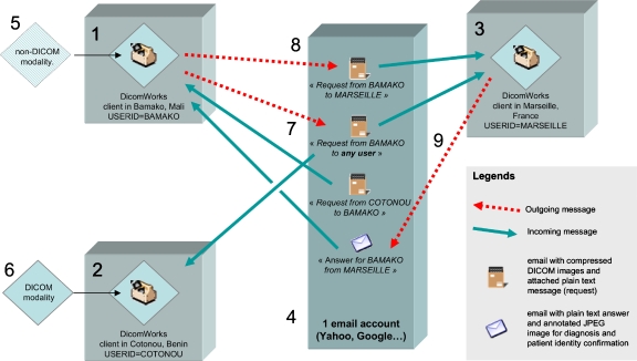
DicomWorks was installed as a part of various teleradiology solutions in the United States (eg, in a network stretching from the Alaskan Arctic to Southern California), in Europe (eg, to connect five regional hospitals in Greifswald, Germany), and in multiple low-cost solutions based on e-mail transmissions in Mali, Algeria, Morocco, Chile, Benin, and Cambodia, as detailed in Figure 4. Special tools were included to facilitate teleradiology applications and make the application compatible with different types of teleradiology servers [post office protocol (POP) and simple mail transfer protocol for e-mail, FTP, and DICOM], whether using built-in interfaces or using external applications that collaborate with DicomWorks as plug-ins (Fig. 1):
Non-DICOM images (BMP or JPEG) can be merged with editable patient data and compressed as JPEG lossy DICOM, thus facilitating their exchange/transport on low-bandwidth networks. Rarely necessary in developed countries, this feature is very important in developing countries where DICOM-compatible US devices are still minority.
Import and export of DICOM images using regular e-mail accounts is the simplest method to share images with colleagues: DicomWorks can check your account using the POP3 protocol and display the images immediately, leaving other messages on the server. The sending of anonymized and compressed images is also achieved by DicomWorks (Fig. 4).
Import and export of DICOM images using FTP.
Import and export using the DICOM protocol are also available thanks to the plug-in technology. They required a permanent static IP address, a secured virtual private network, and DICOM communication software on both the client and server sides.
Fig 4.
An example of an international teleradiology solution installed between Africa (Mali, Benin, Morocco, Algeria) and France (Marseille, Lyon, Frejus) using DicomWorks’ built-in e-mail import and export functions: Requesting physicians (1 and 2) and remote experts (3) only need a DicomWorks client application installed on a standard PC connected to the Internet. They all share a single e-mail account on a simple mail transfer protocol/POP e-mail server (here on a dedicated server in Marseille to avoid security issues), but they can also have individual accounts. In the first case, images are imported from non-DICOM modalities (5) and converted to DICOM-compatible files using DicomWorks on station 1. In the second case, images from a CT scanner are native DICOM and easily sent to DicomWorks using a Store-SCP client application on the PC. Communications use regular Internet connections. Any user can upload a message on the e-mail server and specify a recipient (8) using a predefined list (in local preferences file). Cases can also be left available to all physicians (7), so that unanswered cases can be viewed by several experts. Transmitted images are left in DICOM format, but anonymized, compressed using a lossless algorithm, and attached as a single file to the e-mail message. Images are retrieved using DicomWorks’ built-in POP client that filters messages addresses to its specific “USERID” (fixed and encrypted in local preferences), or to “ALL.” Reviewing is performed using DicomWorks viewing tools. A simple “Answer” button is available to send a response (that may contain a reference JPEG image) to the original sender (9).
DISCUSSION
Users
We developed a competitive and reliable solution to review images inside and outside radiology departments. Its distribution is facilitated by an exemption from payment and a simple and localized interface. Our experience as radiologists led us to include some original features that professional PACS solutions or other open-source/free applications3,8,9 did not provide and could be an obstacle for many, such as opening a CD-ROM with a single mouse click, showing the patients list and the images simultaneously to avoid window manipulations, viewing two series simultaneously and synchronize them, exporting images to digital presentation software (such as Microsoft PowerPoint) in a few steps, editing DICOM tags, sending annotated images by e-mail, or just creating a new CD-ROM of an anonymized study. Surprisingly, these functions were as well appreciated by clinicians (cardiologists, neurologists, surgeons, pathologists, and dermatologists) as by radiologists (Table 1), and this pointed out the interest of a simple and localized program for clinicians, who do not read studies like radiologists and need a simple, familiar, and localized interface, instead of multiple different ones like those they find on DICOM CDs. Image viewing was the most frequently declared usage with 25% of the registered users. It is certainly more important because image-viewing features are fully available without registration, and people who only use DicomWorks for image viewing may never register. Additionally, an important part of registered users is represented by IT technicians who use DicomWorks for solving image manipulation problems (conversions, anonymization) or patient data reconciliation in a PACS environment (batch DICOM tags modifications). As the Internet is now ubiquitary, even in the lowest-developed countries, and as the program is localized and free and runs on the lowest configurations, we think the repartition of the users could be a representation of digital imaging usage in the world. We found no reference in the literature about such repartition for comparison. The majority of the users come from countries where digital imaging modalities are widely available. Although logical, this reveals that a lot of features are missing from commercial systems and that there is a need for low-cost viewing systems even in the most developed countries.
Teleradiology
Table 1 details the geographic origins of the registered users depending on their country’s Human Development Index.10 Four hundred forty two users (1%) specifically registered for teleradiology, with 84% coming from the most developed countries where teleradiology is growing exponentially for multiple reasons: the shortage of specialized physicians in an increasing number of otherwise well-equipped institutions with constantly increasing activity11 or for night calls.12 North America is proportionally more represented than the European Union for teleradiology, with 42% against 29% of the users who registered specifically for teleradiology, whereas both regions have similar proportions when reported to their population (12,909 registered users for about 351 million inhabitants in North America against 15,050 registered users for about 460 million inhabitants in the European Union). This illustrates the difference between North America, where interstate teleradiology has been practiced for many years,13 and the European Union, where this practice is just starting. Also, even if several European countries have issued guidance on the practice of teleradiology, ethic and legal issues remain difficult to resolve in the European Union, as no common regulation is available on this subject. Only 0.1% of total registrations (n = 35,839) came from the least-developed countries, with no request specific to teleradiology. Overall, Table 1 data demonstrate that teleradiology is only developed in places where equipment and medical resources are widely available, and used when people need facilities in addition to a stable routine practice (night hawk, image sharing…). International teleradiology is surprisingly more aimed at commercial overseas interpretation in the highest developed countries13,14 than at humanitarian purposes. Nevertheless, low-cost teleradiology15 is an interesting solution in emerging countries where physicians work in distant or geographically difficult areas and cannot afford to buy expensive commercial teleradiology systems. These experiences remain rare.16,17
Having this in mind, and to help promote teleradiology, we keep developing DicomWorks as a universal, free, and user-friendly DICOM viewer that can be plugged on any teleradiology network or DICOM workflow, regardless of its architecture or communication protocol (wide area network, POP, FTP, secured FTP, etc…). We hope this will help teleradiology network administrators to delegate the client part of their network, including the image review, to free applications, and focus their human and economic resources on the security and reliability of communications because patient data are transmitted through the Internet (building a robust and secure server, establishing stable, fast, and secured communications through the Internet).
CONCLUSION
DicomWorks was developed to provide a free solution to the daily image manipulations problems radiologists have to face. The DICOM viewer provides a customizable and nearly professional solution for image interpretation, education, teaching, and research. Our 5-year experience and the feedback of several thousands of radiologists and clinicians worldwide led us to build an opened platform that can work as a single client interface for reviewing multiple and various image sources, whether local or remote (LAN, e-mail servers, FTP servers, DICOM servers). It is part of multiple teleradiology solutions that can focus their human and economic resources on other critical parts of the teleradiology network. We strongly believe that proprietary teleradiology solutions with dedicated lines and proprietary software will make way for a new generation of teleradiology solutions using secured Internet connections and based on standard (DICOM and Integrating the Healthcare Enterprise) compliant software.
References
- 1.The DICOM Standard. http://www.dclunie.com/dicom-status/status.html. Accessed May 22, 2006
- 2.Puech P, Boussel L: DicomWorks 1.3.5. http://www.dicomworks.com. Accessed May 22, 2006
- 3.OFFIS DICOM Toolkit. http://dicom.offis.de. Accessed May 19, 2006
- 4.Moore SM, Hoffman SA, Beecher DE: DICOM shareware: a public implementation of the DICOM standard. Medical imaging 1994-PACS: design and evaluation. Proc SPIE 2165:772–781, 1994
- 5.van Herk M, Zijp L: ConQuest DICOM Server. http://www.xs4all.nl/~ingenium/dicom.html. Accessed May 19, 2006
- 6.Collins J. Education techniques for lifelong learning: making a PowerPoint presentation. Radiographics. 2004;24:1177–1183. doi: 10.1148/rg.244035180. [DOI] [PubMed] [Google Scholar]
- 7.Yam CS. Using PowerPoint to create high-resolution images for journal publications. AJR Am J Roentgenol. 2005;185:273–276. doi: 10.2214/ajr.185.1.01850273. [DOI] [PubMed] [Google Scholar]
- 8.Escott EJ, Rubinstein D. Free DICOM image viewing and processing software for your desktop computer: what’s available and what it can do for you. Radiographics. 2003;23:1341–1357. doi: 10.1148/rg.235035047. [DOI] [PubMed] [Google Scholar]
- 9.Gillespy T, 3rd, Richardson ML, Rowberg AH. Displaying radiologic images on personal computers: practical applications and uses. J Digit Imaging. 1994;7:101–106. doi: 10.1007/BF03168502. [DOI] [PubMed] [Google Scholar]
- 10.United Nations Development Programme: Human Development Report 2005. http://hdr.undp.org/. Accessed May 22, 2006
- 11.Larson DB, Cypel YS, Forman HP, Sunshine JH. A comprehensive portrait of teleradiology in radiology practices: results from the American College of Radiology’s 1999 Survey. AJR Am J Roentgenol. 2005;185:24–35. doi: 10.2214/ajr.185.1.01850024. [DOI] [PubMed] [Google Scholar]
- 12.Mun SK, Tohme WG, Platenberg RC, Choi I. Teleradiology and emerging business models. J Telemed Telecare. 2005;11:271–275. doi: 10.1258/1357633054893373. [DOI] [PubMed] [Google Scholar]
- 13.Wachter RM. International teleradiology. N Engl J Med. 2006;354:662–663. doi: 10.1056/NEJMp058286. [DOI] [PubMed] [Google Scholar]
- 14.Jarvis L, Stanberry B. Teleradiology: threat or opportunity? Clin Radiol. 2005;60:840–845. doi: 10.1016/j.crad.2005.04.001. [DOI] [PubMed] [Google Scholar]
- 15.Dohrmann PJ. Low-cost teleradiology for Australia. Aust N Z J Surg. 1991;61:115–117. doi: 10.1111/j.1445-2197.1991.tb00186.x. [DOI] [PubMed] [Google Scholar]
- 16.Helitzer D, Heath D, Maltrud K, Sullivan E, Alverson D. Assessing or predicting adoption of telehealth using the diffusion of innovations theory: a practical example from a rural program in New Mexico. Telemed J E Health. 2003;9:179–187. doi: 10.1089/153056203766437516. [DOI] [PubMed] [Google Scholar]
- 17.Javadi M, Subhannachart P, Levine S, et al. Diagnosing pneumonia in rural Thailand: digital cameras versus film digitizers for chest radiograph teleradiology. Int J Infect Dis. 2006;10:129–135. doi: 10.1016/j.ijid.2005.01.007. [DOI] [PMC free article] [PubMed] [Google Scholar]


