

Abstract
Previous studies have shown that Joint Photographic Experts Group (JPEG) 2000 compression is better than JPEG at higher compression ratio levels. However, some findings revealed that this is not valid at lower levels. In this study, the qualities of compressed medical images in these ratio areas (∼20), including computed radiography, computed tomography head and body, mammographic, and magnetic resonance T1 and T2 images, were estimated using both a pixel-based (peak signal to noise ratio) and two 8 × 8 window-based [Q index and Moran peak ratio (MPR)] metrics. To diminish the effects of blocking artifacts from JPEG, jump windows were used in both window-based metrics. Comparing the image quality indices between jump and sliding windows, the results showed that blocking artifacts were produced from JPEG compression, even at low compression ratios. However, even after the blocking artifacts were omitted in JPEG compressed images, JPEG2000 outperformed JPEG at low compression levels. We found in this study that the image contrast and the average gray level play important roles in image compression and quality evaluation. There were drawbacks in all metrics that we used. In the future, the image gray level and contrast effect should be considered in developing new objective metrics.
Key words: Image quality, JPEG, JPEG2000, image compression
INTRODUCTION
Image compression techniques are required for effective storage and transmission of large image data sets in medical digital archives, picture archiving and communications systems, and telemedicine networks and radiology information systems.1,2
Two compression techniques have been widely used for medical image compression that enable a higher data transmission speed and compact data size: the discrete cosine transform (DCT), i.e., Joint Photographic Experts Group (JPEG), and the discrete wavelet transform, i.e., JPEG2000, compression algorithms. Digital image compression3 is divided into two categories, namely, lossless and lossy. Lossless techniques4 enable the complete image to be reconstructed from the compressed data set as a perfect reproduction of the original. However, this method can only reduce the image size by a factor of 2 to 3. Much higher compression ratios are desirable to produce a more substantial compression impact. Lossy techniques enable significantly higher compression levels with less quality degradation.5,6 Seeram reviewed 90 papers published from 1998 to 2004 about the irreversible image compression quality in digital radiology.7 Most studies in his paper indicated that it is conceivable to compress a radiological image to around a ratio of 15:1. High compression ratio (e.g., > 50:1)8 has an advantage in that it saves space but may not maintain image quality. In this work, only the conceivable image compression ratio (ICR) level will be considered.
For some time now, a considerable effort has been made to evaluate digital image compression techniques to permit the image quality required for medical images.8–19 The performances of JPEG and JPEG2000 have been extensively evaluated using subjective and objective metrics. These metrics include receiver operating characteristics or mean opinion score (MOS) for subjective and mean square error (MSE) or peak signal to noise ratio (PSNR) for objective metrics.
JPEG2000 outperformed JPEG at higher compression ratios.6,8–19 However, it may not be valid at lower compression ratios. Although some papers mentioned the benefits of JPEG2000,9–14 two others said the results are indistinguishable.15,16 Some papers even reported that JPEG is superior to JPEG2000 for medical images using MOS, especially in low-compression-ratio areas.8,17–19 They suggested that JPEG demonstrated lower image distortion at lower compression ratios. Siddiqui et al reported that JPEG may outperform JPEG2000 for the compression ratios generally used in medical imaging systems using JNDmetrix (Sarnoff; Princeton, NJ, United States).8 The JNDmetrix is a computational just-noticeable-differences model that simulates known physiological mechanisms in the human visual system, including the contrast sensitivity of the eye, luminance, spatial frequency, and orientation responses of the visual cortex.16 However, they also claimed that the advantage of JPEG at lower compression ratios was not observed when the image quality was measured in PSNR.8,19 They also reported that PSNR poorly indicates image quality.8,19
PSNR is extensively implemented to judge image quality based on the MSE measure. This measurement is a pixel-wise error metric that indicates the quality degradation in the form of the Minkowski metric.19 These indices do not provide any information regarding the type of loss that causes quality deterioration and does not correlate well with subjective quality measurements.5,20–22 The blocking artifacts from the JPEG algorithm were not considered by these metrics.22
Recently, Wang et al.20 and Chen et al.5,21,23,24 suggested evaluating image quality from a local region instead of using pixels. They proposed estimating image spatial information from a local region with an 8 × 820 and a 9 × 921,23,24 window size instead of from pixels. The Q index suggested by Wang et al correlates well to human vision evaluations of compressed images.20 The Moran statistic suggested by Chen et al proved to be a good index for determining the smoothness or sharpness of an image.5,21,23,24 These metrics can be called window-based metrics.
We know that the JPEG suffers from blocking artifacts. JPEG algorithm begins by dividing the original image into 8 × 8-pixel blocks. A DCT is applied individually to each block to generate an 8 × 8 block of coefficients representing energy in a range from lower to higher frequency. Because the coefficients in each block are quantized separately, this leads to artificial horizontal and vertical borders between these blocks. The blocking artifacts are shown obviously in JPEG images, as noted in Figure 1. This artifact is explicit with higher levels of compression but no published papers suggested that it occurs in a low compression region. To diminish the effects of the blocking artifacts on image quality evaluation, a window-based metrics with an 8 × 8 jump window can be used. This jump window estimates image spatial information using nonoverlapping and noncrossing borders. In contrast, a sliding window slides a window on an image to estimate image quality from top-left corner to base-right corner pixel by pixel. The degree of JPEG blocking artifacts can be estimated by comparing the image quality evaluation results using a jump window with those from the sliding windows.
Fig 1.

The blocking artifacts are obviously shown in JPEG image. The “Lena” image was compressed around 20:1 by JPEG (left) and JJ2000 (right).
In this study, we will propose evaluating two lossy image compression algorithms at low compression levels based on three metrics: two window-based objective image quality metrics and a PSNR. First, we will introduce the conventional PSNR, a viewing area Q index, and the structure-sensitive Moran statistics. We will then present the comparison results among different quality indices and windowing techniques when applied to various processed images. Finally, we will compare the performances of the compression methods using these three metrics.
METHODS AND MATERIALS
In the following equation, let O and M represent the original and processed images. Let their pixel values be denoted as IO and IM, respectively.
The Pixel-Based Metric
This class of methods measures quality degradation in the form of Minkowski metric. It can be shown as
 |
1 |
where N is the total number of pixels and α is a constant. Among the variants of α, MSE is the common criterion used when α is equal to 2. MSE measures the image difference by taking the mean of the squared differences between all corresponding pixels. This metric is very sensitive to image degradation but does not correlate well to subjective quality measures.20 For example, when two images are relatively displaced by one pixel, the image quality is the same but the measured MSE will be very large.21
In this study, the PSNR was used as a pixel-based metric. The PSNR is a measure to indicate how “close” one image is to another. This error-measure-based metric is
 |
2 |
where n is the depth of bits in a pixel.
The Window-Based Metrics
Two window-based image quality metrics are used in this study: a Q index proposed by Wang and Bovic20 and the MPR by Chen et al.5 For both metrics, image spatial information is estimated from a local region with an 8 × 8 window size20 instead of a single pixel.
The Q Index
To avoid the drawbacks encountered by the error-based Minkowski metric, a Q index was proposed by Wang and Bovik.20 The quality of the images is estimated as
 |
3 |
where  and σ2 are the mean and variance of the pixel values inside the window, and
 |
4 |
is the covariance between windows of images IO and IM (N = number of pixels in the window). The WO (i) and WM (i) are the gray levels of pixels in the windows of the original (O) and reconstructed images (M).
The dynamic range of Q is [0, 1]. The best value of 1 is achieved when IO and IM are identical. Note that the covariance measurement is dependent upon the relative location of sequential pixels. The Q index is calculated for a window size of 8 × 8 using a sliding window approach.20
The Moran Peak Ratio
The Moran coefficient A for pixels in an m × n window is calculated as:25
 |
5 |
where xi is the gray level of pixel i;  is the mean gray level inside the window; δij = 1 if pixel i and j are equal, and 0 otherwise;
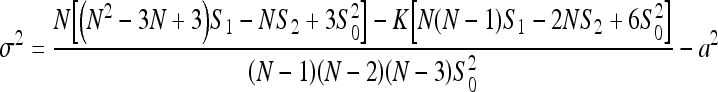
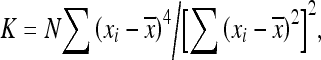
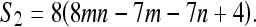
 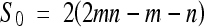 ; m and n are the number of rows and columns in the window; and N is the total number of pixels in the window. The numerator is a measure of the covariance and the denominator is a measure of the variance among the pixels. For a larger A value, there is a greater correlation between pixels and the image is blurred. When the size of N is large enough ( > 25), the variable approximately follows a normal distribution with the mean and variance given by26
 |
6 |
and
 |
7 |
where
 |
8 |
 |
9 |
and
 |
10 |
We can use the standardized normal statistic
 |
11 |
to determine the structural information of an image.
The Moran Z value of each pixel is represented with a centered 8 × 8 (i.e., 64 > 25) window because of the blocking characteristics of JPEG compression. Following that, a Z histogram can be produced by collecting all pixel Z values and sorting them into bins. This Z histogram had been proven to correspond well to the image variation in spatial properties.5,21,23,24 The spatial correlation increases with the image blurring and accompanies the increase in Z value. This Z value will increase in certain areas to form a peak. The MPR can be defined as “the ratio of the highest peak values in the Z histograms between the manipulated and original images.”5
Sliding and Jump Windows
The JPEG algorithm divides the image into many 8 × 8 pixel blocks that are processed independently. The JPEG suffers from blocking artifacts at increasing compression ratios. However, these artifacts have not been reported for images in a lower compression region. We proposed calculating the Q index and MPR by the use of both a nonoverlapping (jump window) and a pixel-by-pixel (sliding window) window, both with a size of 8 × 8. The image quality calculation results may have been affected by the blocking artifact of JPEG when the sliding window was used. These artifacts can be determined by applying a jump window to JPEG images and JPEG2000 images concurrently, followed by a comparison of the two results. Both windows started from the top-left corner and proceed through the whole image, but only the body or the head regions of the images were calculated to avoid areas outside of the patient.
Image Compression Algorithms
Two algorithms were used in this study. ViewMed (version 1.0.0.308. 2005, found at http://www.jpg.com/medical), through the courtesy of the Pegasus Imaging (Tampa, FL, United States), was used for JPEG compression and JJ2000 (version 4.1, available on the Internet at http://jj2000.epfl.ch) was used for JPEG2000 compression. The ICR can be defined as “a measure of the original image size versus the compressed image size.”
Images
For extensive evaluation, we applied the above three metrics to various medical image modalities: computer tomography (CT), magnetic resonance images (MR), computed radiography (CR), and mammography (MM) images. We randomly chose nine CT images, including five body and four head images; seven MM images; five CR chest images; and seven MR T1 and T2 brain images for this study. These CT images were taken from two serial 3-dimensional studies using a GE 9800 scanner with an image size of 512 × 512 and 12 bits deep. The MM images were digitized from film at a size of 2,048 × 2,048 × 12 bits with an Eikonix 1412 CCD camera (through the courtesy of the National Expert and Training Centre for Breast Cancer Screening and the Department of Radiology at the University of Nijmegen, the Netherlands). The CR images were 512 × 512 and 12 bits deep in size, and were cropped from the original size (2,048 × 2,494). The MR images were produced using a GE Signal 1.5 T scanner with an image size of 512 × 512 and 12 bits deep.
RESULTS AND EXPERIMENTS
Images were first compressed using JPEG and JJ2000 at various low compression levels (∼20). Following that, the image qualities of the reconstructed images were evaluated using the above metrics and windows, respectively.
The Peak Signal to Noise Ratio
The PSNR with various compression ratios for six categories of medical images are shown in Figure 2. This figure shows that the quality of JJ2000 is better than JPEG for all modalities using PSNR. The lines in this figure show an average PSNR value versus ICR. The differences of PSNR between JJ2000 and JPEG are great for T2 images, as noted in this figure.
Fig 2.

The PSNR versus ICR for JPEG (broken lines) and JJ2000 (solid lines) of six medical image modalities.
The Q Index
The Q value approaches 1 when the reconstructed images have the best quality. This Q value decreases as ICR increases, which means that the image quality degrades as noted in Figure 3. In this figure, the sliding window was used to calculate the Q values of images. The image quality of JJ2000 is obviously superior to JPEG for CR and CT images. For both MM and MR images, JJ2000 shows an advantage when ICR rises above 15, but it appears indistinct in lower ICR regions.
Fig 3.

The comparison of Q indices of JJ2000 (solid lines) with JPEG (broken lines) by using sliding windows.
The Q index versus ICR results using jump windows are shown in Figure 4. The Q values in Figures 3 and 4 follow the same trend. The image quality of JJ2000 is better than JPEG with Q estimation, even with jump windows, as seen in Figures 3 and 4. The Q indices comparison results between sliding and jump windows on JPEG images are shown in Figure 5. The jump window Q indices are always higher than those of the sliding windows except for CR images, as noted in Figure 5. This effect may mean that there are no blocking artifacts or that the deterioration covers the blocking artifacts. In Figure 5, the differences of Q indices between the sliding and jump windows noticeably increase with ICR. However, the blocking artifacts are not apparent for tomographic images (MR and CT).
Fig 4.

The comparison of Q indices of JJ2000 (solid lines) with JPEG (broken lines) by using jump windows.
Fig 5.

The comparison of Q indices of JPEG images using sliding windows (broken lines) with jump windows (solid lines).
The Moran Peak Ratio
Chen et al5 showed that the Moran statistics could be used as a good quality index for measuring the sharpness or smoothness of an image. The MPR shows different trends for the effects of smoothing or sharpening in an image. For discrete wavelet transformation algorithm, at low-compression-ratios region, the MPR curve is actually shown hollow because of the sharpness effect.6 At higher compression levels, the image qualities deteriorate due mainly to the blurring effects, and the curves ascend linearly with the compression ratio.5,6
The MPR is an approach to 1 when two images are identical and lower than 1 for sharpness and vice versa. We predicted that the blocking artifact of JPEG would result in sharpening the edges of 8 × 8 blocks. The use of jump windows on JPEG and JJ2000 concurrently is meant to detect this artifact.
The MPR of JPEG and JJ2000 were estimated using sliding and jump windows, as shown in Figures 6 and 7, respectively. The blocking artifact plays a distinct role for MPR CR, CT head, CT body, and MM estimation, but not in MR images when comparing Figures 6 and 7. In Figure 7, the MPR curves rise higher than the curves in Figure 6, proving the blocking artifact effects. In contrast, the blurring effect of JPEG on CR chest images is more than the blocking artifact effects for ICR greater than 7, as presented in these two figures. This result is consistent with the measurement of Q. For the CR, MM, and CT body images, JPEG produces more blurring effects on images than JJ2000, as noted in Figure 7. For MR images, JPEG produces more sharpening on images than JJ2000. Using jump windows, it was revealed that the CT head and MM images are indistinguishable between JJ2000 and JPEG with a ICR lower than 15. The sharpening effects on MR T1 and T2 images with JPEG counteracted the blocking effect, as displayed in both Figures 6 and 7. The JJ2000 MPR of MR images are close to 1, which means there was no distortion on the images. JJ2000 is more advantageous than JPEG.
Fig 6.

The comparison of MPR of JJ2000 (solid lines) with JPEG (broken lines) using sliding windows.
Fig 7.

The comparison of MPR of JJ2000 (solid lines) with JPEG (broken lines) using jump windows.
DISCUSSION
The Peak Signal to Noise Ratio
Using the PSNR, JJ2000 performs consistently higher than JPEG for all medical modalities. The PSNR curves of JPEG and JJ2000 approach each other in regions with a higher ICR, as noted in Figure 2. This figure also shows that the differences between JJ2000 and JPEG for CT and MR images are greater than those of the CR and MM images, especially in MR T2.
The images of CR and MM are formed using x-ray projection. X-ray contrasts are smaller than tomographic images. In this study, the CT contrast was not as good as that of the head MR images. The contrast of T2 was the biggest of the six modalities.
A previous study18 evaluated image quality using two observers after image compression and transmission. In their study, they used the Society of Motion Picture and Television Engineers (SMPTE) electronic phantom test pattern (Fig. 8). Results showed that with wavelet compression, the high-contrast resolution of the phantom test pattern did not decrease at 10:1 and 20:1 lossy compression. However, mild and moderate misregistration was found in this region. There was a loss in low-contrast resolution at 1% and 5% modulation, corresponding to 10:1 and 20:1 compression, respectively. We assumed then that the differences of PSNR curves between JPEG and JJ2000 may have been caused by image contrast.
Fig 8.

The SMPTE test pattern shows high contrast resolution regions (white arrow) and low contrast resolution regions (black arrow).
To verify this, a SMPTE test pattern was compressed using JPEG and JJ2000 after adding Gaussian noise (μ = 100, σ = 20) to each pixel to simulate real medical images. The five high-and-low-contrast resolution regions in the phantom test pattern (as marked in Fig. 8) were cropped to estimate PSNR. We found that the differences in PSNR curves between JPEG and JJ2000 in high-contrast areas were higher than those in low-contrast areas, as noted in Figure 9. The contrast affects the results of image compression if the image quality is estimated using PSNR.
Fig 9.

The PSNR of low-contrast-resolution region (a) and high-contrast-resolution region (b) of SMPTE pattern for JPEG (broken lines) and JJ2000 (solid lines) in various ICR.
The Q Index
In Figures 3 and 4, the curves of Q indices can be easily divided into two groups. One group showed high (CR, CT head, and body images) differences of Q indices between JPEG and JJ2000, while the other group showed low (MM and MR T1 and T2 images) differences. The curve trends are the same using either jump or sliding windows. For high gray-level images, the JPEG qualities are much lower than those of the JJ2000, as noted in Figures 3 and 4. We found that the image gray level histogram of six modalities can be divided into two groups as well, as noted in Figure 10. The histograms of MM and MR images have low gray level in contrast to CR and CT images. The Q index, as seen in Eq. 3, depends on the average pixel values and variances in windows. The gray level of pixels plays a role in the Q index estimation and causes variations in image quality.
Fig 10.

The histograms of image gray level for six medical image modalities.
To verify this, an MM image was again manipulated using JPEG and JJ2000 image compression and reconstruction. Following a gray-level shift of 1,000 added to each pixel on the MM image, the Q indices between JJ2000, JPEG, and the high/low gray levels of these MM images were compared. The high-gray level MM images obviously produced larger differences in Q index curves between JJ2000 and JPEG.
The Moran Peak Ratio
We hypothesized in “The Q Index” section that there are no blocking artifacts, or that the deterioration covers the blocking artifacts in CR images produced by JPEG. However, Figures 6 and 7 show that, for CR images, the deterioration in JPEG on CR images outweighed the blocking artifact effects. Comparing Figures 6 and 7 (CR, CT, and MM) reveals the obvious blocking artifact effects, which cannot be neglected, even in low ICR regions. One drawback of the MPR metric is its higher variation due to the lower number of samples in the jump window (see MR T1 and T2 in Fig. 7). It is a fact that MPR is a ratio of the highest peak value in the Z histograms between the manipulated and original images. Each Z value corresponds to a calculation result on an 8 × 8 window. The sample size will decrease and the histogram shape will vary when using the nonoverlapping jump window in contrast to the sliding window. In addition, the Moran Z value calculations were performed only in the image body or head region. For an MR T1 head image, using a jump window decreased the sample size to 1/60 than when using a sliding window. The lower sample size causes higher variation in MPR.
The MPR of JPEG MR images were always lower than those of JJ2000, even when using jump windows. This is because the sharpening effects caused by denoising occurred in high contrast MR images. In contrast Figures 6 and 7 show that for MR images, the MPR of JJ2000 was close to 1. For MR images, it can be concluded that the JJ2000 outperforms JPEG when estimated using MPR.
CONCLUSION
The qualities in low-ICR areas of compressed medical images, including CR, CT, MM, and MR, were extensively examined in this study. Based on our results, JJ2000 (JPEG2000) is superior to JPEG in low-compression regions. We found that the contrast and the image gray level averages play important roles in image compression and quality evaluation.
There are drawbacks in using PSNR, Q, and MPR because these metrics depend on contrast, gray level, or the number of samples. The objective image quality metrics are still attractive because of their cost and convenience and because they are independent of the viewing conditions and individual observers. The gray level and contrast effect on quality estimation should be considered in the development of new objective metrics.
Acknowledgements
The authors would like to thank Mr. Josiah Yoder of Purdue University, Mr. Thomas Leonard of Hillsdale College, and Dr. Maria N. Cusipag of Shu-Zen College of Medicine and Management for their help in editing this paper. This work has been supported by two research grants in Taiwan: NSC 95-2314-B-471-001 from the National Science Council and SZM095301038 from Shu-Zen College of Medicine and Management.
References
- 1.Huang HK. PACS—Picture Archiving and Communication Systems in Biomedical Imaging. New York: VCH Publishers; 1996. [Google Scholar]
- 2.Przelaskowski A. Vector quality measure of lossy compressed medical images. Comput Biol Med. 2004;34:193–207. doi: 10.1016/S0010-4825(03)00058-1. [DOI] [PubMed] [Google Scholar]
- 3.Wong S, Zaremba L, Gooden D, Huang HK. Radiologic image compression—a review. Proc IEEE. 1995;83:194–219. doi: 10.1109/5.364466. [DOI] [Google Scholar]
- 4.Chen ZD, Chang RF, Kuo WJ. Adaptive predictive multiplicative autoregressive model for medical image compression. IEEE Trans Med Imaging. 1999;18:181–184. doi: 10.1109/42.759128. [DOI] [PubMed] [Google Scholar]
- 5.Chen TJ, Chuang KS, Wu J, Chen SC, Hwang IM, Jan ML. Quality degradation in lossy wavelet image compression. J Digit Imaging. 2003;16:210–215. doi: 10.1007/s10278-003-1652-0. [DOI] [PMC free article] [PubMed] [Google Scholar]
- 6.Erickson BJ. Irreversible compression of medical images. J Digit Imaging. 2002;15:5–14. doi: 10.1007/s10278-002-0001-z. [DOI] [PMC free article] [PubMed] [Google Scholar]
- 7.Seeram E. Irreversible compression in digital radiology. A literature review. Radiography. 2006;12:45–59. doi: 10.1016/j.radi.2005.04.002. [DOI] [Google Scholar]
- 8.Siddiqui KM, Johnson JP, Reiner BI, Siegel EL: Discrete cosine transform JPEG compression vs. 2D JPEG2000 compression: JNDmetrix visual discrimination model image quality analysis, Proceedings of SPIE, vol. 5748, Medical Imaging: PACS and Imaging Informatics, 2005, pp 202–207
- 9.Eikelboom RH, Yogesan K, Barry CJ, Constable IJ, Tay-Kearney ML, Jitskaia L, House PH. Methods and limits of digital image compression of retinal images for telemedicine. Invest Ophthalmol Vis Sci. 2000;41:1916–1924. [PubMed] [Google Scholar]
- 10.Brennecke R, Burgel U, Rippin G, Post F, Rupprecht HJ, Meyer J. Comparison of image compression viability for lossy and lossless JPEG and Wavelet data reduction in coronary angiography. Int J Card Imaging. 2001;17:1–12. doi: 10.1023/A:1010644318298. [DOI] [PubMed] [Google Scholar]
- 11.Iyriboz TA, Zukoski MJ, Hopper KD, Stagg PL. A comparison of wavelet and Joint Photographic Experts Group lossy compression methods applied to medical images. J Digit Imaging. 1999;12(2 Suppl 1):14–17. doi: 10.1007/BF03168745. [DOI] [PMC free article] [PubMed] [Google Scholar]
- 12.Ricke J, Maass P, Lopez Hanninen E, Liebig T, Amthauer H, Stroszczynski C, Schauer W, Boskamp T, Wolf M. Wavelet versus JPEG (Joint Photographic Expert Group) and fractal compression. Impact on the detection of low-contrast details in computed radiographs. Invest Radiol. 1998;33:456–463. doi: 10.1097/00004424-199808000-00006. [DOI] [PubMed] [Google Scholar]
- 13.Hui OT, Besar R. Medical image compression using JPEG2000 and JPEG: a comparison study. J Mech Med Biol. 2002;2:313–328. doi: 10.1142/S021951940200054X. [DOI] [Google Scholar]
- 14.Janhom A, Stelt PF, Sanderink GCH. A comparison of two compression algorithms and the detection of caries. Dentomaxillofacial Radiol. 2000;31:257–263. doi: 10.1038/sj.dmfr.4600704. [DOI] [PubMed] [Google Scholar]
- 15.Slone RM, Foos DH, Whiting BR, Muka E, Rubin DA, Pilgram TK, Kohm KS, Young SS, Ho P, Hendrickson DD. Assessment of visually lossless irreversible image compression: comparison of three methods by using an image-comparison workstation. Radiology. 2000;215:543–553. doi: 10.1148/radiology.215.2.r00ap47543. [DOI] [PubMed] [Google Scholar]
- 16.Li F, Sone S, Takashima S, Kiyono K, Yang ZG, Hasegawa M, Kawakami S, Saito A, Hanamura K, Asakura K. Effects of JPEG and wavelet compression of spiral low-dose CT images on detection of small lung cancers. Acta Radiol. 2001;42:156–160. doi: 10.1080/028418501127346657. [DOI] [PubMed] [Google Scholar]
- 17.Przelaskowski A: Hybrid vector measures of compressed medical images, SPIE Symposium Medical Imaging: Image perception and performance, http://www.ire.pw.edu.pl/~arturp/Publikacje/ap_HVM.pdf, 2000
- 18.Kalyanpur A, Neklesa VP, Taylor CR, Daftary AR, Brink AR. Evaluation of JPEG and wavelet compression of body CT images for direct digital teleradiologic transmission. Radiology. 2000;217:772–779. doi: 10.1148/radiology.217.3.r00nv22772. [DOI] [PubMed] [Google Scholar]
- 19.Ebrahimi F, Chamik M, Winkler S. JPEG vs. JPEG2000: an objective comparison of image encoding quality. Proc SPIE Appl Digit Image Proc. 2004;5558:300–308. [Google Scholar]
- 20.Wang Z, Bovic AC. A universal image quality index. IEEE Signal Process Lett. 2002;3:81–84. doi: 10.1109/97.995823. [DOI] [Google Scholar]
- 21.Chen TJ, Chuang KS, Wu J, Chen SC, Hwang IM, Jan ML. A novel image quality index using Moran I statistics. Phys Med Biol. 2003;48:N131–N137. doi: 10.1088/0031-9155/48/8/402. [DOI] [PubMed] [Google Scholar]
- 22.Wang Z, Bovik AC, Evans BL. Blind measurement of blocking artifacts in images. IEEE Int Conf Image Proc. 2000;3:981–984. [Google Scholar]
- 23.Chen TJ, Chuang KS, Chiang YC, Chang JH, Liu RS. A statistical method for evaluation quality of medical images: case study in bit discarding and image compression. Comput Med Imaging Graph. 2004;28:167–175. doi: 10.1016/j.compmedimag.2004.01.003. [DOI] [PubMed] [Google Scholar]
- 24.Chen TJ, Chuang KS, Chang JH, Shiao YH, Chuang CC: A blurring index for medical images. J Digit Imaging 19:118–125, 2006 DOI: 10.1007/s10278-005-8736-y, 2005 [DOI] [PMC free article] [PubMed]
- 25.Chuang KS, Huang HK. Assessment of noise in a digital image using the join-count statistic and Moran test. Phys Med Biol. 1992;37:357–369. doi: 10.1088/0031-9155/37/2/004. [DOI] [PubMed] [Google Scholar]
- 26.Cliff AD, Ord JK. Spatial process: models and applications. London: Pion; 1981. [Google Scholar]