Abstract
One year after the emergence of the 2009 influenza (H1N1) pandemic, most cases have been relatively mild; however, the possibility of enhanced viral growth ability by reassortment with seasonal viruses cannot be overlooked. Here, we show that reassortant viruses containing a seasonal H1 HA and swine-origin NA and M genes have enhanced virus growth over their wild-type parental viruses. The emergence of such viruses in nature could, therefore, represent a threat.
Keywords: Influenza virus, swine-origin, pandemic, seasonal, reassortment
The influenza virus genome comprises eight single-stranded RNA segments of negative polarity, which allows genetic reassortment among different viruses when they co-infect a single cell. Reassortment is important for influenza virus evolution, contributing to the generation of novel viruses, such as those that cause influenza pandemics (Laver & Webster, 1973).
In March 2009, a swine-origin influenza A virus (S-OIV) of the subtype H1N1 emerged in North America, spreading rapidly through the human population and establishing the first influenza pandemic of the twenty-first century (Smith et al., 2009). On the first anniversary of its emergence, most cases have been relatively mild (WHO, 2010). However, there is concern that, as the virus adapts to the human host, virulence may increase (Maines et al., 2009), although this has not yet been observed. The introduction of known pathogenicity markers, such as the PB1-F2 protein and the E627K and D701N mutations in the PB2 gene, as well as the introduction of human virus-like features in the S-OIV NS1, all failed to increase S-OIV virus growth in cell culture, or virulence in animal models (Hai et al., 2010; Hale et al., 2010a, b; Herfst et al., 2010). Nevertheless, the possibility of acquisition of enhanced replicative ability or pathogenicity through reassortment with contemporary seasonal H1N1 and H3N2 viruses cannot be overlooked (Herfst et al., 2010).
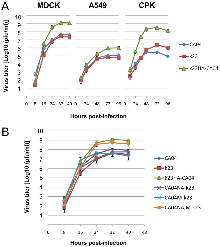
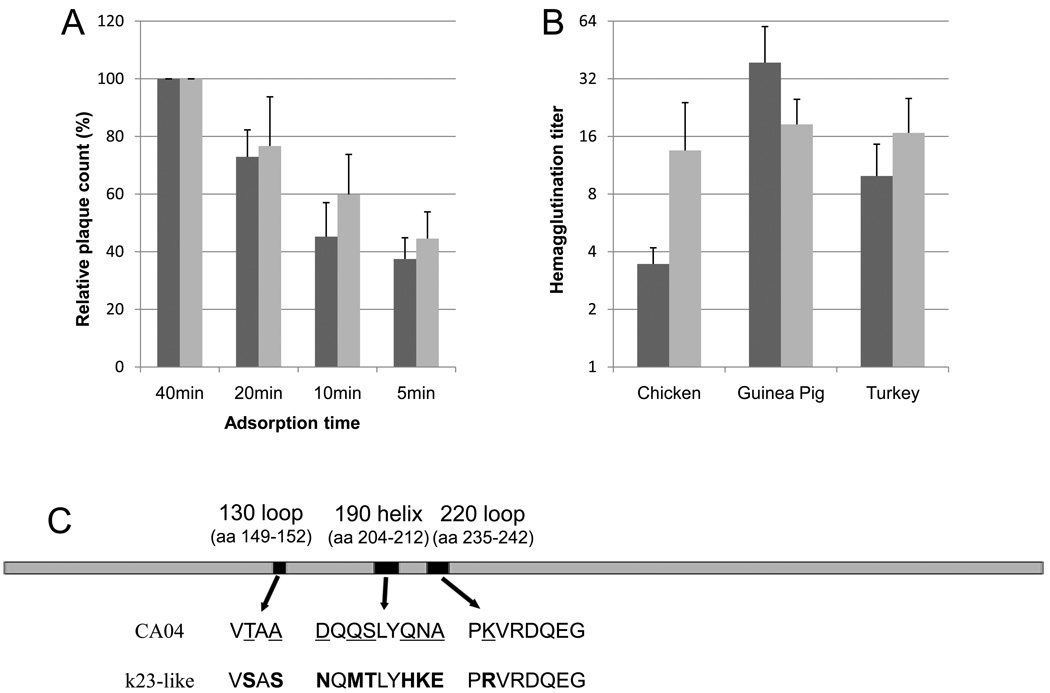
To determine if reassortment between S-OIV and seasonal H1N1 influenza viruses results in viruses with increased growth capacity, we produced reassortants by reverse genetics (Neumann et al., 1999), and analyzed their growth in cell culture. In preliminary experiments, we found that a reassortant virus containing the HA gene from A/Kawasaki/UT-SAI-k23/2008 (H1N1) (k23) on an A/California/04/2009 (H1N1) (CA04) background produced plaques in Madin-Darby canine kidney (MDCK) cells that were distinctly larger than those of either parental virus (Table 1). This prompted us to compare the growth properties of this reassortant (designated k23HA-CA04) with that of the parental viruses (k23 and CA04) in three different cell lines: MDCK, A549, and the porcine kidney cell line CPK (Komaniwa et al., 1981). Interestingly, k23HA-CA04 had significantly enhanced growth over either parental virus in these three cell lines (Fig. 1A). We then asked whether the enhanced growth of k23HA-CA04 was due to a difference in the receptor binding properties of the k23 and CA04 viruses. To this end, we devised a “virus wash away assay”, which consists of a plaque assay, in which the adsorption times are progressively shortened. The relative reduction in the number of plaques provides a measure of the affinity between the viruses and cells. The results of this assay indicated that CA04 has slower attachment to cells than does k23HA-CA04, as evidenced by the fewer plaques produced when adsorption times were limited to 5 or 10 minutes (t-test, p < 0.05) (Fig. 2A).
Table 1.
Rescue efficiency, stock titers and plaque size of viruses.
| Virus | Rescue efficiency (log10PFU/ml)* |
Titer of stock virus grown in MDCK cells (log10PFU/ml) |
Plaque size in MDCK cells (mm)† |
|---|---|---|---|
| CA04 | 4.26 | 7.88 | 0.26 ± 0.08 |
| k23 | 2.48 | 7.68 | 0.58 ± 0.22 |
| k23HA-CA04 | 6.04 | 9.00 | 1.57 ± 0.35 |
| CA04NA-k23 | 5.27 | 8.08 | 1.01 ± 0.24 |
| CA04M-k23 | 4.26 | 7.72 | 0.91 ± 0.29 |
| CA04NA,M-k23 | 6.00 | 8.73 | 1.41 ± 0.28 |
| Nw3487 | 5.23 | 8.70 | 1.01 ± 0.36 |
| k23HA-Nw3487 | 6.15 | 9.30 | 1.76 ± 0.31 |
| Nw3487NA,M-k23 | 5.95 | 8.73 | 1.25 ± 0.34 |
| Ut42 | 4.15 | 7.68 | 0.29 ± 0.09 |
| k23HA-Ut42 | 6.08 | 9.08 | 1.42 ± 0.36 |
| Ut42NA,M-k23 | 6.00 | 8.83 | 1.27 ± 0.31 |
Measured by determining the viral titer in the supernatant of transfected 293T cells at 48 hours post-infection, by plaque assay using MDCK cells.
Values represent mean ± standard deviations, measured at 48 hours post-infection; n=100. The plaque size of CA04NA,M-k23 differed significantly from that of CA04NA-k23 and CA04M-k23 (p < 0.001).
FIG. 1.
(A) Viral growth of CA04, k23, and k23HA-CA04 in MDCK, A549, and CPK cells. (B) Viral growth of CA04, k23, k23HA-CA04, CA04NA-k23, CA04M-k23, and CA04NA,M-k23 in MDCK cells. Cells were infected with viruses at a multiplicity of infection of 0.001. Virus yields at the indicated time points were determined by plaque assay in MDCK cells. Data represent the mean of three independent infections ± standard deviations.
FIG. 2.
(A) Virus wash away assay. CA04 (dark grey) and k23HA-CA04 (light grey) were subjected to plaque assay in MDCK cells, where the adsorption times were progressively shortened from 40 to 5 minutes. Cells were maintained at 37°C during the adsorption period. Data represent the relative plaque count obtained for the indicated adsorption periods, normalized to values of the 40 minute adsorption time (100%). Data represent the mean of three independent experiments ± standard deviations. Plaque counts of CA04 and k23HA-CA04 differed significantly with adsorption times of 5 and 10 minutes (t-test, p < 0.05). (B) Relative hemagglutination titers of CA04 and k23HA-CA04. Virus stocks were adjusted to a titer of 7.6×10⁷ pfu/ml, before a standard hemagglutination assay using chicken, guinea pig, and turkey erythrocytes was performed. CA04 is shown in dark grey; k23HA-CA04 is shown in light grey. Data represent the mean of three independent experiments ± standard deviations. (C) Schematic diagram of the chimera CA04 HA. Black columns represent sequences encoding the structures that form the rim of the HA RBS (130 loop, 190 helix and 220 loop). Mutations were introduced into the pPol1-CA04HA plasmid to substitute the original CA04 (underlined) with k23-like (bold) amino acids in the encoded HA protein.
To gain insight into the relative binding activities of the CA04 and k23 hemagglutinins, we performed hemagglutination assays (HA) with the two viruses, using red blood cells (RBC) from three different species: chicken, turkey, and guinea pig. As shown in Fig. 2B, the reassortant k23HA-CA04 showed higher hemagglutination activity on chicken and to some extent on turkey RBCs, whereas CA04 had slightly higher activity on guinea pig RBCs. While an accurate interpretation of the relative hemagglutination activities of these viruses may be complex, the results do show a difference in receptor binding between the two viruses. Glycan array analyses of the receptor specificity of the S-OIV have yielded contrasting results; while Maines et al. (2009) found that S-OIV exhibited dose-dependent binding to a single α2-6 glycan, with minimal binding to α2-3 glycans, Childs et al. (2009) observed broader specificity, with binding to both α2-6 and α2-3-linked receptors. It should be noted, however, that these are results from in vitro assessments of HA binding to synthetic glycans, and that the actual availability of the various species of carbohydrate in different cell types, and their exploitation by influenza viruses, is still poorly understood.
To further investigate the importance of receptor-binding to the growth of k23HA-CA04 and CA04, we produced a virus containing chimeric CA04 HA, in which the amino acid sequences corresponding to the structures that form the rim of the receptor binding site (RBS), the 130 loop, 190 helix and 220 loop (Gamblin et al., 2004), were replaced by those of k23 HA (Fig. 2C). Using a forward primer that introduced the mutations A448T and G454T, and a reverse primer that introduced the mutation A707G, we performed a PCR reaction using the pPol1-CA04 HA plasmid as a template. The PCR product was then purified and used as a megaprimer in a back-to-back PCR reaction to amplify the entire pPol1-CA04 HA plasmid. This second PCR product was digested with HindIII and DpnI and ligated, thus introducing k23 HA-like mutations into the CA04 130 loop and 220 loop regions of pPol1-CA04 HA. Finally, we used a new set of primers containing BsmBI sites to introduce k23 HA-like mutations into the region corresponding to the 190 helix, thus generating the plasmid pPol1-CA04HA(k23RBS). By reverse genetics, we produced a virus with this chimeric HA, and all remaining genes from CA04 (designated CA04HA[k23RBS]-CA04). The virus could be rescued, but it failed to grow to higher titers, or produce larger plaques in MDCK cells than wild-type CA04 (data not shown). Although the possibility of structural constraints cannot be overlooked, this result suggests that the receptor binding region alone is not responsible for the enhanced growth of k23HA-CA04.
The reassortant k23HA-CA04 showed remarkable enhanced growth over either parental virus. We therefore sought to identify the genes of CA04 that, together with the k23 HA, were responsible for this increased growth. To this end, we used reverse genetics to generate a series of single gene reassortants, containing each of the CA04 genes on a k23 genetic background. We found that viruses containing either NA or M from CA04 on a k23 background (designated CA04NA-k23 and CA04M-k23) had better rescue efficiency (as measured by determining viral titers in the supernatant of transfected 293T cells) than k23, and produced larger plaques than either CA04 or k23, although neither reassortants reached the levels of k23HA-CA04 (Table 1). Therefore, we generated another reassortant, containing both the NA and M genes from CA04 on a k23 background; this reassortant (designated CA04NA,M-k23) had high rescue efficiency, produced plaques significantly larger than those produced by CA04NA-k23 and CA04M-k23 (p < 0.001) (Table 1), and had enhanced growth in MDCK cells (Fig 1B), approaching the characteristics of k23HA-CA04, indicating that cooperation between the HA, NA, and M genes is essential for enhanced virus growth.
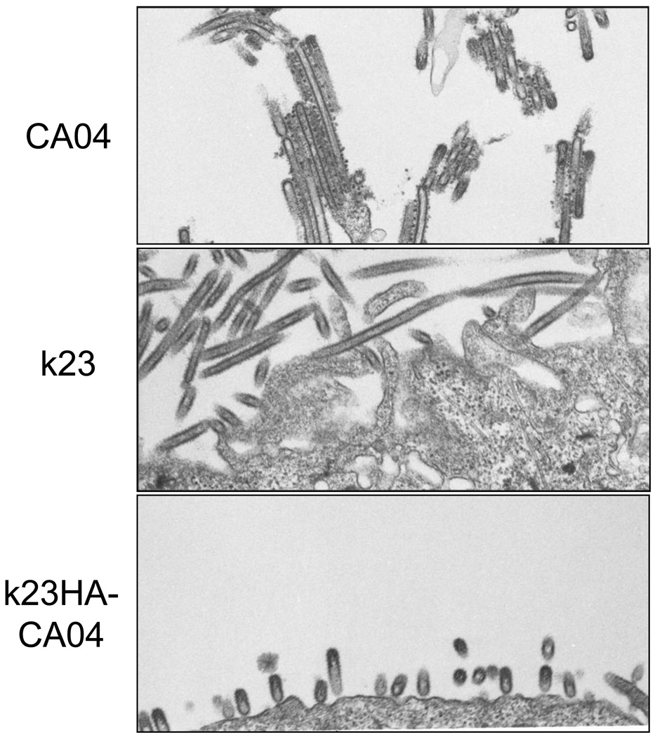
The HA, NA, and M gene products play central roles in the assembly and budding of influenza viruses at the plasma membrane (Nayak et al., 2009). Electron microscopic analysis of the viruses showed that, while CA04 and k23 were predominantly filamentous viruses, k23HA-CA04 particles were distinctly shorter, with a predominance of quasi-spherical virus particles (Fig. 3). This finding may suggest that a cooperation between the k23 HA and the CA04 NA and M proteins may lead to more efficient viral morphogenesis and, thereby, contribute to increased viral production in cell culture.
FIG. 3.
Ultrathin section electron micrographs of CA04, k23 and k23HA-CA04 viruses. MDCK cells were infected with the viruses at a multiplicity of infection of 0.1. Samples were fixed 20 hours post-infection and analyzed as previously described (Noda et al., 2006).
To determine whether these findings also apply to other S-OIVs, we used A/Norway/3487/2009 (H1N1) (Nw3487) and A/Utah/42/2009 (H1N1) (Ut42) to produce reassortants containing the k23 HA on the S-OIV background (k23HA-Nw3487, k23HA-Ut42), and NA and M genes from the S-OIV on the k23 background (Nw3487NA,M-k23; Ut42NA,M-k23). These experiments repeated the pattern seen with CA04, since both the introduction of the seasonal HA into the Nw3487 and Ut42 background and the introduction of NA and M from Nw3487 and Ut42 into the k23 background led to the generation of viruses with higher rescue capability, increased plaque size, and enhanced virus growth in MDCK cells, relative to the parental viruses (Table 1), thus confirming that the finding is conserved among S-OIVs.
In summary, here we demonstrated that reassortment between S-OIV and seasonal H1N1 viruses generates reassortants with enhanced growth properties, when there is a combination of a seasonal HA, and swine-origin NA and M genes.
The virus wash away assay and relative hemagglutination activity experiments indicate that differences in the efficiency of virus attachment to cells may play a role in the enhanced growth kinetics of the reassortants in this study. Moreover, considering the cooperation between HA, NA, and M for virus morphogenesis (Nayak et al., 2009), it is also possible that a suitable combination of these genes may somehow facilitate this process, thus contributing to viral fitness.
Influenza virions have a pleomorphic morphology that ranges from spherical to filamentous. Previous studies have mapped the determinants of virion morphology mainly to the M1 protein, although M2 has also been shown to play a role (Bourmakina & García-Sastre, 2003; Iwatsuki-Horimoto et al., 2006; Liu et al., 2002; Roberts et al., 1998). Moreover, spherical virion morphology is associated with high virus growth (Liu et al., 2002). Our findings are in agreement with this latter report, since the high growth properties of our reassortants also correlated with spherical virion morphology. However, further investigation is needed to fully understand the functional basis for the enhanced growth of these reassortants.
It is interesting that, while the introduction of well-known virulence markers to S-OIV failed to influence the replication of the viruses (Hai et al., 2010; Hale et al., 2010a, b; Herfst et al., 2010), the introduction of the HA gene from a seasonal virus greatly enhanced viral growth. This highlights how little we know of the molecular mechanisms that control the replicative ability of influenza viruses.
Our findings have important implications for human health, as they may indicate a reemergence potential for the “old” H1 HA, especially after the human population acquires immunity to the new S-OIV HA. The seasonal H1 HA, which descends from the 1918 Spanish influenza virus HA, already reemerged once, after over two decades, causing the so-called “Russian flu” (Zakstelskaja et al., 1978). It is, therefore, of fundamental importance that continuous surveillance and containment measures be implemented, in response to this risk.
Acknowledgments
We thank Susan Watson for scientific editing. This work was supported by a contract research fund from the Ministry of Education, Culture, Sports, Science and Technology of Japan, by the Program of Founding Research Centers for Emerging and Reemerging Infectious Diseases, and is supported in part by Grants-in-Aid for Specially Promoted Research and for Scientific Research, by ERATO (Japan Science and Technology Agency), by the National Institute of Allergy and Infectious Diseases Public Health Service research grants, USA, and by the Center for Research on Influenza Pathogenesis (CRIP) funded by the National Institute of Allergy and Infectious Diseases (Contract HHSN266200700010C).
Footnotes
Publisher's Disclaimer: This is a PDF file of an unedited manuscript that has been accepted for publication. As a service to our customers we are providing this early version of the manuscript. The manuscript will undergo copyediting, typesetting, and review of the resulting proof before it is published in its final citable form. Please note that during the production process errors may be discovered which could affect the content, and all legal disclaimers that apply to the journal pertain.
REFERENCES
- Bourmakina SV, García-Sastre A. Reverse genetics studies on the filamentous morphology of influenza A virus. J. Gen. Virol. 2003;84:517–527. doi: 10.1099/vir.0.18803-0. [DOI] [PubMed] [Google Scholar]
- Childs RA, Palma AS, Wharton S, Matrosovich T, Liu T, Chai W, Campanero-Rhodes MA, Zhang Y, Eickmann M, Kiso M, Hay A, Matrosovich M, Feizi T. Receptor-binding specificity of pandemic influenza A (H1N1) 2009 virus determined by carbohydrate microarray. Nat. Biotechnol. 2009;27:797–799. doi: 10.1038/nbt0909-797. [DOI] [PMC free article] [PubMed] [Google Scholar]
- Gamblin SJ, Haire LF, Russen RJ, Stevens DJ, Xiao B, Ha Y, Yasisht N, Steinhauer DA, Daniels RS, Elliot A, Wiley DC, Skehel JJ. The structure and receptor binding properties of the 1918 influenza hemagglutinin. Science. 2004;303:1838–1842. doi: 10.1126/science.1093155. [DOI] [PubMed] [Google Scholar]
- Hai R, Schmolke M, Varga ZT, Manicassamy B, Wang TT, Belser JA, Pearce MB, García-Sastre A, Tumpey TM, Palese P. PB1-F2 expression by the 2009 pandemic H1N1 influenza virus has minimal impact on virulence in animal models. J. Virol. 2010;84:4442–4450. doi: 10.1128/JVI.02717-09. [DOI] [PMC free article] [PubMed] [Google Scholar]
- Hale BG, Steel J, Manicassamy B, Medina RA, Ye J, Hickman D, Lowen AC, Perez DR, García-Sastre A. Mutations in the NS1 C-terminal tail do not enhance replication or virulence of the 2009 pandemic H1N1 influenza A virus. J. Gen. Virol. 2010a;91:1737–1742. doi: 10.1099/vir.0.020925-0. [DOI] [PMC free article] [PubMed] [Google Scholar]
- Hale BG, Steel J, Medina RA, Manicassamy B, Ye J, Hickman D, Hai R, Schmolke M, Lowen AC, Perez DR, García-Sastre A. Inefficient control of host gene expression by the 2009 pandemic H1N1 influenza A virus NS1 protein. J. Virol. 2010b;84:6909–6922. doi: 10.1128/JVI.00081-10. [DOI] [PMC free article] [PubMed] [Google Scholar]
- Herfst S, Chutinimitkul S, Ye J, de Wit E, Munster VJ, Schrauwen EJ, Bestebroer TM, Jonges M, Meijer A, Koopmans M, Rimmelzwaan GF, Osterhaus AD, Perez DR, Fouchier RA. Introduction of virulence markers in PB2 of pandemic swine-origin influenza virus does not result in enhanced virulence or transmission. J. Virol. 2010;84:3752–3758. doi: 10.1128/JVI.02634-09. [DOI] [PMC free article] [PubMed] [Google Scholar]
- Iwatsuki-Horimoto K, Horimoto T, Noda T, Kiso M, Maeda J, Watanabe S, Muramoto T, Fujii K, Kawaoka Y. The cytoplasmic tail of the influenza A virus M2 protein plays a role in viral assembly. J. Virol. 2006;80:5233–5240. doi: 10.1128/JVI.00049-06. [DOI] [PMC free article] [PubMed] [Google Scholar]
- Komaniwa H, Fukusho A, Shimizu Y. Micro method for performing titration and neutralization test of hog cholera virus using established porcine kidney cell strain. Natl. Inst. Anim. Health Q. (Tokyo) 1981;21:153–158. [PubMed] [Google Scholar]
- Laver WG, Webster RG. Studies on the origin of pandemic influenza. 3. Evidence implicating duck and equine influenza viruses as possible progenitors of the Hong Kong strain of human influenza. Virology. 1973;51:383–391. doi: 10.1016/0042-6822(73)90437-6. [DOI] [PubMed] [Google Scholar]
- Liu T, Muller J, Ye Z. Association of influenza virus matrix protein with ribonucleoproteins may control viral growth and morphology. Virology. 2002;304:89–96. doi: 10.1006/viro.2002.1669. [DOI] [PubMed] [Google Scholar]
- Maines TR, Jayaraman A, Belser JA, Wadford DA, Pappas C, Zeng H, Gustin KM, Pearce MB, Viswanathan K, Shriver ZH, Raman R, Cox NJ, Sasisekharan R, Katz JM, Tumpey TM. Transmission and pathogenesis of swine-origin 2009 A(H1N1) influenza viruses in ferrets and mice. Science. 2009;325:484–487. doi: 10.1126/science.1177238. [DOI] [PMC free article] [PubMed] [Google Scholar]
- Nayak DP, Balogun RA, Yamada H, Zhou ZH, Barman S. Influenza virus morphogenesis and budding. Virus Res. 2009;143:147–161. doi: 10.1016/j.virusres.2009.05.010. [DOI] [PMC free article] [PubMed] [Google Scholar]
- Neumann G, Watanabe T, Ito H, Watanabe S, Goto H, Gao P, Hughes M, Perez DR, Donis R, Hoffmann E, Hobom G, Kawaoka Y. Generation of influenza A viruses entirely from cloned cDNAs. Proc. Natl. Acad. Sci. U S A. 1999;96:9345–9350. doi: 10.1073/pnas.96.16.9345. [DOI] [PMC free article] [PubMed] [Google Scholar]
- Noda T, Sagara H, Yen A, Takada A, Kida H, Cheng RH, Kawaoka Y. Architecture of ribonucleoprotein complexes in influenza A virus particles. Nature. 2006;439:490–492. doi: 10.1038/nature04378. [DOI] [PubMed] [Google Scholar]
- Roberts PC, Lamb RA, Compans RW. The M1 and M2 proteins of influenza A virus are important determinants in filamentous particle formation. Virology. 1998;240:127–137. doi: 10.1006/viro.1997.8916. [DOI] [PubMed] [Google Scholar]
- Smith GJ, Vijaykrishna D, Bahl J, Lycett SJ, Worobey M, Pybus OG, Ma SK, Cheung CL, Raghwani J, Bhatt S, Peiris JS, Guan Y, Rambaut A. Origins and evolutionary genomics of the 2009 swine-origin H1N1 influenza A epidemic. Nature. 2009;459:1122–1125. doi: 10.1038/nature08182. [DOI] [PubMed] [Google Scholar]
- WHO. [accessed September 17, 2010];Geneve, Switzerland: WHO; Pandemic (H1N1) 2009 - Update 89. 2010 www.who.int/csr/don/2010_02_26/en/index.html.
- Zakstelskaja LJ, Yakhno MA, Isacenko VA, Molibog EV, Hlustov SA, Antonova IV, Klitsunova NV, Vorkunova GK, Burkrinskaja AG, Bykovsky AF, Hohlova GG, Ivanova VT, Zdanov VM. Influenza in the USSR in 1977: recurrence of influenzavirus A subtype H1N1. Bull. World Health Organ. 1978;56:919–922. [PMC free article] [PubMed] [Google Scholar]


