
Figure 3.
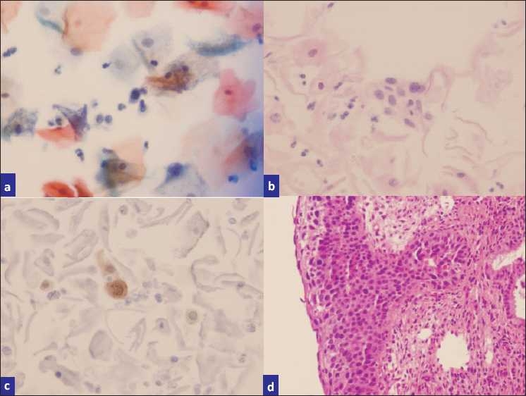
a) ASC-H (rare single cells with hyperchromatic nuclei and high N:C ratios), b) H and E stained cell block sections, c) p16-stained sections highlighting scattered high-grade cells, d) biopsy showing CIN III with extensive endocervical glandular involvement.