This case illustrates the importance of eliciting the use of alternative and non-prescription medicines when taking histories and also considering lead poisoning in the differential of unexplained abdominal pain.
Case
A previously well 26-year-old Asian man presented acutely with a two-day history of severe, colicky, central upper abdominal pain, associated with vomiting and constipation. There were no precipitating factors and the pain was not relieved by simple analgesia. He was on no prescribed medication and denied taking any other drugs. Clinical examination was unremarkable, as was an abdominal radiograph. Routine haematology and biochemistry blood tests, including blood film, were normal apart from a raised Alanine Transaminase (ALT) at 222 IU/mL. Although in great distress initially, his symptoms resolved spontaneously within two days and he was discharged.
Two weeks later he presented with identical symptoms, having been symptom-free in the interim. ALT was again moderately raised at 180 IU/mL. Gastroscopy and ultrasound of the abdomen were both normal. An abdominal CT scan was normal apart from slight terminal ileal thickening. However, a subsequent colonoscopy showed a normal terminal ileum; histology revealed only mild non-specific inflammation and both ZN staining and TB culture of the biopsies were negative. Chest X-ray, Mantoux test and Yersinia serology were also negative. During this admission he exhibited behavioural disturbance, was transiently disoriented and threatened suicide. Although these features settled, a psychiatric assessment was undertaken. It transpired the patient had recently consulted his GP about marital difficulties due to erectile dysfunction, for which the GP had declined the patient's request for Viagra. Again all of his symptoms settled spontaneously, his ALT normalized and he was discharged.
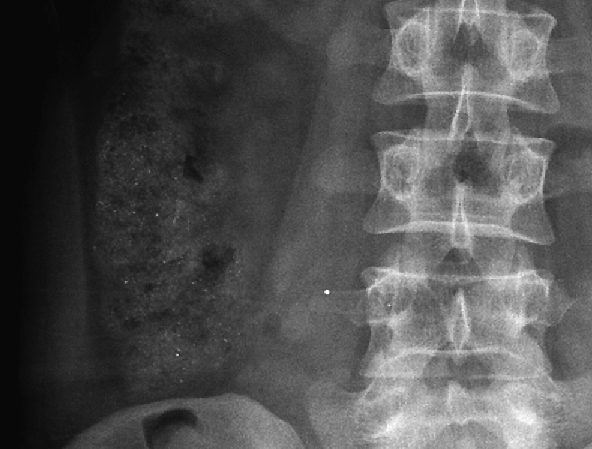
Shortly after discharge however, he was re-admitted with further abdominal pain and again displaying erratic behaviour. On this occasion an abdominal radiograph showed some high density specks in the colon (Figure 1). ALT was raised at 390 but again returned to normal soon after admission. A comprehensive panel of blood tests screening for causes of liver disease was entirely negative and magnetic resonance cholangiopancreatography (MRCP) revealed a normal liver and biliary system. In view of the pain, constipation and psychological symptoms, urine and blood were sent for porphyria testing. Although the urine porphyrin:creatinine ratio was markedly raised at 230 (normal range 0–35), the absence of raised urinary porphobilinogen during an acute attack meant that acute intermittent porphyria was extremely unlikely. The differential for raised urinary porphyrins is wide, encompassing a range of disease processes that affect haem synthesis either in the liver or blood ( Table 1). However, the episodic nature of his symptoms and ALT derangement and their spontaneous resolution in hospital led us to suspect an environmental toxin. Lead toxicity fitted the clinical picture well and laboratory testing revealed a blood lead level of 7.62 µmol/L (normal range 0.00–0.60), corresponding to severe intoxication at a level sufficient to explain his abdominal symptoms and his encephalopathic features. On further discussion with the patient, he admitted to having used several doses of a drug for erectile dysfunction (Kamagra), which had been obtained via a friend from an Internet site based in India. The timing of ingestion corresponded both to his symptoms and to the presence of metallic particles in the colon on X-ray; this was therefore presumed to be the source of lead exposure.
Figure 1.

Abdominal radiograph showing high density specks in the colon
Table 1.
Secondary causes of porphyrinuria8
| Secondary causes | Examples |
|---|---|
| Toxins | Alcohol, hexachlorobenzene, polyhalogenated biphenyls, dioxins (TCDD), vinyl chloride, carbon tetrachloride, benzene, chloroform, lead, mercury, arsenic |
| Liver damage | Cirrhosis, hepatitis, cholestasis, fatty liver, drug-induced liver damage |
| Drug effects | Some sedatives, anaesthetics and sulfa-containing antibiotics |
| Haematological disease | Hemolytic, sideroachrestic, sideroblastic, aplastic anaemias, ineffective erythropoiesis; pernicious anaemia, thalassemia, leukaemia, erythroblastosis |
| Malignancy | Particularly those affecting the hepato-pancreato-biliary system |
| Hereditary hyperbilirubinaemias | |
| Infectious diseases | Infectious mononucleosis, poliomyelitis |
| Disturbances of iron metabolism | Haemochromatosis, haemosiderosis |
| Pregnancy | |
| Diabetes mellitus |
Chelation therapy was considered, but as his symptoms had resolved, he was simply followed up with advice to avoid further ingestion of Kamagra tablets. At 6 weeks post discharge the lead level had fallen to 4.47umol/L and at 6 months to 1.85 umol/L, a roughly exponential decline reflecting lead's long half-life and thus supporting the story of discontinued exposure. No further rises in liver enzymes were seen and to date the patient has remained asymptomatic.
Discussion
The toxic effects of lead are mainly a result of interference with a wide range of enzymes, and as such many body systems may be affected ( Table 2). Presenting complaints vary according to the level of lead in the blood and the time course of the subject's exposure.1,2 At low blood levels, non-specific symptoms of malaise, anorexia and irritability predominate. At higher levels, gastrointestinal symptoms are common, with abdominal pain accompanied by vomiting and constipation or diarrhoea. Anaemia also occurs frequently, with basophilic stippling of erythrocytes on blood film. Peripheral neuropathies may be seen, and as blood levels rise further neuro-cognitive defects progressing to frank encephalopathy, seizures and even death can occur.1,3
Table 2.
End organ damage caused by lead poisoning
| Gastrointestinal | ‘Lead colic’ |
| Anorexia, constipation, nausea and vomiting | |
| Hepatitis | |
| Neurological | Peripheral neuropathy |
| Cerebral oedema | |
| Behavioural change and acute psychosis | |
| With higher levels: encephalopathy, seizures, coma and eventually death | |
| Haematological | Anaemia |
| Haemolysis | |
| Renal | Fanconi-like syndrome |
| Chronic interstitial nephropathy | |
| Cardiovascular | Myocarditis |
| Hypertension (chronic exposure) | |
| Developmental (paediatric) | Neurodevelopmental delay and learning difficulties |
| Growth retardation | |
| Hearing impairment | |
| Reproductive system | Men: low sperm count and abnormal sperm morphology |
| Women: miscarriage, prematurity, low birth weight and contamination of breast milk | |
| Other | Gout |
| Gum discolouration |
Most commonly the picture is of chronic lead intoxication, often due to occupational exposure over a long period of time ( Table 3). These cases usually present insidiously with non-specific symptoms affecting multiple organ systems. In contrast, we have described a case of acute intoxication following a single, short episode of ingestion.
Table 3.
Sources of lead poisoning
| Occupational | Lead mining and smelting, plumbing, auto mechanics, glass manufacturing, construction work, battery manufacturing and recycling, welding, plastic manufacturing, production of paints and pigments |
| Paint | Commonly in children through ingestion of lead-containing paint used to decorate toys; also via inhalation in workers involved in stripping of leading-containing paints |
| Soil | Mainly a result of pesticide use or contamination from lead-containing petrol |
| Water | Contamination of water from lead in soil, atmospheric lead or plumbing fixtures |
| Products | Including many traditional cosmetics (transdermal exposure) as well as traditional and counterfeit medicines |
Such cases present a diagnostic challenge due to the relative paucity of positive clinical findings and as such often lead to delayed diagnosis. In some cases appendicectomies and even laparotomies have been carried out before eventually arriving at the diagnosis of lead poisoning.4,5
The pattern of abdominal pain suffered by our patient is characteristic, with paroxysms of severe, colicky abdominal pain. The rise in ALT that we observed with symptomatic episodes has also been reported elsewhere as the only routine biochemical abnormality in lead poisoning.6 As shown in Figure 1, lead particles themselves may be visualized on an abdominal radiograph shortly after ingestion.
Treatment of lead intoxication is by chelation with edentate disodium calcium or DMSA. This is effective at reducing blood levels acutely andimproving symptoms, though its role in the treatment of asymptomatic individuals with elevated blood lead levels is less certain, as the majority of the whole body lead content is stored in soft tissue and bone, where its half-life is in the order of decades.1,3
Cases of lead poisoning have declined overall in the Western world due to the regulation of industrial practices.3 However, increasing numbers of cases are being reported as a result of the contamination of herbal and counterfeit medications.1,6 Ayurvedic medicines are commonly implicated, with more than 80 reported cases of lead poisoning from their use; analytic studies have found metals in 30–65% of Ayurvedic products sold outside the United States, and 20% of those sold in Boston, USA.1 Lead may be present as a result of contamination from soil or manufacturing processes, or be included intentionally, either as an active ingredient or to add weight to preparations.1
Cases relating to counterfeit or non-licensed medications are less well-documented, but as use of the Internet for the purchase of such items grows, reports are increasing. Kamagra, the medication taken by our patient, is a preparation of sildenafil produced in India. It is unlicensed in the UK but illegal distribution of genuine and counterfeit preparations is acknowledged as a major problem by the Medicines and Healthcare products Regulatory Agency. The dangers associated with Kamagra are illustrated by reports on the Internet of adverse events and even death following its use. Although the cause of such adverse events is often unknown, contamination of counterfeit medications with lead containing paint has been reported on several occasions by the United Nations Interregional Crime and Justice Research Institute Programme on Counterfeiting.7
Although rare, clinicians should be aware of lead toxicity as a potential cause of abdominal pain, particularly when the symptoms are disproportionate to the clinical findings. Suspicion should be especially high where there is a history of non-prescription medication use, which patients are often reluctant to disclose.
Footnotes
DECLARATIONS —
Competing interests None declared
Funding None
Ethical approval Written informed consent to publication has been obtained from the patient or next of kin
Guarantor TB
Contributorship TB: data collection, research and literature review, manuscript author; MJ: lead clinician, manuscript revision and oversight
Reviewer Riaz Dor
Acknowledgements
None
References
- 1.Karri SK, Saper RB, Kales SN . Lead encephalopathy due to traditional medicines . Curr Drug Saf 2008. ;3 :54 –9 [DOI] [PMC free article] [PubMed] [Google Scholar]
- 2.Kosnett MJ. Lead. In: Brent J. Critical Care Toxicology: Diagnosis and Management of the Critically Poisoned Patient. Oxford: Gulf Professional Publishing; 2005 [Google Scholar]
- 3.Ogawa M, Nakajima Y, Kubota R, Endo Y. Two cases of acute lead poisoning due to occupational exposure to lead. Clin Toxicol (Phila) 2008;46:332–5 [DOI] [PubMed] [Google Scholar]
- 4.Shiri R, Ansari M, Ranta M, Falah-Hassani K. Lead poisoning and recurrent abdominal pain. Ind Health 2007;45:494–6 [DOI] [PubMed] [Google Scholar]
- 5.Mohammadi S, Mehrparvar AH, Aghilinejad M. Appendicectomy due to lead poisoning: a case report. J Occup Med Toxicol 2008,3:23. [DOI] [PMC free article] [PubMed] [Google Scholar]
- 6.Shamshirsaz AA, Yankowitz J, Rijhsinghani A, Greiner A, Holstein SA, Niebyl JR. Severe lead poisoning caused by use of health supplements presenting as acute abdominal pain during pregnancy. Obstet Gynecol 2009;114:448–50 [DOI] [PubMed] [Google Scholar]
- 7.United Nations Interregional Crime and Justice Research Institute Programme on Counterfeiting. See http://counterfeiting.unicri.it/risks.php?c_=4 .
- 8.Lord JS, Bralley JA. Toxicants and detoxification. In: Laboratory Evaluations in Functional and Integrative Medicine. Duluth, GA: Metametrix; 2008 [Google Scholar]


