Abstract
This study presents a software technology to transform paper-based 12-lead electrocardiography (ECG) examination into (1) 12-lead ECG electronic diagnoses (e-diagnoses) and (2) mobile diagnoses (m-diagnoses) in emergency telemedicine. While Digital Imaging and Communications in Medicine (DICOM)-based images are commonly used in hospitals, the development of computerized 12-lead ECG is impeded by heterogeneous data formats of clinically used 12-lead ECG instrumentations, such as Standard Communications Protocol (SCP) ECG and Extensible Markup Language (XML) ECG. Additionally, there is no data link between clinically used 12-lead ECG instrumentations and mobile devices. To realize computerized 12-lead ECG examination procedures and ECG telemedicine, this study develops a DICOM-based 12-lead ECG information system capable of providing clinicians with medical images and waveform-based ECG diagnoses via Picture Archiving and Communication System (PACS). First, a waveform-based DICOM-ECG converter transforming clinically used SCP-ECG and XML-ECG to DICOM is applied to PACS for image- and waveform-based DICOM file manipulation. Second, a mobile Structured Query Language database communicating with PACS is installed in physicians’ mobile phones so that they can retrieve images and waveform-based ECG ubiquitously. Clinical evaluations of this system indicated the following. First, this developed PACS-dependent 12-lead ECG information system improves 12-lead ECG management and interoperability. Second, this system enables the remote physicians to perform ubiquitous 12-lead ECG and image diagnoses, which enhances the efficiency of emergency telemedicine. These findings prove the effectiveness and usefulness of the PACS-dependent 12-lead ECG information system, which can be easily adopted in telemedicine.
Key words: ECG, DICOM, PACS, telemedicine
Introduction
The use of 12-lead electrocardiography (ECG) generating paper-based output is extensive in clinical practice. While electronic medical image examination and management via Picture Archiving and Communication System (PACS) has been widely employed, paper-based ECG examination and diagnoses are commonly used in Taiwan. The major problem in developing 12-lead ECG e-diagnoses is the heterogeneous 12-lead ECG data formats, such as binary Standard Communications Protocol (SCP) ECG and text-based Extensible Markup Language (XML) ECG, which are not capable of integrating waveform- and image-based medical data. Additionally, the ECG waveforms stored in SCP-ECG and XML-ECG files are compressed or encrypted by many vendor-specific 12-lead ECG instruments in clinical practice. Unlike the extensive development of medical images with unified and open Digital Imaging and Communications in Medicine (DICOM) format1, the applications of waveform-based 12-lead ECG are limited. Despite that the data structure of DICOM-ECG was announced by National Electrical Manufactures Association in 2000,2 most 12-lead ECG manufactures still use vendor-specific SCP-ECG or XML-ECG data format. In 2002, the international academic organization OPEN-ECG3 provided technical references to International Standards Organization (ISO)-defined SCP-ECG4 decoding for ECG waveform extraction and data structure interpretation of Food and Drug Administration (FDA) XML-ECG,5 which are different from commonly used SCP-ECG and XML-ECG in clinical practice.
With the technical assistance of OPEN-ECG, several successful applications on data format conversion among ISO-defined SCP-ECG, FDA XML-ECG, and DICOM-ECG were presented from 2003 through 2008.6–8 Meanwhile, the conversion between clinically used compatible SCP-ECG/XML-ECG and DICOM-ECG was investigated in Hsieh and colleagues’ studies.9–11 In 2007, the first use of DICOM-based 12-lead ECG instrument by Motaras was reported.12 In order to improve work efficiency, service quality, and file management, hospital administrators and clinicians must integrate heterogeneous ECG file formats and information systems. In this study, we present an e-technology to acquire 12-lead ECG waveform data and perform signal processing from clinically used paper-based HP SCP-ECG and Philips XML-ECGECG, which are presently used at Taoyuan Armed Forces General Hospital in Taiwan. A 12-lead ECG information system installed in PACS is used to realize information system integration and to enhance waveform-based and pixel-based data manipulation.
According to the Emergency Room (ER) of Taoyuan Armed Forces General Hospital in Taiwan, approximately 10% of ER cardiovascular patients needed to be codiagnosed by on-site ER physicians and off-site cardiologists via tele-consultation during the year of 2008. However, phone discussions between on-site ER physicians and off-site cardiologists can lead to diagnostic disagreement and treatment delay under the condition where the reference to patients’ medical images or ECGs was lacking. Because 12-lead ECG and chest X-ray are the major two diagnostic tools in heart disease confirmation, the development of mobile systems that can ubiquitously access ECGs and chest X-ray is in great demand. In many fields other than cardiovascular medicine, several mobile systems capable of providing off-site clinicians with remote DICOM-based image diagnoses have been successfully used.13–15 While there are several successful ECG telemedicine applications on device design,16 mobile ECG signal acquisition,17 as well as ECG signal transmission between an ambulance and a hospital,18 there are few clinical applications on ubiquitous 12-lead ECG m-diagnoses. Until now, there is no mobile information system which delivers 12-lead ECG for the need of emergency telemedicine. The major goal of this study is to realize 12-lead ECG e-diagnoses and m-diagnoses through the use of a DICOM-based 12-lead ECG information system capable of integrating heterogeneous waveform-based ECGs and pixel-based images.
Methods
The PACS-Dependent 12-Lead ECG Information System
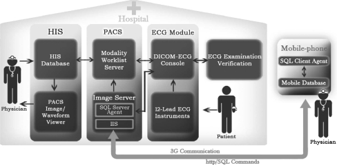
Figure 1 illustrates the 12-lead ECG information system in PACS to realize 12-lead ECG e-diagnoses and m-diagnoses in clinical practice. The system is mainly composed of four parts. This first part is the ECG module, which is responsible for 12-lead ECG waveform extraction, noise removal, and DICOM-ECG conversion. The second part is PACS, which is modified to enable the integration of waveform-based ECG and pixel-based image stored in image server. ECG modality is also added in the worklist server of PACS to verify patients’ ECG scheduling and patients’ information. The third part is Hospital Information System (HIS), used at hospitals to manage patients’ information, medical records, and physicians’ orders and to exchange information with modality worklist server of PACS for the verification of patients’ information, ECG scheduling, and other medical workup. The fourth part is mobile database, which is installed in the mobile phones of cardiologists away from hospitals for ECG and image diagnoses via PACS.
Fig 1.
PACS-dependent 12-lead ECG information system. This 12-lead ECG information system is used in PCAS to share the same abilities of file archiving, manipulation, and communication with DICOM-based images. This developed system is used to provide clinicians with immediate electronic medical procedures and ubiquitous 12-lead ECG and image diagnoses for patients with heart diseases. An ECG console as shown in ECG module is set up to acquire ECG files generating from clinically used ECG instruments, such as SCP-ECG and XML-ECG, to transform the data into DICOM-ECG, and then to transmit them to image server in PACS for storage. Patients’ information and ECG examination schedules can be retrieved and monitored via the connection between modality work-list server of PACS and HIS. On-site clinicians can verify patients’ medical information in ECG console. On-site cardiologists can interpret patients’ 12-lead ECG and image reports from HIS directly. Ubiquitous 12-lead ECG and image diagnoses are also realized by enabling the connection between PACS and mobile database installed in the mobile phones of the off-site cardiologists.
ECG Module
In ECG module, an ECG console is set up to connect with clinically used 12-lead ECG instruments and PACS via hospital intranet. The major tasks of ECG console include (1) 12-lead ECG waveform extraction, (2) noise reduction, and (3) DICOM-ECG transformation from paper-based 12-lead ECG instruments with SCP and XML data formats. Additionally, a graphic user interface is placed in ECG console to communicate with PACS for the verification of ECG examination scheduling and patients’ information.
12-Lead ECG Waveform Extraction
SCP-ECG is a binary-based format consisting of specific sections. However, the HP SCP-ECG adopted in clinical applications has varied in several important sections: Section 0 representing section indicator, Section 8 representing ECG measurement and diagnostic interpretation, and Section 128 representing 12-lead ECG waveform storage.9 In Section 0, HP SCP-ECG uses 10-byte data structure instead of ISO-defined 16 bytes to locate the physical site of other sections. In Section 8, HP SCP-ECG stores both ECG measurement and computer-assisted diagnostic interpretation, which differs from ISO SCP-ECG where ECG measurement and diagnostic interpretation were stored in Sections 7 and 8, respectively. In Section 128, the HP SCP-ECG waveform data are compressed by a fixed Huffman table rather than ISO-defined two-table Huffman encoding rules. Table 1 presents the required attributes of HP SCP-ECG for DICOM-ECG conversion. The ECG waveform data in Section 128 were decompressed by a fixed Huffman table based on our previous investigation as shown in Table 2.
Table 1.
Extracted Attributes from SCP-ECG for DICOM-ECG Conversion
| Section | Necessary Attributes | ||
|---|---|---|---|
| Section 1 | Patient ID | Acquisition date | Acquisition time |
| Section 8 | Heart rate | PR interval | RR interval |
| QRS duration | QT interval | QTc | |
| P Axis | QRS axis | Computer-assisted interpretation | |
| Section 128 | 12-Lead ECG Waveform raw data | ||
Table 2.
Twelve-Lead ECG Waveform Decoding Rule in Clinically used SCP-ECG
| Bit Length | Amplitude of Waveform | |
|---|---|---|
| Negative values | Positive values | |
| 3 | −2,−1 | 0,1 |
| 5 | −4,−3 | 2,3 |
| 7 | −12~−5 | 4~11 |
| 11 | −140~−13 | 12~139 |
| 16 | −2,048~−141 | 140~2,047 |
XML-ECG Processing
Philips XML-ECG, which provides hospitals with originally noise-interfered ECG waveform, is a text-based file comprising several tags representing 12-lead ECG measurement. The required clinical information for DICOM-ECG conversion is listed in Table 3. The noise-interfered ECG waveform data are extracted from the “waveform” tag of Philips 12-lead ECG XML files, with a frequency band ranging from 0.05 to 150 Hz at a sampling rate of 500 Hz. The stationary wavelet transform (SWT),19 which uses Haar function as the mother wavelet, is applied to ECG signals with eight levels decomposition as listed in Eq. 1. In Eq. 1, a denotes the approximate coefficient, d denotes detail coefficient, Φ denotes the scaling function, Ψ represents the mother wavelet, and j and k denote decomposition level and translation in time, respectively. The commonly occurred ECG baseline wander can be corrected by setting a as zero. The de-nosing soft threshold, th, is modified and applied on detail coefficients to remove high-frequency EMG as shown in Eq. 2, where N represents the data length. The thresholds of detail coefficients below or above level 3 are adjusted by the level factor j. The coefficients after the threshold adjustment based on Eq. 2 are processed by Inverse wavelet transform to restore time-domain signal with noise reduction and correct waveform shape, as shown in Eq. 3.20
 |
1 |
 |
2 |
 |
3 |
Table 3.
Required Attributes of XML-ECG for DICOM-ECG Conversion
| Parent Tag | Sub Tag | Meaning |
|---|---|---|
| <waveforms> | <parsedwaveforms> | 12-lead ECG waveform raw data |
| <patient> | <patientid> | PatientID |
| <measurements> | date | Acquisition data |
| time | Acquisition time | |
| <globalmeasurements> | ||
| <meanventrate> | Heart rate | |
| <meanprint> | PR interval | |
| <meanqrsdur> | QRS interval | |
| <meanqtint> | QT interval | |
| <meanqtc> | QTc | |
| <pfrontaxis> | P axis | |
| <qrsfrontaxis> | QRS axis | |
| <tfrontaxis> | T axis | |
| <interpretations> | <statement> | Computer-assisted interpretation |
DICOM-ECG
DICOM-ECG is a binary-based format presented by several elements. Each element consists of number-represented tag, data type, data length, and data values. The extracted SCP-ECG and XML-ECG data, such as patient ID, ECG study ID, examination date, examination time, ECG sampling rate, and waveform data, are transformed into the corresponding DICOM elements based on the definition of DICOM 3.0 Suppl. 30.2 Additionally, computer-assisted interpretation from Section 8 of HP SCP-ECG or Philips XML-ECG is added into user-defined elements. As the final step, DICOM-used service object pair (SOP) is added to the DICOM-ECG file which can be manipulated under PACS. Table 4 lists the DICOM-ECG tags used in this study.
Table 4.
The Necessary DICOM Tags of DICOM-ECG
| Tag’s name | Tag’s number | |
|---|---|---|
| SOP | Meta Element Group Length | (0002,0000) |
| File Meta Information Version | (0002,0001) | |
| Media Storage SOP Class UID | (0002,0002) | |
| Media Storage SOP Instance UID | (0002,0003) | |
| Transfer Syntax UID | (0002,0010) | |
| Implementation Class UID | (0002,0012) | |
| Implementation Version Name | (0002,0013) | |
| Modlity | SOP Class UID | (0008,0016) |
| SOP Instance UID | (0008,0018) | |
| Study Date | (0008,0020) | |
| Content Date | (0008,0023) | |
| AcquisitionDateTime | (0008,002A) | |
| Study Time | (0008,0030) | |
| Content Time | (0008,0033) | |
| Accession Number | (0008,0050) | |
| Modality | (0008,0060) | |
| Manufacturer | (0008,0070) | |
| ReferringPhysicianName | (0008,0090) | |
| Patient | Patient’s Name | (0010,0010) |
| Patient’s ID | (0010,0020) | |
| Patient’s Birth Date | (0010,0030) | |
| Patient’s Sex | (0010,0040) | |
| Study | Study Instance UID | (0020,000D) |
| Series Instance UID | (0020,000E) | |
| StudyID | (0020,0010) | |
| Series Number | (0020,0011) | |
| Instance (form...Image) Number | (0020,0013) | |
| AcquisitionContextSequence | (0040,0555) | |
| ECG | NumberOfWaveformChannel | (003A,0005) |
| NumberOfWaveformSamples | (003A,0010) | |
| SamplingFrequency | (003A,001A) | |
| WaveformBitsAllocated | (5400,1004) | |
| WaveformSequence | (5400,0100) | |
| User-defined (ECG interpretation) | (5401,0001)~(5401,0013) |
The PACS
Traditional PACS is designed for DICOM-based image retrieval. In this study, a PACS enabling waveform and image manipulation is developed and described as follows:
Image/Waveform Database
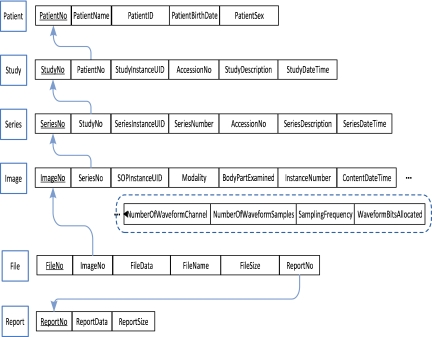
A DICOM file including either image or waveform format is first stored in the PACS image server and then processed to extract the necessary elements to the six corresponding relations of database listed in Figure 2. The simplified relations shown in Figure 2 is installed in mobile phones for data communication between PACS and mobile phones. As shown in Figure 2, the entity Patient represents patients’ information and other related attributes. The entity Study represents patients’ examination access number, examination date, examination time, and other related attributes. The entity Series represents a series of studies with the examination modality, examination specification, and related attributes. The entity Image represents the image-based DICOM file, its acquisition specification, and related attributes. In Figure 2, four attributes in image relations, including waveform channel, waveform sampling rate, total number of waveform data, and waveform bit allocation, are added to access the waveform-based ECG and image simultaneously. In addition, the entity File and the entity Report are used to locate the physical site of the DICOM files and basic text-based structure reports in PACS, respectively.
Fig 2.
A database schema for data delivery between PACS and mobile database.
ECG Modality Worklist Server
The major two functions of the modality worklist server include: (1) to obtain patient information, examination items, and examination schedule from HIS and (2) to notify HIS when an examination is already finished. To enable PACS to recognize 12-lead ECG examination, an “ECG” modality is added. As indicated in Figure 3, a 12-lead ECG examination order is stored in two relations “worklist” and “item” in HIS database for PACS ECG work-list server queries. The worklist relation in HIS has the attributes of patients’ information, examination access number, examination schedule, and examination status flag. The item relation in HIS has the attributes of examination items, study modality, and a status flag for the availability of DICOM-ECG. The flag in worklist relation indicates the examination status, with different codes standing for “examination will be performed,” “examination is performing,” and “examination is finished,” respectively, which can be observed by clinicians. The status flag in item relation indicates whether the DICOM file is stored in the image server.
Fig 3.
Two relations added in HIS to realize computerized 12-lead ECG procedure.
Data Delivery Between Mobile Database and PACS
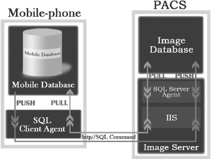
A Microsoft-based mobile phone that displays 65536 colors with VGA resolution 400 × 800 pixels and CPU speed at 500 MHz is used to retrieve ECG and images stored in PACS. To visualize and manipulate DICOM-based ECG and images, several VB.NET scripts are installed in the mobile phone. To facilitate data delivery between mobile phones and PACS, several Structured Query Language (SQL)-based scripts are also installed in the mobile phone. Figure 4 displays the mechanism of remote data access for DICOM-based file delivery. The database shown in Figure 2 and SQL client agent are installed in mobile phones. The mobile phones can deliver SQL query commands via Hypertext Transfer Protocol (HTTP) to the Internet Information Server (IIS) installed in the image database server of PACS. The SQL server agent installed in IIS of PACS will “pull” the query results back to SQL client agent and store the data in mobile database. As the data can be also “pushed” to PACS via the transaction in mobile database, the PACS can be updated synchronically.
Fig 4.
The mechanism of data delivery between mobile database and PACS. SQL server agent with Internet Information server and SQL client agent are installed on PACS image server and mobile database, respectively, to enable the data delivery between PACS and remote mobile phones. The mobile database can issue SQL query commands via http to SQL server agent and then receive the results. Meanwhile, the PACS image server will be updated synchronically with mobile database if the transaction takes place.
Results
ECG Signal Processing
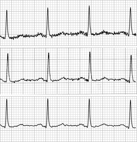
Figure 5 shows the effectiveness of ECG signal processing based on SWT described in Eqs. 1–3. An original noise-interfered lead II ECG extracted from Philips XML-ECG is shown in the top panel of Figure 5. The Philips-processed ECG waveform, which is the scanned reproduction of paper ECG, is shown in the middle panel of Figure 5. The SWT-processed ECG is shown in the bottom panel of Figure 5. It should be noted that the SWT-processed ECG can keep original ECG characteristics without distortion in amplitude, duration, and shape when compared with the paper ECG and the original ECG.
Fig 5.
ECG signal processing. The comparisons of ECG signal processing between Philips paper-based ECG and SWT-processed ECG are shown. An original noise-interfered ECG signal is shown in the top panel. The Philips processed ECG is shown in the middle panel. The SWT-processed ECG is shown in the bottom panel. Both Philips processed ECG and the SWT-processed ECG can keep the characteristics of the original ECG in amplitude, duration, and shape.
The Demonstration of 12-Lead ECG E-Checkup
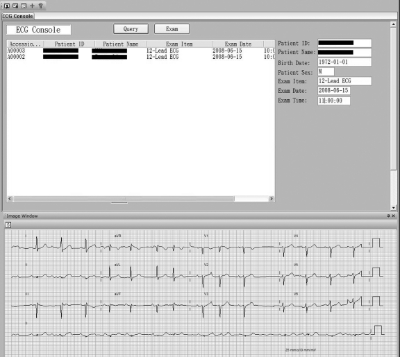
With the use of PACS-dependent 12-lead ECG information system, paperless patients’ ECG checkup notice and ECG record retrieval can be realized. Consequently, problems such as the misplacement of paper ECG checkup notice and the blurred paper ECG reports can be effectively improved. Figure 6 displays the electronic flow of 12-lead ECG examination. An interface set up in the ECG console of ECG room is used for ECG scheduling verification. The clinicians in the ECG room can verify ECG checkup schedules via the PACS ECG work-list server to confirm the patient’s and checkup information, which are shown on the left panel of Figure 6. After the verification procedure is completed, the 12-lead ECG measurement is performed, and a new DICOM-ECG file is generated and transmitted to the PACS image server. Clinicians use the ECG console on the right panel and on the bottom panel of Figure 6 to ensure the DICOM-ECG file is stored in the image server of PACS. The PACS sends a signal to notify HIS to change the ECG examination status from “examination will be performed” to “examination is finished.” Once the 12-lead ECG measurement is performed and stored in the PACS image server, ECG reports are ready to be browsed.
Fig 6.
The illustration of 12-lead ECG computerized procedures. The traditional paper-based 12-lead ECG examination procedure is replaced by the electronic procedure using PACS-dependent 12-lead ECG information system. Information such as patients’ ID, examination items, and schedules are first verified in ECG console by clinicians as shown on the left panel. After ECG examination is performed, clinicians ensure that the DICOM-ECG is successfully stored in the image server as shown on the right panel and on the bottom panel.
The Demonstration of 12-Lead ECG E-Diagnosis
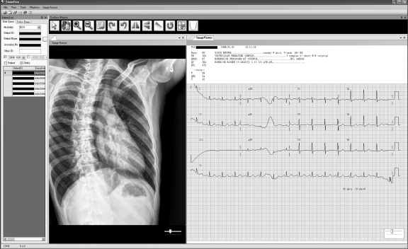
Cardiovascular physicians give diagnoses after browsing 12-ECG records directly from HIS by activating the PACS image viewer, which can visualize the 12-lead ECG waveform. By placing the waveform interpreter in the conventional PACS image viewer, the 12-lead ECG waveform and image-based medical images can be visualized at the same time to enhance data manipulation as demonstrated in Figure 7.
Fig 7.
The illustration of 12-lead ECG and chest X-ray E-diagnoses. Twelve-lead ECG and chest X-ray are two critical references to the diagnoses of heart diseases. An integrated waveform- and image-based PACS viewer is developed for remote physicians to interpret 12-lead ECG and images simultaneously, which enhances the efficiency of heterogeneous data retrieval and review.
The Demonstration of 12-Lead ECG and Image M-Diagnoses in Emergency Telemedicine
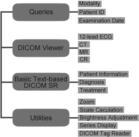
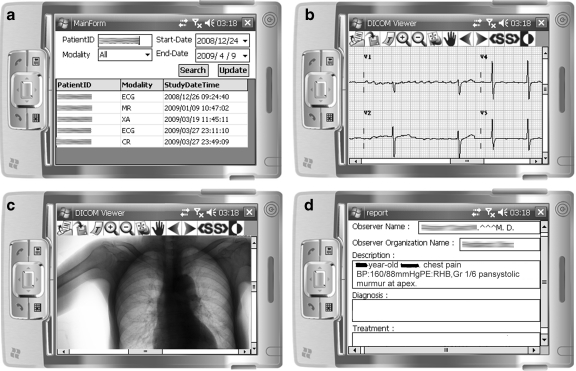
Figure 8 presents the manipulation functions installed in mobile phones to allow off-site cardiologists to conduct 12-lead ECG and image m-diagnose. The manipulation functions consist of four parts: (1) patients’ record query with various modalities, (2) DICOM-based waveform and image visualization, (3) basic text-based DICOM structure report reader, and (4) utilities such as zooming, brightness adjustment, and scale measurement. A series of screen shots of 12-lead ECG m-diagnoses applied in clinical practice are illustrated in Figure 9a–d. Figure 9a shows the mobilized patients’ record queries including the present and the previous ECGs and other modality workups, such as chest X-ray and MR. Figure 9b displays DICOM-based 12-lead ECG visualization with an enlarged scale. Figure 8c demonstrates chest X-ray visualization in the same DICOM viewer. Figure 9d displays the reading of a basic text-based DICOM structured report of patients’ information, symptoms, and lab tests, which can be referenced by off-site cardiologists.
Fig 8.
Manipulation functions of mobile 12-lead ECG/image information system. There are four major functions of mobile 12-Lead ECG/Image information system installed in mobile phones, including: (1) patients’ record queries, (2) DICOM-based waveform/ image visualization, (3) basic text-based structure report reading, and (4) image manipulation utilities.
Fig 9.
The demonstration of 12-lead ECG /image M-diagnoses. a–d Series of ECG and image manipulation in tele-cardiology. a The patient’s record queries including various modality examinations such as ECG, SR, X-ray, and MR. b Enlarged 12-lead ECG browsing. c Chest X-ray browsing. d The patient’s medical record in a basic text-based DICOM structure report.
Clinical Evaluation of M-Diagnosis in Emergency Tele-Cardiology
The 12-lead ECG m-diagnosis developed in this study is specifically designed to meet the demand of ER tele-cardiology at Taoyuan Armed Forces General Hospital in Taiwan. The 12-lead ECG m-diagnoses are used when on-site ER physicians need immediate tele-consultation with off-site cardiologists.
Traditionally, phone discussions between on-site ER physicians and off-site cardiologists can be inefficient and ineffective without having off-site cardiologists browsing ECG and chest X-ray. To give the correct diagnoses and provide timely and proper treatment, the off-site cardiologist has to go to the hospital first and then read paper-based ECG and image reports. With the use of this developed mobile system, the off-site cardiologist has immediate access to patients’ ECG and chest X-ray ubiquitously. Consequently, this tele-consultation and m-diagnosis are more efficient and results in more accurate diagnoses as compared with the conventional phone tele-consultation. With this M-diagnosis, the off-site cardiologist does not need to return to the hospital unless he/she must perform on-site treatment.
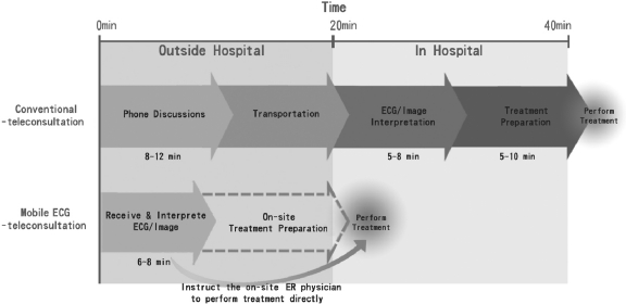
Figure 10 presents the improved time efficiency with the use of this mobile system in 25 case studies of patients with heart diseases in Taiwan. On average, off-site cardiologists spend 20 min to return to hospital without the use of this mobile system. After arriving at the hospital, the off-site cardiologists spend an average of 5–8 min to read ECG and image reports and an average of another 5–10 min to have the treatment prepared. With the use of this mobile system, off-site cardiologists only need an average of 6 to 8 min to receive and read ECG and chest X-ray and then determine the effective treatment strategies, which can all be conducted remotely. Additionally, the off-site cardiologist may instruct on-site physician to perform proper treatment immediately or to have treatment prepared before the off-site cardiologist arrives at the hospital.
Fig 10.
Clinical evaluations of 12-lead ECG/image M-diagnoses. With the help of developed mobile information system, the efficiency of medical diagnostic and treatment flows in emergency telemedicine can be greatly improved. According to the findings of 25 emergency case studies of patients with heart diseases at Taoyuan Armed Forces General Hospital in Taiwan, the use of mobile information system can first minimize the traveling time of the remote cardiologist unless on-site treatment by the remote cardiologist is necessary. This system can also reduce an average of 5–8 min for the remote cardiologist to request and receive medical data and another 5–10 min of treatment preparation time before the remote cardiologist has to return to the hospital to perform on-site treatment.
Discussion
To demonstrate its usefulness in e-diagnoses and m-diagnoses, this developed ECG system was evaluated from both the perspectives of system performance and clinical application, respectively. From the perspective of system performance, we evaluated the following: (1) the efficiency of 12-lead ECG operation under PACS and (2) the speed of mobile data link. From the perspective of clinical application, three senior cardiologists and four emergency physicians evaluated the accuracy and the efficiency of e-diagnoses and m-diagnoses. These evaluations are summarized in the following subsections.
ECG Waveform Verification
To validate the accuracy of processed ECG waveforms, three cardiologists compared the waveforms of computerized ECG and paper ECG of their patients. The amplitude, duration, and shape of ECG in P, QRS complex, and T parts were examined by selecting one or more lead signals per 12-lead ECG report. Among 1,000 HP SCP-ECG copies whose frequency band ranges from 0.5 to 40 Hz, the decoded ECG waveforms using Table 2’s decoding rule were found to be exactly the same as the waveforms visualized on the paper ECG. Among 2,000 Philips XML-ECG copies which needed noise-removal processing, approximately 40 copies showed QRS wave amplitude differences between SWT-processed ECG and paper ECG. Specifically, in these copies, R was found to be greater than one small grid, and Q or S was found to be greater than 0.5 small grid on clinically used ECG paper. While comparing these 40 copies of SWT-based ECG with original ECG waveforms, we found that the differences of R, Q, S amplitudes between SWT-processed ECG and original ECG were smaller than 0.5 small grid, which met the ECG waveform accuracy criteria set by the three cardiologists who evaluated this study. As shown in Figure 5, the R amplitude difference between paper ECG and SWT-processed ECG is approximately one small grid. It should be noted that the R wave amplitudes on the original ECG and the SWT-processed ECG are almost identical.
System Evaluation of PACS-Dependent 12-Lead ECG
Low cost This study develops an e-technology to transform paper-based 12-lead ECG to computerized ECG, which can be performed in PACS. That is, hospitals do not need to purchase additional hardware and software for 12-lead ECG archiving, manipulation, and communication.
Data Integration/System Integration This study enables the integration of two commonly used ECG formats adopted in clinical practice. Additionally, heterogeneous waveform-based ECG and pixel-based medical images are integrated in PACS to enhance data manipulation and system maintenance.
Utilization PACS can process and manage ECGs and images simultaneously without any delay.
Application This developed ECG system can provide clinicians with raw 12-lead ECG waveform to develop advanced clinical applications in the future, such as clinical database-supported 12-lead ECG on-line and mobile e-learning, 12-lead ECG and relative cardiovascular image knowledgebase, and 12-lead ECG tele-diagnoses on the ambulance.
Speed Evaluation of 12-Lead ECG and Image in Telemedicine
The effectiveness of medical signal and image transmission by using 3G/3.5G mobile network has been validated in recent study.21 Generally, the 3G mobile network that allows data transmission in the speed of 1.5~3.6 Mbps can be used effectively to retrieve the DICOM-based 12-lead ECG and DICOM image. The speed evaluation of remote DICOM file retrieval between a mobile phone and PACS is listed in Table 5. It should be noted that retrieving a 65 kb DICOM-ECG, a 135 kb MR, a 205 kb CT, and a 5.2 Mb CR will take 5, 10, 9, and 181 s, respectively, at the 2 Mbps downloading rate.
Table 5.
Speed Evaluation of DICOM-ECG and DICOM-Based Image Transmission Between PACS and Mobile Phone Via 3G Wireless Communication
| Modality | Pixel | Bit depth | Size | Quantity | Time (s) |
|---|---|---|---|---|---|
| ECG (12-lead ECG) | 65 Kb | 1 | 5 | ||
| CR (Cheat X-ray) | 2420 × 2430 | 12 | 5.2 Mb | 1 | 185 |
| MR | 256 × 256 | 12 | 136 Kb | 1 | 10 |
| MR | 256 × 256 | 12 | 136 Kb | 14 | 19 |
| CT | 512 × 512 | 12 | 205 Kb | 1 | 9 |
| CT | 512 × 512 | 12 | 205 Kb | 6 | 38 |
Clinical Evaluation of 12-Lead ECG E-Diagnoses
The use of DICOM-ECG in PACS has two major benefits in clinical operation. First, time efficiency on ECG retrieval and disease diagnoses is strengthened by the faster electronic ECG examination procedure as compared with the traditional ECG examination which generates paper-based output. Second, the interoperability of 12-lead ECG records among hospitals may be facilitated with commonly used DICOM protocol in further studies.
Clinical Evaluation of 12-Lead ECG M-Diagnoses
In the 12-lead ECG emergency telemedicine, this mobile system can facilitate physicians’ timely responsiveness to patient care and strengthen their sense of responsibility while being away from the hospital.
The use of mobile ECG diagnoses can enhance the efficiency of medical treatment as shown in Figure 10.
Diagnostic disagreement between on-site ER physicians and off-site cardiologists can be reduced with the use of this mobile system.
Twelve-lead ECG and chest X-ray are critical references for cardiologists to determine diagnoses and treatment strategies in the emergency medicine. This system includes both 12-lead ECG and chest X-ray, with which off-site cardiologists can efficiently give correct diagnoses and proper treatment by reading patients’ ECG and X-ray simultaneously and ubiquitously in an integrated medical system, which is illustrated in Figures 7 and 9c.
Conclusion
In this study, we developed an e-technology to enable the integration of waveform-based 12-lead ECG and pixel-based medical images in a unified DICOM format. By using this e-technology, we also developed PACS-dependent 12-lead ECG information to realize electronic procedures of 12-lead ECG examinations and to facilitate 12-lead ECG/medical image tele-consultation in clinical practice. Clinical evaluations made by cardiologists have proved the usefulness of this system. First, it can reduce diagnostic disagreements and enhance time efficiency in e-diagnoses and m-diagnoses. Secondly, this system can improve the management of 12-lead ECG and images in a unified database of PACS. In conclusion, the adoption of this PACS-dependent 12-lead ECG information can enable the integration of heterogeneous information systems in hospitals, and the use of DICOM-ECG will facilitate the interoperability of 12-lead ECG records among hospitals.
Acknowledgements
The author would like to thank the National Science Council of Taiwan for financially supporting this research under contracts NSC 97-2221-E-155-024 and NSC 95-2221-E-155-087.
References
- 1.American College of Radiology, National Electrical Manufacturers Association: Digital Imaging and Communications in Medicine DICOM: Version 3.0. Number PS3, 1999
- 2.DICOM 3.0 Supplement 30. Waveform Interchange, Nat. Elect. Manufacturers Assoc.: ARC-NEMA, Digital Imaging and Communications, NEMA, Washington D.C., 2000
- 3.Open-ECG Web Page. http://www.openecg.net
- 4.Health informatics—Standard communication protocol—Computer-assisted electrocardiography. ICS: 35.240.80 IT applications in health care technology; reference number EN 1064:2005+A1, 2007
- 5.Brown B, Kohls M, Stockbridge N: FDA-XML data format design specification, revision C. www.openecg.net, 1–27, 2003
- 6.Sakkalis V, Chiarugi F, Kostomanolakis S, Chronaki CE, Tsiknakis M, Orphanoudakis SC. A gateway between the SCP-ECG and the DICOM supplement 30 waveform standard. Comput Cardiol. 2003;30:25–28. doi: 10.1109/CIC.2003.1291081. [DOI] [Google Scholar]
- 7.Schloegl A, Chiarugi F, Cervesato E, Apostolopoulos E, Chronaki CE. Two-way converter between the HL7 aECG and SCP-ECG data formats using BioSig. Comput Cardiol. 2007;34:253–256. doi: 10.1109/CIC.2007.4745469. [DOI] [Google Scholar]
- 8.Ettinger MJB, Lipton JA, Wijs MCJ, Putten N, Nelwan SP. An open source toolkit in DICOM. Comput Cardiol. 2008;35:441–444. [Google Scholar]
- 9.Chiang CC, Yang YC, Tzeng WC, Tzeng WL, Hsieh JC. An SCP compatible electrocardiogram database for signal transmission, storage and analysis. Comput Cardiol. 2004;31:621–624. doi: 10.1109/CIC.2004.1443015. [DOI] [Google Scholar]
- 10.Hsieh JC. A novel DICOM-based 12-lead electrocardiogram documentary system. J Electrocardiol. 2007;40(6):S83. [Google Scholar]
- 11.Hsieh JC, Yu KC, Lo SC, Hung CC, Yeh CH: A novel computerized 12-lead electrocardiograph system for clinical applications. 19th International Conference of Biosignal, Brno, Czech, ID88, 2008
- 12.Hilbel T, Brown BD, Bie J, Lux RL, Katus HA. Innovation and advantage of the DICOM-ECG standard for viewing, interchange and permanent archiving of the diagnostic electrocardiogram. Comput Cardiol. 2007;34:633–636. doi: 10.1109/CIC.2007.4745565. [DOI] [Google Scholar]
- 13.Andrade R, Wangenheim A, Bortoluzzi MK. Wireless and PDA: a novel strategy to access DICOM-compliant medical data on mobile devices. Int J Med Inform. 2003;71:157–163. doi: 10.1016/S1386-5056(03)00093-5. [DOI] [PubMed] [Google Scholar]
- 14.Nakataa N, Suzukib N, Fukuda Y, Fukuda K. Accessible web-based collaborative tools and wireless personal PACS: feasibility of group work for radiologists. Int Congr Ser. 2004;1268:260–264. doi: 10.1016/j.ics.2004.03.125. [DOI] [Google Scholar]
- 15.Tang FH, Law MY, Lee AC, Chan LWC. A mobile phone integrated health care delivery system of medical images. J Digit Imaging. 2004;17(3):217–225. doi: 10.1007/s10278-004-1015-5. [DOI] [PMC free article] [PubMed] [Google Scholar]
- 16.Hu F, Jiang M, Xiao Y. Low-cost wireless sensor networks for remote cardiac patients monitoring applications. Wirel Commun Mob Comput. 2008;8:513–529. doi: 10.1002/wcm.488. [DOI] [Google Scholar]
- 17.Hadzievski L, Boiovic B, Vukcevic V, Belicev P, Pavlovic S, Vasiljevic-Pokrajcic Z, Ostojic M. A novel mobile transtelephonic system with synthesized 12-lead ECG. IEEE Trans Inform Technol Biomed. 2004;8(4):428–438. doi: 10.1109/TITB.2004.837869. [DOI] [PubMed] [Google Scholar]
- 18.Giovas P, Papadoyannis D, Thomakos D, Papazachos G, Rallidis M, Soulis D, Stamatopoulos C, Mavrogeni S, Katsilambros N. Transmission of electrocardiograms from a moving ambulance. J Telemed Telecare. 1998;4(s1):5–7. doi: 10.1258/1357633981931533. [DOI] [PubMed] [Google Scholar]
- 19.Nason GP, Silverman BW. The Stationary Wavelet Transform and Some Statistical Applications, in Lecture Notes in Statistics: Wavelets and Statistics, vol. New York: Springer; 1995. pp. 281–299. [Google Scholar]
- 20.Hsieh JC, Hung CC, Lo SC, Yu KC, Yeh CH: The Clinical Application of Stationary Wavelet Transform Based 12-Lead ECG Noise Elimination, 19th International Conference of Biosignal. Brno, Czech, ID87, 2008
- 21.Komnakos D, Vouyioukas D, Maglogiannis I, Constantinou P. Performance evaluation of an enhanced uplink 3.5G system for mobile healthcare applications. Int J Telemed Appl. 2008;2008:1–11. doi: 10.1155/2008/417870. [DOI] [PMC free article] [PubMed] [Google Scholar]


