

Abstract
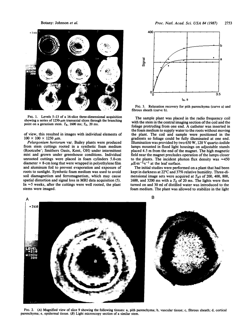
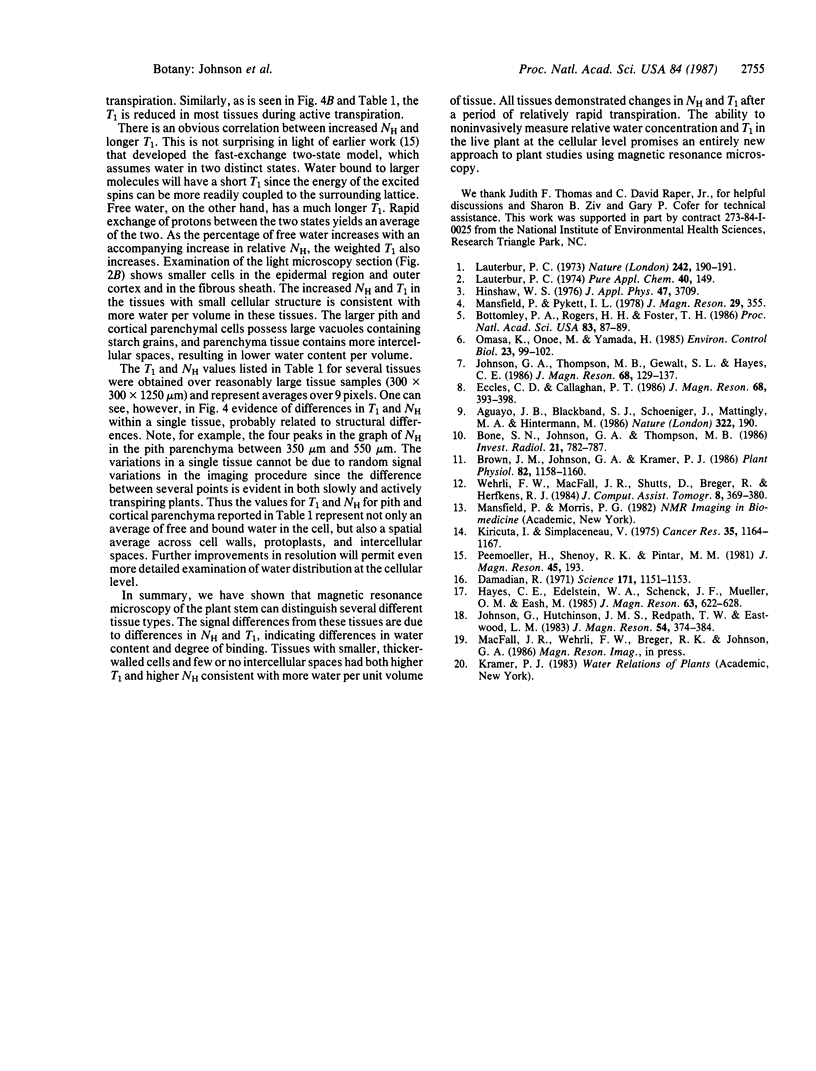
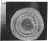
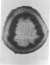
Differences in water content and degree of binding in the various stem tissues of Pelargonium hortorum were observed by magnetic resonance imaging. 1H images were obtained with a resolution of 100 microns in the transverse plane and a slice thickness of 1250 microns. It was possible to distinguish the principal tissues of the stem by differences in their proton density or apparent water content and spin lattice relaxation time (T1) or degree of water binding. Measurements were made while the plant was slowly and actively transpiring. In the slowly transpiring plant, T1 of various tissues ranged from an average of 659 to 865 ms with a proton density variation of from 72 to 100%. In the actively transpiring plant, T1 ranged from an average of 511 to 736 ms, and the proton density was reduced, ranging between 62 and 88% of the peak value found in the slowly transpiring plant. The fibrous sheath surrounding the vascular tissue and the epidermal region was found to have the highest spin density and T1. Both tissues are comprised of relatively small thick-walled cells. Cortical and pith parenchyma are composed of larger, thinner-walled cells with numerous intercellular spaces and lower spin density and T1. The differences are attributed to the higher water content by volume in the tissue composed of smaller, more compactly arranged cells. The resolution obtained in this work enables clear definition of tissues in the living plant and quantitative information concerning differences in the distribution and extent of binding of water.
Full text
PDF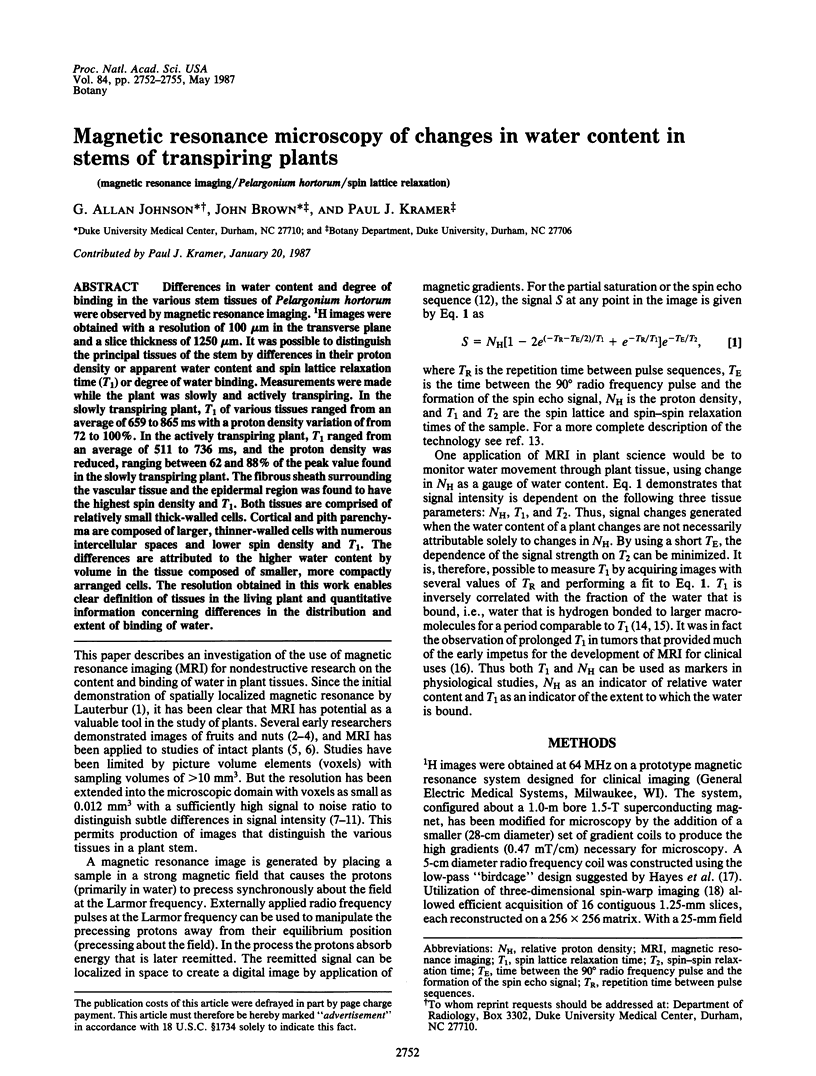
Images in this article
Selected References
These references are in PubMed. This may not be the complete list of references from this article.
- Aguayo J. B., Blackband S. J., Schoeniger J., Mattingly M. A., Hintermann M. Nuclear magnetic resonance imaging of a single cell. Nature. 1986 Jul 10;322(6075):190–191. doi: 10.1038/322190a0. [DOI] [PubMed] [Google Scholar]
- Bone S. N., Johnson G. A., Thompson M. B. Three-dimensional magnetic resonance microscopy of the developing chick embryo. Invest Radiol. 1986 Oct;21(10):782–787. doi: 10.1097/00004424-198610000-00003. [DOI] [PubMed] [Google Scholar]
- Bottomley P. A., Rogers H. H., Foster T. H. NMR imaging shows water distribution and transport in plant root systems in situ. Proc Natl Acad Sci U S A. 1986 Jan;83(1):87–89. doi: 10.1073/pnas.83.1.87. [DOI] [PMC free article] [PubMed] [Google Scholar]
- Brown J. M., Johnson G. A., Kramer P. J. In Vivo Magnetic Resonance Microscopy of Changing Water Content in Pelargonium hortorum Roots. Plant Physiol. 1986 Dec;82(4):1158–1160. doi: 10.1104/pp.82.4.1158. [DOI] [PMC free article] [PubMed] [Google Scholar]
- Damadian R. Tumor detection by nuclear magnetic resonance. Science. 1971 Mar 19;171(3976):1151–1153. doi: 10.1126/science.171.3976.1151. [DOI] [PubMed] [Google Scholar]
- Kiricuta I. C., Jr, Simplăceanu V. Tissue water content and nuclear magnetic resonance in normal and tumor tissues. Cancer Res. 1975 May;35(5):1164–1167. [PubMed] [Google Scholar]
- Wehrli F. W., MacFall J. R., Shutts D., Breger R., Herfkens R. J. Mechanisms of contrast in NMR imaging. J Comput Assist Tomogr. 1984 Jun;8(3):369–380. doi: 10.1097/00004728-198406000-00001. [DOI] [PubMed] [Google Scholar]