Abstract
The aim of the study was to investigate the stabilising effect of dynamic interspinous spacers (IS) in combination with interlaminar decompression in degenerative low-grade lumbar instability with lumbar spinal stenosis and to compare its clinical effect to patients with lumbar spinal stenosis in stable segments treated by interlaminar decompression only. Fifty consecutive patients with a minimum age of 60 years were scheduled for interlaminar decompression for clinically and radiologically confirmed lumbar spinal stenosis. Twenty-two of these patients (group DS) with concomitant degenerative low-grade lumbar instability up to 5 mm translational slip were treated by interlaminar decompression and additional dynamic IS implantation. The control group (D) with lumbar spinal stenosis in stable segments included 28 patients and underwent only interlaminar decompression. The mean follow-up was 46 months in group D and 44 months in group DS. A visual analogue scale (VAS), Oswestry Disability Index (ODI) and walking distance were evaluated pre- and postoperatively. The segmental instability was evaluated in flexion-extension X-rays. The implantation of an IS significantly reduced the lumbar instability on flexion-extension X-rays. At the time of follow-up walking distance, VAS and ODI showed a significant improvement in both groups, but no statistical significance between groups D and DS. Four patients each in groups D and DS had revision surgery during the period of evaluation. The stabilising effect of dynamic IS in combination with interlaminar decompression offers an opportunity for an effective treatment for degenerative low-grade lumbar instability with lumbar spinal stenosis.
Introduction
In 1954 Verbiest [1] was the first to explain the pathology of spinal stenosis. He declared that lumbar spinal stenosis refers to a pathological condition resulting in the narrowing of the spinal canal and compression of the neurological structures. Lumbar spinal stenosis is therefore a clinical condition and not a radiological finding or diagnosis. The most common cause of lumbar spinal stenosis is degenerative disc disease; therefore, especially elderly people in increasing numbers require spinal decompression surgery [2]. Further reasons for lumbar spinal stenosis can be disc herniation [3, 4], hypertrophy of the ligamentum flavum, spondylolisthesis, disc bulge, degenerative facet joint arthritis and thickened laminae [3, 5, 6]. The compression of the neurological structures leads to a reduction in walking distance, weakness, numbness and tingling. The symptoms increase in lumbar extension and are relieved in lumbar flexion [7]. On the other hand degenerative spondylolisthesis, described by Newman [8] in 1955, causes segmental instability with sagittal and axial malalignment, which induces local back pain. The primary levels of lumbar instabilityaffected are L4–5, followed by L3–4, L5–S1, L2–3 and L1–2 [1, 9, 10]. With the population continuously aging, the incidence of surgical decompression will rise. When conservative physical therapy fails, decompression of the spinal canal is recommended to improve walking distance and relieve pain.
To avoid postoperative segmental instability, interlaminar decompression surgery with only partial laminectomy, undercutting and preservation of the facet joint is preferred. During the last few years there has been a growing tendency towards small interventions in spinal surgery [7].
The idea and function of interspinous spacers (IS) is the enlargement of the neuroforamen, load relief for the intervertebral disc, reduction of extension, limitation of segmental movement and segmental buffering [11]. Very little clinical data are available about interspinous spacers, especially dynamic spacers, exerting a stabilising effect in low-grade lumbar instability.
The retrospective cohort study design does not usually interfere with the regular clinical diagnostic and treatment decision making process. Clinicians are not asked to compromise their clinical judgment and can treat patients as they would usually [12].
In this retrospective cohort study we investigated the stabilising effect of dynamic IS in combination with interlaminar decompression in degenerative low-grade lumbar instability with lumbar spinal stenosis and compared its clinical effect to patients with lumbar spinal stenosis in stable segments treated with pure interlaminar decompression.
Material and methods
Between 2002 and 2007, 50 consecutive patients, 36 women and 14 men, with lumbar spinal stenosis were included in this cohort study with retrospective evaluation of clinical and radiological data. The minimum age of the patients was 60 years (mean: 72 years; SD: 7.4; range: 60–84 years). The patients suffered either from isolated lumbar spinal stenosis or from lumbar spinal stenosis combined with degenerative low-grade lumbar instability up to 5 mm translational slip on functional X-rays analogous to grade 1 spondylolisthesis. Patients were scheduled for decompression of lumbar spinal stenosis up to three segments due to spinal claudication mainly with leg pain and reduced walking distance. In this time period 234 patients with a minimum age of 60 years were treated by decompression of lumbar spinal stenosis and dorsal fusion of the lumbar spine for degenerative lumbar instability. Criteria of exclusion were age under 60 years, severe segmental lumbar instability more than grade 1 spondylolisthesis, previous lumbar surgery, acute fracture, paraplegia, cauda equina syndrome, local infection or tumours and metastases. Compression of the spinal cord was confirmed by preoperative magnetic resonance imaging (MRI). Patient selection to concomitant interspinous spacer implantation was preoperatively defined by a radiologically verified degenerative low-grade lumbar instability up to 5 mm translational slip on functional X-rays, confirmed by the surgeon through intraoperative mechanical testing for segmental instability by pulling the spinous process. Twenty-eight patients with isolated lumbar spinal stenosis underwent interlaminar decompression (group D: control group) with undercutting and preservation of the facet joint. In 22 patients with combined degenerative low-grade lumbar instability and spinal stenosis a dynamic IS (DIAM™, “Device for Intervertebral Assisted Motion”, Medtronic, Fridley, MN, USA) was additionally implanted after interlaminar decompression (group DS). All surgical interventions in both groups were performed by two surgeons.
We examined the mean duration of preoperative symptoms relating to spinal stenosis until surgical intervention and the body mass index (BMI). A visual analogue scale (VAS) for leg pain, Oswestry Disability Index (ODI) and walking distance were evaluated preoperatively and at follow-up. Walking distance was grouped in ≤ 50, ≤ 500, ≤ 2,000 and > 2,000 m (means unlimited walking distance) before the operation and at follow-up.
For evaluation of segmental movements we measured the translational slip as well as the intervertebral angle on flexion and extension X-rays (flexion-extension angle). The segmental instability was measured with the AGFA Impax ES software (Mortsel, Belgium) measurement tools on preoperative and follow-up digital radiographs. The digital measurements were independently performed by two orthopaedic surgeons and one radiologist and the mean of the three values was taken for further calculations.
All values are given as mean and range. We used the paired samples t test for statistics and considered a p value < 0.05 to be significant. Normality was compared between the groups by using the Mann-Whitney U test. For binomial distribution the McNemar test was used. Non-parametric variables between the two groups were analysed by the chi-square test. All statistical calculations were performed using SPSS 11.5.
Results
The two groups showed a homogeneous distribution of preoperative score results and walking distance. The mean follow-up of clinical and radiological measurements was 46 months (range: 24–98 months) in group D and 44 months (range: 24–60 months) in group DS (Table 1).
Table 1.
Clinical and radiological characteristics of the study population
| Mean | Group D | Group DS | pa | |||
|---|---|---|---|---|---|---|
| Age | 71 | 73 | 0.6b | |||
| Gender M/F | 7/21 | 7/15 | 0.01c | |||
| Preoperative pain latency (months) | 28 | 32 | 0.5b | |||
| Follow-up (months) | 46 | 44 | 0.9b | |||
| BMI | 28 | 31 | 0.1b | |||
| OP at 1 level | 16 | 16 | ||||
| OP at 2 levels | 9 | 4 | ||||
| OP at 3 levels | 3 | 1 | ||||
| Walking distance (m) | Preoperative | Postoperative | Preoperative | Postoperative | ||
| ≤50 | 15 (54%) | 5 (18%) | 11 (52%) | 4 (19%) | ||
| >50 but ≤500 | 13 (46%) | 9 (32%) | 8 (38%) | 6 (29%) | ||
| >500 but ≤2,000 | 0 (0%) | 7 (25%) | 2 (10%) | 9 (43%) | ||
| >2,000 | 0 (0%) | 7 (25%) | 0 (0%) | 2 (10%) | ||
| VAS | 9 | 4 | 9 | 3 | 0.7 | |
| ODI | 65% | 27% | 63% | 23% | 0.7 | |
| Translational slip (mm) | 1.5 | 1.8 | 2.3 | 1.2 | 0.07 | 0.03 |
| Flexion-extension angle (°) | 5.4 | 5.4 | 6.2 | 3.7 | 0.9 | < 0.001 |
| Revision OP | 4 (14.3%) | 4 (18.2%) | 0.007c | |||
BMI body mass index, OP operation, VAS visual analogue scale, ODI Oswestry Disability Index)
at test
bMann-Whitney U test
cMcNemar test
Patients from group D declared a mean preoperative pain latency of 28 months (range: 3–108 months) and patients from group DS of 32 months (range: 6–108 months) (Table 1).
The mean BMI in group D was 28 (range: 20–39) and 31 (range: 22–47) in group DS.
In group D 16 patients had decompression in one segment, nine patients in two segments and three patients in three segments (Table 1). In group D the most frequent single-stage decompression was at the segment L4–5 in 12 patients (43%), two-stage decompressions were most frequent from L3 to L5 in six patients (21%) and three-stage decompressions were done in two patients (7%) at stage L2 to L5. In group DS interlaminar decompression in one segment was carried out on 16 patients, two segments on four patients and three segments in only one patient, whereas ISs were implanted at one level in 20 patients and three levels in one patient (Table 1). In group DS the most frequent single level interlaminar decompression and same level IS implantation was also at the segment L4–5 in 16 patients (76%); there were no implanted ISs at two levels but one (5%) at three levels from L2 to L5.
The mean preoperative VAS score was 9 (range: 4–10) in group D and also 9 (range: 5–10) in group DS. The VAS results at follow-up improved significantly to 4 in group D (range: 1–10; p < 0.001) and to 3 in group DS (range 1–8; p < 0.001). There was no significant difference between groups D and DS (Table 1).
The preoperative ODI showed a mean of 65% in group D (range: 22–86) and 63% in group DS (range: 27–93). The ODI results at follow-up decreased significantly to 27% in group D (range: 0–62; p < 0.001) and 23% in group DS (range: 2–46; p < 0.001). We could not find a significant difference between groups D and DS (Table 1).
Patients reported a mean preoperative walking distance of 152 m in group D (range: 50–500 m) and 202 m in group DS (range: 50–1,000 m). At follow-up a general increase in walking distance could be achieved with 2,166 m in group D (range: 50–5,000 m) and 1,462 m in group DS (range: 50–5,000 m). The results showed a significant improvement in walking distance at follow-up in both groups (group D: p < 0.001; group DS: p < 0.001). There was no significant difference between groups D and DS (Table 1).
Radiological outcome
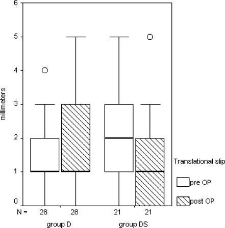
In group D the differences of the translational slip between flexion and extension radiographs showed a preoperative mean of 1.5 mm (range: 0–4 mm) and at follow-up a mean of 1.8 mm (range: 0–5 mm) (Table 1, Fig. 1). In group DS a mean preoperative translational slip of 2.3 mm (range: 0–5 mm) was measured and at follow-up a mean of 1.2 mm (range: 0–5 mm) which corresponded to a significant reduction of translational slip (p = 0.03) (Table 1, Fig. 1).
Fig. 1.
Box plots show the medians and interquartile ranges of groups D and DS in translational slip pre- and postoperatively. A slight tendency of increasing translational slip can be observed postoperatively in group D, whereas a significant stabilising effect of dynamic IS reducing the postoperative translational slip can be seen in group DS
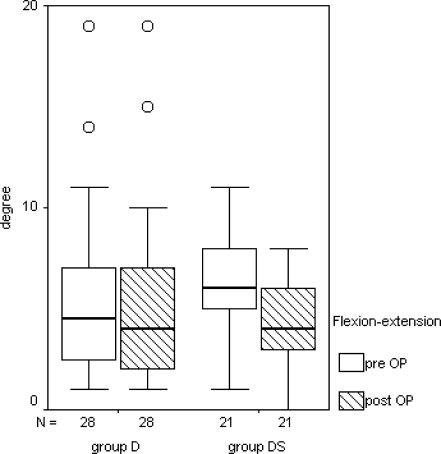
The mean preoperative segmental flexion-extension angle in group D was 5.4° (range: 1–19°) and did not change during the process (Table 1, Fig. 2). The mean preoperative segmental flexion-extension angle in group DS was 6.2° (range: 0–11°) and was significantly reduced at the follow-up with a mean of 3.7° (range: 0–7°; p < 0.001) (Table 1, Fig. 2).
Fig. 2.
Box plots show the medians and interquartile ranges of groups D and DS in pre- and postoperative flexion-extension angle. No difference in flexion-extension angle can be seen in group D, whereas a significant stabilising effect of dynamic IS reducing the postoperative flexion-extension angle in group DS can be observed
In group D four (14.3%) of 28 patients and also four (18.2%) of 22 patients in group DS were not satisfied with the operative outcome and had to undergo revision surgery (Table 1). The first revision operation was one year after primary surgery because of an implant infection to remove the interspinous spacer. The other revision operations were done for recurrence of pain two years after primary intervention at the earliest and four years postoperatively at the latest. Three patients in group D and three patients in group DS needed dorsal fusion for ongoing instability. One patient in group D had revision of the interlaminar decompression at the same level. One patient from group DS was lost to follow-up because of death from unrelated causes.
Discussion
In our daily clinical routine we frequently encounter patients with lumbar spinal stenosis combined with degenerative low-grade lumbar instability. These patients were normally scheduled for interlaminar decompression when conservative therapy failed as recommended in the literature [2, 3, 9, 13–17]. Because posterolateral intercorporal fusion was not indicated in these patients, we performed a dynamic IS implantation at the unstable segments to avoid progressive instability or even stabilise the segment. The idea of our retrospective cohort study was to compare the clinical and radiological results of the patients with lumbar spinal stenosis and concomitant degenerative low-grade instability up to 5 mm translational slip treated with a dynamic IS and interlaminar decompression (group DS) with those patients who had only interlaminar decompression in stable segments with spinal stenosis (group D). Comparing the results of groups D and DS there was no significant difference in the clinical outcome between the groups but a significant stabilising effect of ISs in the radiological outcome.
The combination of interlaminar decompression with IS implantation is a relatively new and effective method in the treatment of spinal stenosis combined with a degenerative low-grade lumbar instability up to grade 1 spondylolisthesis. ISs offer the advantage of small and short surgical intervention with low perioperative risk for the patient, and implantation even under local anaesthesia is possible. This can be seen as an advantage especially for elderly patients reducing the perioperative risk.
In the literature there is a controversial discussion of IS to replace microsurgical decompression in patients with lumbar stenosis and continuous claudication. Several studies [4, 10, 11, 18–24] of different products of IS used for the treatment of degenerative disc disease, degenerative spondylolisthesis and spinal stenosis have been assessed with some good but also some dissatisfying results, depending on the interspinous device and indication criteria for implantation. The interspinous device does not replace microsurgical decompression in patients with massive stenosis and continuous claudication, but offers a safe, effective and less invasive alternative in selected patients with spinal stenosis [10].
Zucherman et al. [22] reported, in a prospective randomised multicentre study, a significant improvement in neurogenic intermittent claudication treated by an interspinous process decompressive system. The two year results of 175 patients with a stiff interspinous spacer showed an improvement in clinical results with VAS and ODI at one and two years follow-up [23]. This spacer does not result in any significant radiographic changes to the lumbar spine. There were no differences between the mean disc heights, curvature of the spine or angulation of the spine after the spacer implantation and the control group compared at one and two years. There was also no difference in the degree of spondylolisthesis between the spacer group and the control groups [23].
Verhoof et al. [21] reported a high failure rate of the same stiff spacer after short-term follow-up in patients with spinal stenosis caused by degenerative spondylolisthesis. Postoperative radiographs and MRI did not show any changes in the percentage of slip or spinal dimensions. The results of Zucherman et al. [22, 23] and Verhoof et al. [21] concerning the stabilising effect of this device showed that the stiff interspinous spacer did not improve a segmental instability or reduce the degree of a spondylolisthesis. Sobottke et al. [24] reviewed the clinical and radiological results of X-Stop™, Wallis™ and DIAM™ spacers and described an improvement of the radiographic parameters of foraminal height, width and cross-sectional area for the X-Stop™ implant greater than the Diam and Wallis implants; however, there was no significant difference among the three regarding symptom relief. The interspinous implant did not worsen low-grade spondylolisthesis [24]. In our opinion stiff ISs offer an adequate therapy option for lumbar spinal stenosis in stable segments but in unstable segments the repetitive contact between the stiff spacer and the bone leads to bone resorption and loosening of the spacer. Our results with the dynamic IS in low-grade lumbar instability showed a statistically significant reduction in segmental translational slip and flexion-extension angle, which can be explained by the flexibility of the spacer and its redressing fixation with the tethers around the supra-adjacent and subadjacent spinous processes. The preservation of the supraspinous ligament is essential for the stabilising effect of the spacer. Another reason for a long-term stabilising effect of the dynamic IS can be explained by the latticed surface of the spacer where the surrounding soft tissue scar can adhere and induce a long-lasting stabilising effect. Kim et al. [19] assessed the IS as an additional treatment to laminectomy and microdiscectomy and noted no alteration in pre- to postoperative disc height or sagittal alignment and no difference in VAS or MacNab outcome scores between the groups treated with (31 patients) or without (31 patients) the IS.
The one year results of the Coflex™ spacer compared with posterior lumbar interbody fusion (PLIF) in two groups of patients with spinal stenosis and mild segmental instability showed significant improvement of VAS and ODI score in both groups, but the Coflex group showed less stress on the superior adjacent level than the PLIF group [25]. This observation would also point to the use of IS for mild segmental instability in elderly patients, especially the dynamic IS should absorb the stress on the adjacent spinous process because of its dynamic effect.
There are relatively new implants, which offer new ideas in the treatment of lumbar spinal stenosis. For stabilising a low-grade lumbar instability, dynamic ISs seem to have an advantage over stiff IS and show a lower failure rate comparing our follow-up results to those of Verhoof et al. [21].
In our opinion the combination of interlaminar decompression and dynamic IS offers an effective opportunity for patients with spinal stenosis and low-grade lumbar instability up to grade 1 spondylolisthesis. Further studies will be necessary to prove the long-term effect of ISs in stabilising a low-grade lumbar instability, and further long-term evaluations are necessary to prove if ongoing segmental loosening can be prevented by interspinous spacers.
Acknowledgments
Conflict of interest The authors declare that they have no conflict of interest.
References
- 1.Verbiest H. A radicular syndrome from developmental narrowing of the lumbar vertebral canal. J Bone Joint Surg Br. 1954;36-B:230–237. doi: 10.1302/0301-620X.36B2.230. [DOI] [PubMed] [Google Scholar]
- 2.Spratt KF, Keller TS, Szpalski M, Vandeputte K, Gunzburg R. A predictive model for outcome after conservative decompression surgery for lumbar spinal stenosis. Eur Spine J. 2004;13:14–21. doi: 10.1007/s00586-003-0583-2. [DOI] [PMC free article] [PubMed] [Google Scholar]
- 3.Fritz JM, Delitto A, Welch WC, Erhard RE. Lumbar spinal stenosis: a review of current concepts in evaluation, management, and outcome measurements. Arch Phys Med Rehabil. 1998;79:700–708. doi: 10.1016/S0003-9993(98)90048-X. [DOI] [PubMed] [Google Scholar]
- 4.Yoshida M, Shima K, Taniguchi Y, Tamaki T, Tanaka T. Hypertrophied ligamentum flavum in lumbar spinal canal stenosis. Pathogenesis and morphologic and immunohistochemical observation. Spine. 1992;17:1353–1360. doi: 10.1097/00007632-199211000-00015. [DOI] [PubMed] [Google Scholar]
- 5.Fujiwara A, An HS, Lim TH, Haughton VM. Morphologic changes in the lumbar intervertebral foramen due to flexion-extension, lateral bending, and axial rotation: an in vitro anatomic and biomechanical study. Spine. 2001;26:876–882. doi: 10.1097/00007632-200104150-00010. [DOI] [PubMed] [Google Scholar]
- 6.Verbiest H. Chapter 16. Neurogenic intermittent claudication in cases with absolute and relative stenosis of the lumbar vertebral canal (ASLC and RSLC), in cases with narrow lumbar intervertebral foramina, and in cases with both entities. Clin Neurosurg. 1973;20:204–214. doi: 10.1093/neurosurgery/20.cn_suppl_1.204. [DOI] [PubMed] [Google Scholar]
- 7.Inufusa A, An HS, Lim TH, Hasegawa T, Haughton VM, Nowicki BH. Anatomic changes of the spinal canal and intervertebral foramen associated with flexion-extension movement. Spine. 1996;21:2412–2420. doi: 10.1097/00007632-199611010-00002. [DOI] [PubMed] [Google Scholar]
- 8.Newman PH. Spondylolisthesis, its cause and effect. Ann R Coll Surg Engl. 1955;16:305–323. [PMC free article] [PubMed] [Google Scholar]
- 9.Jenis LG, An HS. Spine update. Lumbar foraminal stenosis. Spine. 2000;25:389–394. doi: 10.1097/00007632-200002010-00022. [DOI] [PubMed] [Google Scholar]
- 10.Kuchta J, Sobottke R, Eysel P, Simons P. Two-year results of interspinous spacer (X-Stop) implantation in 175 patients with neurologic intermittent claudication due to lumbar spinal stenosis. Eur Spine J. 2009;18:823–829. doi: 10.1007/s00586-009-0967-z. [DOI] [PMC free article] [PubMed] [Google Scholar]
- 11.Wilke HJ, Drumm J, Haussler K, Mack C, Steudel WI, Kettler A. Biomechanical effect of different lumbar interspinous implants on flexibility and intradiscal pressure. Eur Spine J. 2008;17:1049–1056. doi: 10.1007/s00586-008-0657-2. [DOI] [PMC free article] [PubMed] [Google Scholar]
- 12.Bryant DM, Willits K, Hanson BP. Principles of designing a cohort study in orthopaedics. J Bone Joint Surg Am. 2009;91(Suppl 3):10–14. doi: 10.2106/JBJS.H.01597. [DOI] [PubMed] [Google Scholar]
- 13.Amundsen T, Weber H, Nordal HJ, Magnaes B, Abdelnoor M, Lilleâs F. Lumbar spinal stenosis: conservative or surgical management?: a prospective 10-year study. Spine. 2000;25:1424–1435. doi: 10.1097/00007632-200006010-00016. [DOI] [PubMed] [Google Scholar]
- 14.Arbit E, Pannullo S. Lumbar stenosis: a clinical review. Clin Orthop Relat Res. 2001;384:137–143. doi: 10.1097/00003086-200103000-00016. [DOI] [PubMed] [Google Scholar]
- 15.Benz RJ, Ibrahim ZG, Afshar P, Garfin SR. Predicting complications in elderly patients undergoing lumbar decompression. Clin Orthop Relat Res. 2001;384:116–121. doi: 10.1097/00003086-200103000-00014. [DOI] [PubMed] [Google Scholar]
- 16.Ciol MA, Deyo RA, Howell E, Kreif S. An assessment of surgery for spinal stenosis: time trends, geographic variations, complications, and reoperations. J Am Geriatr Soc. 1996;44:285–290. doi: 10.1111/j.1532-5415.1996.tb00915.x. [DOI] [PubMed] [Google Scholar]
- 17.Hurri H, Slätis P, Soini J, Tallroth K, Alaranta H, Laine T, Heliövaara M. Lumbar spinal stenosis: assessment of long-term outcome 12 years after operative and conservative treatment. J Spinal Disord. 1998;11:110–115. doi: 10.1097/00002517-199804000-00003. [DOI] [PubMed] [Google Scholar]
- 18.Floman Y, Millgram MA, Smorgick Y, Rand N, Ashkenazi E. Failure of the Wallis interspinous implant to lower the incidence of recurrent lumbar disc herniations in patients undergoing primary disc excision. J Spinal Disord Tech. 2007;20:337–341. doi: 10.1097/BSD.0b013e318030a81d. [DOI] [PubMed] [Google Scholar]
- 19.Kim KA, McDonald M, Pik JH, Khoueir P, Wang MY. Dynamic intraspinous spacer technology for posterior stabilization: case-control study on the safety, sagittal angulation, and pain outcome at 1-year follow-up evaluation. Neurosurg Focus. 2007;22:E7. [PubMed] [Google Scholar]
- 20.Kondrashov DG, Hannibal M, Hsu KY, Zucherman JF. Interspinous process decompression with the X-STOP device for lumbar spinal stenosis: a 4-year follow-up study. J Spinal Disord Tech. 2006;19:323–327. doi: 10.1097/01.bsd.0000211294.67508.3b. [DOI] [PubMed] [Google Scholar]
- 21.Verhoof OJ, Bron JL, Wapstra FH, Royen BJ. High failure rate of the interspinous distraction device (X-Stop) for the treatment of lumbar spinal stenosis caused by degenerative spondylolisthesis. Eur Spine J. 2008;17:188–192. doi: 10.1007/s00586-007-0492-x. [DOI] [PMC free article] [PubMed] [Google Scholar]
- 22.Zucherman JF, Hsu KY, Hartjen CA, Mehalic TF, Implicito DA, Martin MJ, Johnson DR, 2nd, Skidmore GA, Vessa PP, Dwyer JW, Puccio S, Cauthen JC, Ozuna RM. A prospective randomized multi-center study for the treatment of lumbar spinal stenosis with the X STOP interspinous implant: 1-year results. Eur Spine J. 2004;13:22–31. doi: 10.1007/s00586-003-0581-4. [DOI] [PMC free article] [PubMed] [Google Scholar]
- 23.Zucherman JF, Hsu KY, Hartjen CA, Mehalic TF, Implicito DA, Martin MJ, Johnson DR, 2nd, Skidmore GA, Vessa PP, Dwyer JW, Puccio ST, Cauthen JC, Ozuna RM. A multicenter, prospective, randomized trial evaluating the X STOP interspinous process decompression system for the treatment of neurogenic intermittent claudication: two-year follow-up results. Spine. 2005;30:1351–1358. doi: 10.1097/01.brs.0000166618.42749.d1. [DOI] [PubMed] [Google Scholar]
- 24.Sobottke R, Schlüter-Brust K, Kaulhausen T, Röllinghoff M, Joswig B, Stützer H, Eysel P, Simons P, Kuchta J. Interspinous implants (X Stop, Wallis, Diam) for the treatment of LSS: is there a correlation between radiological parameters and clinical outcome? Eur Spine J. 2009;18:1494–1503. doi: 10.1007/s00586-009-1081-y. [DOI] [PMC free article] [PubMed] [Google Scholar]
- 25.Kong DS, Kim ES, Eoh W. One-year outcome evaluation after interspinous implantation for degenerative spinal stenosis with segmental instability. J Korean Med Sci. 2007;22:330–335. doi: 10.3346/jkms.2007.22.2.330. [DOI] [PMC free article] [PubMed] [Google Scholar]


