
Fig. 5.
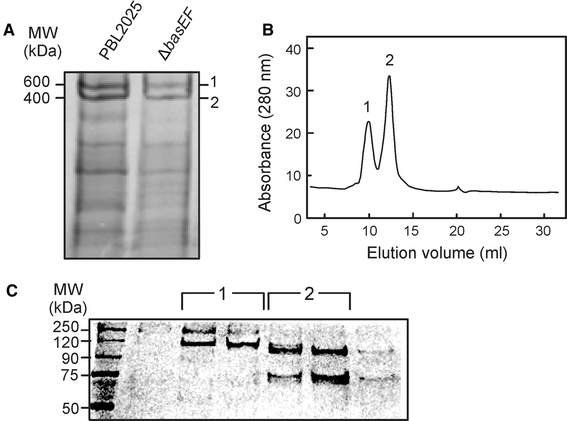
Analysis of the sugar binding proteins extracted from membranes derived from S. solfataricus wild-type and ΔbasEF cells and purified with ConA lectin affinity purification. Isolated ConA fractions were analyzed on Blue-Native PAGE (a). Wild-type ConA fraction was analyzed by size exclusion chromatography (b), resulting in two defined peaks, which were analyzed by SDS-PAGE (c) and protein bands were identified by mass spectrometry