Abstract
Background
Fast treadmill training improves walking speed to a greater extent than training at a self-selected speed after stroke. It is unclear whether fast treadmill walking facilitates a more normal gait pattern after stroke, as has been suggested for treadmill training at self-selected speeds. Given the massed stepping practice that occurs during treadmill training, it is important for therapists to understand how the treadmill speed selected influences the gait pattern that is practiced on the treadmill.
Objective
The purpose of this study was to characterize the effect of systematic increases in treadmill speed on common gait deviations observed after stroke.
Design
A repeated-measures design was used.
Methods
Twenty patients with stroke walked on a treadmill at their self-selected walking speed, their fastest speed, and 2 speeds in between. Using a motion capture system, spatiotemporal gait parameters and kinematic gait compensations were measured.
Results
Significant improvements in paretic- and nonparetic-limb step length and in single- and double-limb support were found. Asymmetry of these measures improved only for step length. Significant improvements in paretic hip extension, trailing limb position, and knee flexion during swing also were found as speed increased. No increases in circumduction or hip hiking were found with increasing speed.
Limitations
Caution should be used when generalizing these results to survivors of a stroke with a self-selected walking speed of less than 0.4 m/s. This study did not address changes with speed during overground walking.
Conclusions
Faster treadmill walking facilitates a more normal walking pattern after stroke, without concomitant increases in common gait compensations, such as circumduction. The improvements in gait deviations were observed with small increases in walking speed.
According to the American Heart Association, stroke is the leading cause of long-term disability.1 Not surprisingly, the locomotor deficits observed following stroke have a profound impact on functional independence2 and have been positively correlated with fall risk and energy cost and negatively correlated with participation in the community.3–5 Thus, there is a growing body of literature exploring the mechanisms underlying poststroke gait abnormalities and possible rehabilitation interventions.
Treadmill training (with or without body-weight support) has emerged as an intervention that improves walking speed in people who have had a stroke.6–12 In addition, one of the proposed benefits of treadmill training is that it facilitates practice of a more normal walking pattern.13–16 Another benefit of treadmill training is that it allows massed stepping practice.17 Thus, treadmill training may result in repetitive, intensive practice of a more normal gait pattern after stroke. This gait practice is useful for people poststroke because they often have substantial gait deviations. Specific deviations include spatiotemporal asymmetry among stance duration, the duration of double-limb support,18 and step length19 of the paretic and nonparetic limbs.9,20 Joint motions (kinematics) during walking also often are impaired following stroke. These impairments in the sagittal plane include decreased knee flexion during swing and decreased hip extension during terminal stance on the paretic leg.18,19,21 Abnormal kinematics in the frontal plane include limb circumduction and hip hiking on the paretic side during the swing phase of gait.19,22,23
Traditionally, treadmill training studies in people poststroke have relied on slow speeds (for a review, see Dobkin13). Recently, however, “fast” treadmill training has emerged as an intervention that may improve walking speed to a greater extent than traditional programs that train at slower speeds poststroke.8,10 In the 2 clinical trials that examined the effect of treadmill training speed on outcomes, participants were trained while walking on a treadmill at speeds that were faster than their self-selected speed. Despite the fact that the determination of what constituted a “fast speed” differed between the 2 studies (in one study, it was defined as walking as fast as possible8; in the other study, the walking speed was set at 1.0 m/s for all individuals in the “fast” group10), greater improvements in overground walking speed were observed when participants trained at a speed faster than their self-selected speed. In addition, both stride length and cadence increased to a greater extent following fast treadmill training poststroke.8
One of the questions that remain, however, is whether fast treadmill walking facilitates a more normal gait pattern after stroke, as has been suggested for treadmill training at self-selected speeds.13–16 It is possible that the facilitatory effect of treadmill walking can be enhanced by walking faster. On the other hand, it also is possible that fast treadmill walking in people poststroke may increase the gait deviations commonly observed, particularly compensatory strategies such as circumduction. Detrimental changes in the gait pattern related to fast treadmill walking could be especially problematic given the massed stepping practice that occurs with treadmill training.17 Two studies have examined spatiotemporal gait deficits during a single session of fast treadmill walking in people poststroke.21,24 In these studies, participants walked 25% or 30% faster than their self-selected walking speed. Both studies examined swing time asymmetry and showed no change at the fast speed compared with the self-selected speed. One study examined stance time asymmetry and also demonstrated no change at the fast speed.24 Because these studies examined only one speed that was faster than the participants' comfortable speed, it is not clear whether changes in gait deficits would have been observed at even faster speeds or speeds somewhere in between.
The appropriate selection of training parameters, such as treadmill speed, is thought to be critical to achieving the optimal benefit from treadmill training.13,16 What is unclear from both fast treadmill training intervention studies and single-session studies is what speed increase is appropriate. Therapists must balance many competing interests when selecting a treadmill training speed for a patient with stroke. Limitations in cardiovascular health or balance may prevent patients from walking as fast as possible on the treadmill. In addition, speeds that are too fast may lead to unwanted changes in the gait pattern. Thus, in developing a rationale for the proper selection of fast treadmill training speeds, we sought to characterize the impact of systematic increases in gait speed on common gait deviations and compensations in individuals with stroke. We were particularly interested in how speed affects not only spatiotemporal gait deficits, which have been the focus of previous studies, but also sagittal- and frontal-plane gait kinematics, because they also are commonly impaired after stroke. In addition, we were interested in whether gait deviations were influenced only by walking on the treadmill at the fastest possible speed or also by smaller speed increases. We hypothesized that spatiotemporal gait parameters and sagittal-plane kinematics would improve with each incremental speed increase, but that kinematic compensations such as circumduction would increase significantly only at the fastest possible speed. Given the massed stepping practice that occurs during treadmill training, it is important for therapists to understand how the treadmill training speed selected influences the gait pattern that is practiced on the treadmill.
Method
Participants
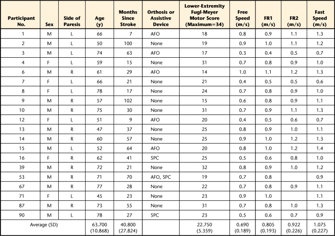
Twenty participants with chronic stroke were recruited. All individuals signed an informed consent statement. To be included, participants must have sustained a single stroke at least 6 months prior to study participation and had to be able to walk independently at multiple speeds with or without an ankle-foot orthosis (AFO) or assistive device. Exclusion criteria included uncontrolled blood pressure or diabetes, cardiovascular or arthritic dysfunction exacerbated by exercise, and active cancer. All participants underwent clinical testing that included the lower-extremity portion of the Fugl-Meyer Assessment25 and the timed Six-Meter Walk Test (6MWT) at self-selected speed.
Instrumentation and Procedure
Participants walked on a split-belt treadmill instrumented with 2 independent 6-degree-of-freedom AMTI force platforms* from which ground reaction force data were collected at 2,000 Hz. Kinematic data were collected with an 8-camera Vicon MX motion capture system† at 100 Hz using a modified Cleveland Clinic marker set. All participants held on to the handrail and wore a safety harness around the chest for fall prevention only; it did not provide body-weight support. Participants' blood pressure, heart rate, and rate of perceived exertion (RPE) were monitored. If participants used an AFO for community mobility, they were permitted to use this orthotic device during the testing, but no assistive devices were permitted (Table).
Table.
Participant Characteristics, Demographics, and Walking Speedsa
FR1 and FR2 were intermediate speeds between free and fast speeds. Ankle-foot orthoses were worn during testing. Assistive devices were used by the participants in the community, but not during testing. AFO=ankle-foot orthosis, SPC=single-point cane, M=male, F=female, L=left, R=right.
The participants were acclimated to the treadmill as necessary. Their overground self-selected walking speed with AFO and without assistive device was measured during the 6MWT and was used to set the free speed while walking on the treadmill. Their fastest speed (fast speed) was determined by slowly increasing the treadmill speed until: (1) the participant reported that he or she could not tolerate any further increase, or (2) the researcher determined that it was unsafe to increase the speed. Two intermediate speeds (FR1 and FR2) between the free and fast speeds comprised the other conditions. These intermediate speeds were chosen to be as equally distributed as possible between the free and fast speeds within the precision of the treadmill speed controls. Most importantly, the intermediate speeds represent speeds that a therapist might choose for treadmill training for an individual. For each speed, the treadmill speed was increased to the target speed over a period of seconds, and data from two 20-second trials were collected. The order in which the speeds were presented was randomized.
Data Analysis
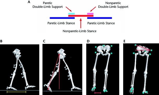
Visual 3D‡ was used for data processing. Foot-strike and lift-off were determined for each limb individually using an automatic algorithm in Visual 3D. Foot-strike was identified when the vertical ground reaction force exceeded 20 N for at least 8 frames, and lift-off was identified when the vertical ground reaction force dropped below 20 N for at least 8 frames. All gait events were checked visually for accuracy. Spatiotemporal measurements were calculated for each leg, at each speed. Temporal measures included single- and double-limb support time. Single-limb support time was the time from contralateral toe-off to heel-strike. Double-limb support time was the time from ipsilateral foot-strike to contralateral toe-off and was labeled as paretic or nonparetic double-limb support based on the limb that was in the stance-to-swing transition (Fig. 1A). Temporal measurements were normalized by stride time and expressed as a percentage of the stride cycle. One spatial measurement, step length, was calculated as the sagittal distance between the right and left heel markers at foot-strike (Fig. 1B). Step length was labeled paretic or nonparetic based on the leading leg.
Figure 1.
Gait parameters. (A) Schematic representation of stance, swing, and double-limb support phases of a typical gait cycle. (B) Step length is measured as the sagittal distance from one foot's heel marker to the contralateral foot's heel marker at heel-strike. (C) Trailing limb angle is defined as the angle between the laboratory's vertical axis and a vector created between the greater trochanter and the fifth metatarsal head at toe-off. (D) Circumduction is calculated as the maximal lateral difference between the location of the bottom heel marker during the stance phase and that same heel marker during the swing phase that immediately followed. (E) Hip hiking is calculated as an angle in the frontal plane between the pelvis' resting position during static standing and its maximal deviation from that position during the stance phase.
Peak hip extension, trailing limb angle, and peak knee flexion also were calculated for each leg and each speed. Peak hip extension was defined as the greatest hip extension angle captured during the stance phase, and peak knee flexion was defined as the greatest knee flexion angle captured during the swing phase. Trailing limb angle was defined as the angle between the laboratory's vertical axis and a vector created between the greater trochanter and the fifth metatarsal head at toe-off26–30 (Fig. 1C).
Paretic-limb swing-phase compensation measurements, including circumduction and hip hiking, were calculated at each speed. Circumduction was calculated in the frontal plane as the maximum difference between the bottom heel marker during stance and the same marker during the immediately subsequent swing phase19 (Fig. 1D). Hip hiking was defined as the angle in the frontal plane between the pelvis position during static standing and its maximal deviation from that position during each paretic-limb swing phase (Fig. 1). Symmetry of the temporal and spatial parameters was determined using the formula31:
 |
Spatiotemporal asymmetry was determined for each participant based on his or her symmetry values during the free walking condition. A participant was determined to have an asymmetric step length if his or her asymmetry value fell outside 1 standard deviation of perfect symmetry.27 Temporal asymmetry was designated if this value fell outside 2 standard deviations of data for people who are healthy. All data were averaged across strides for a given speed.
Data normality was confirmed using the Kolmogorov-Smirnov test for normality. Repeated-measures analyses of variance (ANOVAs) were used to evaluate each variable with respect to speed (step length, single-limb support, double-limb support, trailing limb angle, hip extension, knee flexion, circumduction, and hip hiking). When the ANOVA yielded significant results, post hoc analyses were completed between free speed and FR1, free speed and FR2, and free and fast speeds using a Bonferroni correction for multiple comparisons. For these additional comparisons, the corrected P value that represented statistical significance was P≤.0167. Values for both the paretic and nonparetic legs were compared for the spatiotemporal variables. For the kinematic variables, only data from the paretic leg were compared across speeds. All statistics were completed using Statistica version 6.0.§
Role of the Funding Source
This work was supported by grant 0765314U from the American Heart Association for expenses related to data collection, analysis, and dissemination of results. In addition, instrumentation grants NIH S10 RR022396-01 (National Center for Research Resources) and DOD W911NF-05–1-0097 supported the purchase of the motion capture system and instrumented treadmill, and grant NIH K01 HD050582 provided partial support of the authors' time (D.S.R., C.M.M.).
Results
The Table contains the participant characteristics. Across participants, the average free speed was 0.690 m/s (SD=0.189). The average increase between free speed and FR1 was 0.115 m/s, from FR1 to FR2 was 0.117 m/s, and from FR2 to fast speed was 0.153 m/s. Two participants were able to complete only one speed between the free and fast speeds; thus, their data were not included in the analysis.
Spatiotemporal Gait Parameters
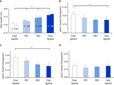
Figure 2 illustrates the results for the spatial gait parameter of step length. Participants showed an increase in both paretic- and nonparetic-limb step length with increasing speed (main effect of speed for both legs, F3,51=55.8, P<.0001, Fig. 2A). From free speed to fast speed, the average increase in paretic-limb step length was 0.104 m, and the average increase in nonparetic-limb step length was 0.134 m. Post hoc testing revealed a significant increase in paretic- and nonparetic-limb step length between free speed and each of the other 3 speeds (P<.01 for all comparisons). In addition, step length asymmetry changed with increasing speed; however, not all participants demonstrated a step length asymmetry at baseline (14 out of 20 participants had asymmetric step lengths at their free walking speed). As a group, these individuals showed a significant progression toward improved symmetry with increased speed (main effect of speed, F3,33=3.41, P=.02, Fig. 2B). Post hoc testing revealed a significant improvement in step length symmetry between free and fast speeds (P=.003). After Bonferroni correction, the differences between free speed and FR1 (P=.04) and between free speed and FR2 (P=.06) were not statistically significant. People with stroke can show step length asymmetries in either direction (eg, for some patients, the paretic leg takes a longer step compared with the nonparetic leg, and for other patients, the opposite is true).18–20 Therefore, we also evaluated the changes in step length asymmetry in these 2 groups separately. Participants who took longer paretic-limb steps at their free speed (n=7) showed improved step length asymmetry with increasing speed (main effect of speed, F3,12=7.88, P=.003). Post hoc testing revealed statistically significant differences between free and fast speeds (P<.001). After Bonferroni correction, the differences between free speed and FR1 (P=.08) and between free speed and FR2 (P=.04) were not statistically significant (Fig. 2C). In contrast, participants who took a shorter paretic-limb step (n=7) showed no changes in symmetry with speed (Fig. 2D).
Figure 2.
Step length. (A) Paretic- and nonparetic-limb step length averaged across all participants (N=20). P=paretic leg, NP=nonparetic leg. (B) Step length asymmetry results across all speeds for all participants with step length asymmetry (n=14). (C) Step length asymmetry results across all speeds for participants with a longer paretic-leg step (n=7) at free speed. (D) Step length asymmetry results across all speeds for participants with a shorter paretic step (n=7) at free speed. FR1 and FR2 were intermediate speeds between free and fast speeds. Asterisk indicates significant difference among speeds. “Abs” refers to absolute value of the quantity in the parentheses.
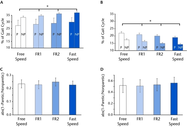
Figure 3 illustrates the results for the temporal gait parameters of percentage of single- and double-limb support. Percentage of single-limb support on both legs increased with increasing speed (paretic leg: F3,51=16.9, nonparetic leg: F3,51=28.6, P<.0001 for both legs, Fig. 3A). From free speed to fast speed, the average increase in paretic single-limb support was 2.7%, and the average increase in nonparetic single-limb support was 3.4%. Post hoc testing revealed a significant increase in all speeds for both legs (P<.05). Percentage of double-limb support on both the paretic and nonparetic legs decreased with increasing speed (F3,51=24.8, P<.0001 for both legs, Fig. 3B). From free speed to fast speed, the average decrease in paretic double-limb support was 3.0%, and the average decrease in nonparetic double-limb support was 3.0%. Post hoc testing revealed a significant increase among all speeds for both legs (P<.01). As for step length, we also were interested in the changes in temporal asymmetry with increasing speed for those participants with asymmetric support times at baseline. Fifteen participants had asymmetric single-limb support times at their free speed, and 12 participants had asymmetric double-limb support times at their free speed. These participants showed no improvement in asymmetry with increased speed (Figs. 3C and 3D).
Figure 3.
Temporal gait parameters. (A) Paretic and nonparetic single-limb support time as a percentage of the gait cycle, averaged across all participants (N=20). (B) Paretic and nonparetic double-limb support time as a percentage of the gait cycle, averaged across all participants (N=20). (C) Single-limb support time symmetry across all speeds for all participants with step length asymmetry (n=15). (D) Double-limb support time symmetry across all speeds for all participants with step length asymmetry (n=12). Error bars represent ± standard error across participants. For all symmetry graphs, 0=perfect symmetry. FR1 and FR2 were intermediate speeds between free and fast speeds. P=paretic leg, NP=nonparetic leg. Asterisk indicates significant difference among speeds. “Abs” refers to absolute value of the quantity in the parentheses.
Kinematic Gait Parameters
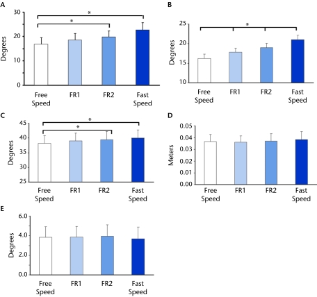
Figure 4 illustrates the results for the kinematic parameters. Paretic hip extension angle increased significantly as speed increased (main effect of speed F3,51=13.9, P<.0001, Fig. 4A), with an average increase in angle of 5.57 degrees from free speed to fast speed. Post hoc testing showed a significant increase in hip extension angle between free speed and FR2 and between free and fast speeds (P<.01 for each comparison.) Paretic peak trailing limb angle increased significantly as speed increased (main effect of speed F3,51=82.2, P<.0001, Fig. 4B), with an average increase in angle of 4.81 degrees from free speed to fast speed. Post hoc testing showed a significant increase in trailing limb angle between free speed and each of the other 3 speeds (P<.001 for all comparisons).
Figure 4.
Kinematic gait parameters for the paretic leg across all speeds, averaged across all participants: (A) peak hip extension, (B) peak trailing limb position, (C) peak knee flexion, (D) peak circumduction during swing, (E) peak hip hiking during swing. Error bars represent ± standard error across participants. FR1 and FR2 were intermediate speeds between free and fast speeds. Asterisk indicates significant difference among speeds.
Paretic peak knee flexion during swing increased slightly with increasing speed, and this increase was significant (main effect of speed, F3,51=4.6, P=.006, Fig. 4C), with an average increase in angle of 1.80 degrees from free speed to fast speed. The increase in peak knee flexion occurred between free speed and FR2 (P<.01) and between free and fast speeds (P<.01). Unlike trailing limb position and peak knee flexion, increasing speed had no impact on paretic- leg circumduction (Fig. 4D) or hip hiking (Fig. 4E).
Discussion
This study investigated the influence of increasing treadmill walking speed on the gait patterns of people with chronic stroke in order to further our understanding of the gait patterns that are reinforced during fast treadmill training. The results show that some gait deviations improve with faster treadmill speeds, whereas others are unaffected. Furthermore, the positive improvements that are observed occur with each incremental speed increase and without concomitant increases in swing-phase gait compensations. These results support the idea that facilitation of a more normal gait pattern through treadmill walking poststroke can be enhanced by walking faster on the treadmill. Thus, fast treadmill walking improves the gait pattern being repetitively and intensively practiced during treadmill training.
Spatiotemporal Parameters
Increasing treadmill speed influenced both paretic-limb step length and step length asymmetry. Step length increased for both legs with each incremental increase in speed. Increased step length (rather than increased cadence) has been associated with increased walking speed following a treadmill and overground walking program in people poststroke.6 Specifically, improvements of 0.1 m in paretic- and nonparetic-limb step length have been associated with significant improvements in walking speed and endurance following training.6 In the present study, changes in step length exceeded this value between the free and fast speeds, but not between the free speed and FR1 or FR2. Future research is needed to interpret the clinical meaningfulness of the smaller improvements in step length observed with the smaller increases in speed. Incremental increases in walking speed also were related to improvements in step length asymmetry, specifically in the participants who normally take a longer paretic-limb step. This finding means that walking at even a slightly faster speed allows these individuals repeated practice of a more symmetrical step length pattern. Step length asymmetry has been associated with slower walking speed and reduced paretic-leg propulsive force during walking poststroke.26,27 Thus, practice of a more symmetrical step length pattern may have beneficial effects on other gait deficits after stroke.
Single-limb support time increased and double-limb support time decreased on both legs with each increase in treadmill speed. These changes are important because single-limb support time generally is shorter in people with stroke compared with people without neurological deficits, whereas double-limb support time generally is longer.32 Furthermore, the paretic-limb changes of 2.7% to 3.0% that we report here exceed the within-session minimal detectible change (MDC) values for these variables in people with chronic stroke.33 This finding demonstrates that these changes are not simply a result of the typical stride-to-stride variability that is characteristic of people following a stroke. Previous studies have examined stance and swing time asymmetry during a single faster treadmill walking speed poststroke and shown no change with increased speed.21,24 Our results extend the previous findings to the measure of double-limb support and to treadmill walking at multiple faster speeds. Our results illustrate that regardless of the magnitude of speed increase, temporal asymmetry is not improved by faster treadmill walking after stroke, despite the change in individual single- and double-limb support times.
Kinematic Parameters
When people with stroke walk at progressively faster treadmill speeds, both hip extension angle and trailing limb angle increase, as demonstrated by our results. The changes from free speed to every other speed in hip extension and trailing limb angle that we report greatly exceed the within-session MDCs for people with chronic stroke,33 again illustrating that these changes are not merely a result of typical stride-to-stride variability. In addition, previous research suggests that the changes in hip extension between free and fast speeds are clinically meaningful.34 In a study of body-weight–supported treadmill training of people with chronic stroke by Mulroy and colleagues,34 participants who were the best responders (improvements in walking speed of 0.8 m/s or greater) showed improvements in hip extension similar to the increases between the free and fast speeds found in the current study. These findings suggest that hip extension increases found with fast walking are likely to be clinically meaningful because they are associated with meaningful changes in walking speed after training.
Trailing limb angle (the angle between the laboratory's vertical axis and a vector connecting the greater trochanter and the fifth metatarsal head) captures hip extension and knee and ankle angle at terminal stance in a single measure. This angle is biomechanically important for forward propulsion of the body. A better trailing limb angle at transition from stance to swing enables a greater component of the ground reaction force to be generated anteriorly and, thus, facilitate forward propulsion.26,27,30 In addition, greater anterior ground reaction force is thought to facilitate improved knee flexion during swing.35 Thus, an increase in trailing limb angle may be important for these aspects of walking in people poststroke.
There was a significant improvement in peak knee flexion with increased walking speed in our group of people with stroke. However, this increase was, on average, only 1.8 degrees between the free and fast speeds. Thus, although there was a statistically significant difference between speeds, the magnitude of the increase was relatively small and does not exceed the within-session MDC.33 This finding is consistent with a qualitative report that walking 30% faster than self-selected speed had little effect on peak knee flexion poststroke.21 The results of this study suggest that increased treadmill walking speed has a limited effect on knee flexion during swing poststroke.
The gait deficits of circumduction and hip hiking did not change with increasing treadmill speed in this group of people with chronic stroke. It has been suggested that circumduction and hip hiking are compensations for decreased paretic knee flexion during swing in people poststroke.19,22,23 If faster walking speeds exaggerate “stiff-knee gait,”36 one of the concerns is that circumduction and hip hiking may increase at faster speeds to compensate for this decreased paretic knee flexion. The fact that these compensations did not increase with increasing speed is consistent with our finding that knee flexion did not worsen and even improved slightly with faster speed. Thus, the hypothesis that fast treadmill walking will increase the gait deviations of circumduction and hip hiking is not supported by the results.
How Fast Is Fast Enough?
Our results demonstrate that improvements in gait deficits during treadmill walking in people poststroke may be achieved at speeds below a person's maximal speed. This finding is important because treadmill training at maximal speed is not always possible or safe after stroke. Secondary diagnoses common in those who have had a stroke (eg, heart disease, bone and joint abnormalities, hypertension) or the risk or fear of falling might limit the walking speed patients can achieve on the treadmill. In addition, many patients with stroke may not be able to sustain their maximal walking speed for sufficient time to effect a meaningful change from treadmill training. Thus, the finding that maximal walking speed is not required to achieve improvements in poststroke gait is powerful, as it could substantially expand the poststroke population that would benefit from treadmill training at a speed faster than self-selected speed.
Even small increases in speed result in improvements in the gait pattern practiced during treadmill training. For example, trailing limb position increased by approximately 2 degrees with every 0.1-m/s increase in speed (Fig. 4). These results may help in decision making regarding speed selection for poststroke treadmill training. The physical therapist often must balance competing needs, such as safety and endurance, with the desire to have a patient walk faster during treadmill training. Our results suggest that although a faster speed is better, even small increases in speed produce beneficial changes in terms of the gait pattern being practiced.
Limitations
There are several limitations to the findings of our study. First, the sample size was small, and the sample contained individuals with a primarily intermediate to high level of ambulatory function poststroke. According to Perry and colleagues' classification, 2 individuals in our sample would be considered household ambulators, 10 would be considered limited community ambulators, and 8 would be considered unlimited community ambulators.3 Therefore, although these data do represent the response of people poststroke across a range of walking speed abilities, the number of individuals in the slowest category (<0.4 m/s) was small. Future research is needed to determine whether our results can be substantiated in a larger group of participants with slow (<0.4 m/s) self-selected walking speeds.3 In addition, although the range of lower-extremity Fugl-Meyer motor scores in our study is similar to scores found in other studies investigating walking in people with chronic stroke,19,37,38 care should be taken when generalizing the results to those with lower-extremity Fugl-Meyer motor scores lower than 14.
Second, participants were permitted to ambulate both with their AFO and while holding on to a handrail during the testing. It is well known that handrail support may affect the walking patterns observed on the treadmill.21 Chen et al21 reported an increase in paretic single-limb support time and overall temporal symmetry with the introduction of a handrail during treadmill walking. Interestingly, Kuys et al39 found similar improvements in overground hemiparetic walking patterns when they compared a group of people who trained by walking overground with a group who trained on a treadmill with handrail support. Thus, despite potential complications associated with the use of the handrail on a treadmill, there is evidence suggesting that beneficial effects may result, including carryover to overground walking in patients with hemiparesis. Specifically related to the design of the current study, studies of fast treadmill training following stroke have allowed handrail support10,17 and AFO use,8,17 and it is not uncommon for handrail support and AFOs to be used during clinical physical therapy treatments. To date, no information is available on how handrail use may change as an individual with hemiparesis is required to walk at faster speeds.
Finally, the goal of this study was to characterize gait parameter changes with fast treadmill walking. It is important to note that the improvements observed with fast walking on the treadmill may not translate to overground walking.
Conclusions
The results of this study demonstrate that, in addition to greater improvements in walking speed,8,10 faster treadmill walking results in massed practice of a more normal walking pattern after stroke without concomitant increases in common gait compensations such as circumduction. Because treadmill training is a common treatment intervention, this information can assist therapists in assessing how selection of treadmill training speed will influence a patient's gait deviations. Furthermore, because improvements in gait deviations with faster walking can be observed with small changes in walking speed, even when the therapist is unable to increase speed to a patient's maximum, specific improvements of the gait pattern can be expected.
The Bottom Line
What do we already know about this topic?
Survivors of stroke typically have spatiotemporal gait abnormalities, and these deficits have been found to have a profound impact on functional independence. Literature continues to emerge in support of treadmill training at various speeds, both with and without body-weight support, as a method of treatment for these deficits.
What new information does this study offer?
This study demonstrates an improvement in many typical gait asymmetries after stroke when people walk on a treadmill at a faster speed than self-selected speed, without increases in swing-phase compensations.
If you're a patient, what might these findings mean for you?
Walking even slightly faster than your comfortable pace while on a treadmill can have a positive impact on some of the abnormalities in your walking pattern after stroke.
Footnotes
All authors provided concept/idea/research design and data analysis. Dr Tyrell, Dr Roos, and Dr Reisman provided writing and data collection. Dr Tyrell and Dr Reisman provided project management and clerical support. Dr Reisman provided fund procurement. Dr Rudolph and Dr Reisman provided facilities/equipment. Dr Tyrell, Dr Rudolph, and Dr Reisman provided consultation (including review of manuscript before submission).
The study protocol was approved by the University of Delaware Human Subjects Review Board.
Platform presentations of this research were given at the Combined Sections Meetings of the American Physical Therapy Association; February 6–9, 2008; Nashville, Tennessee, and February 6–9, 2009; Las Vegas, Nevada. This research also was presented at the 18th International Society for Posture and Gait Research Conference; July 14–18, 2007; Burlington, Vermont.
This work was supported by grant 0765314U from the American Heart Association for expenses related to data collection, analysis, and dissemination of results. In addition, instrumentation grants NIH S10 RR022396-01 (National Center for Research Resources) and DOD W911NF-05-1-0097 supported the purchase of the motion capture system and instrumented treadmill, and grant NIH K01 HD050582 provided partial support of the authors' time (D.S.R., C.M.M.).
Advanced Mechanical Technology Inc, 176 Waltham St, Watertown, MA 02472.
Vicon Motion Systems, 5419 McConnell Ave, Los Angeles, CA 90066.
C-Motion Inc, 20030 Century Blvd, Suite 104A, Germantown, MD 20874.
StatSoft Inc, 2300 E 14th St, Tulsa, OK 74104.
References
- 1. Lloyd-Jones D, Adams RJ, Brown TM, et al. Heart disease and stroke statistics—2010 update: a report from the American Heart Association. Circulation. 2010;121:e46–e215 [DOI] [PubMed] [Google Scholar]
- 2. Schmid A, Duncan PW, Studenski S, et al. Improvements in speed-based gait classifications are meaningful. Stroke. 2007;38:2096–2100 [DOI] [PubMed] [Google Scholar]
- 3. Perry J, Garrett M, Gronley JK, Mulroy SJ. Classification of walking handicap in the stroke population. Stroke. 1995;26:982–989 [DOI] [PubMed] [Google Scholar]
- 4. Lamb SE, Ferrucci L, Volapto S, et al. Risk factors for falling in home-dwelling older women with stroke: the Women's Health and Aging Study. Stroke. 2003;34:494–501 [PubMed] [Google Scholar]
- 5. Reisman DS, Rudolph KS, Farquhar WB. Influence of speed on walking economy poststroke. Neurorehabil Neural Repair. 2009;23:529–534 [DOI] [PMC free article] [PubMed] [Google Scholar]
- 6. Ada L, Dean CM, Hall JM, et al. A treadmill and overground walking program improves walking in persons residing in the community after stroke: a placebo-controlled, randomized trial. Arch Phys Med Rehabil. 2003;84:1486–1491 [DOI] [PubMed] [Google Scholar]
- 7. Barbeau H, Visintin M. Optimal outcomes obtained with body-weight support combined with treadmill training in stroke subjects. Arch Phys Med Rehabil. 2003;84:1458–1465 [DOI] [PubMed] [Google Scholar]
- 8. Pohl M, Mehrholz J, Ritschel C, Ruckriem S. Speed-dependent treadmill training in ambulatory hemiparetic stroke patients: a randomized controlled trial. Stroke. 2002;33:553–558 [DOI] [PubMed] [Google Scholar]
- 9. Silver KH, Macko RF, Forrester LW, et al. Effects of aerobic treadmill training on gait velocity, cadence, and gait symmetry in chronic hemiparetic stroke: a preliminary report. Neurorehabil Neural Repair. 2000;14:65–71 [DOI] [PubMed] [Google Scholar]
- 10. Sullivan KJ, Knowlton BJ, Dobkin BH. Step training with body weight support: effect of treadmill speed and practice paradigms on poststroke locomotor recovery. Arch Phys Med Rehabil. 2002;83:683–691 [DOI] [PubMed] [Google Scholar]
- 11. Visintin M, Barbeau H, Korner-Bitensky N, Mayo NE. A new approach to retrain gait in stroke patients through body weight support and treadmill stimulation. Stroke. 1998;29:1122–1128 [DOI] [PubMed] [Google Scholar]
- 12. Werner C, Von Frankenberg S, Treig T, et al. Treadmill training with partial body weight support and an electromechanical gait trainer for restoration of gait in subacute stroke patients: a randomized crossover study. Stroke. 2002;33:2895–2901 [DOI] [PubMed] [Google Scholar]
- 13. Dobkin B. An overview of treadmill locomotor training with partial body weight support: a neurophysiologically sound approach whose time has come for randomized clinical trials. Neurorehabil Neural Repair. 1999;13:157–165 [Google Scholar]
- 14. Harris-Love ML, Forrester LW, Macko RF, et al. Hemiparetic gait parameters in overground versus treadmill walking. Neurorehabil Neural Repair. 2001;15:105–112 [DOI] [PubMed] [Google Scholar]
- 15. Hassid E, Rose D, Commisarow J, et al. Improved gait symmetry in hemiparetic stroke patients induced during body weight-supported treadmill stepping. J Neurol Rehabil. 1997;11:21–26 [Google Scholar]
- 16. Hesse SA. Treadmill training with partial body weight support in hemiparetic patients: further research needed. Neurorehabil Neural Repair. 1999;13:179–181 [Google Scholar]
- 17. Moore JL, Roth EJ, Killian C, Hornby TG. Locomotor training improves daily stepping activity and gait efficiency in individuals poststroke who have reached a “plateau” in recovery. Stroke. 2009. November 12 [Epub ahead of print] [DOI] [PubMed] [Google Scholar]
- 18. Olney SJ, Griffin MP, McBride ID. Temporal, kinematic, and kinetic variables related to gait speed in subjects with hemiplegia: a regression approach. Phys Ther. 1994;74:872–885 [DOI] [PubMed] [Google Scholar]
- 19. Chen G, Patten C, Kothari DH, Zajac FE. Gait differences between individuals with post-stroke hemiparesis and non-disabled controls at matched speeds. Gait Posture. 2005;22:51–56 [DOI] [PubMed] [Google Scholar]
- 20. Roth EJ, Merbitz C, Mroczek K, et al. Hemiplegic gait: relationships between walking speed and other temporal parameters. Am J Phys Med Rehabil. 1997;76:128–133 [DOI] [PubMed] [Google Scholar]
- 21. Chen G, Patten C, Kothari DH, Zajac FE. Gait deviations associated with post-stroke hemiparesis: improvement during treadmill walking using weight support, speed, support stiffness, and handrail hold. Gait Posture. 2005;22:57–62 [DOI] [PubMed] [Google Scholar]
- 22. Kerrigan DC, Frates EP, Rogan S, Riley PO. Hip hiking and circumduction: quantitative definitions. Am J Phys Med Rehabil. 2000;79:247–252 [DOI] [PubMed] [Google Scholar]
- 23. Neckel ND, Blonien N, Nichols D, Hidler J. Abnormal joint torque patterns exhibited by chronic stroke subjects while walking with a prescribed physiological gait pattern. J Neuroeng Rehabil. 2008;5:19. [DOI] [PMC free article] [PubMed] [Google Scholar]
- 24. Hesse SA, Werner C, Paul T, et al. Influence of walking speed on lower limb muscle activity and energy consumption during treadmill walking of hemiparetic patients. Arch Phys Med Rehabil. 2001;82:1547–1550 [DOI] [PubMed] [Google Scholar]
- 25. Fugl-Meyer AR, Jaasko L, Leyman I, et al. The post-stroke hemiplegic patient, 1: a method for evaluation of physical performance. Scand J Rehabil Med. 1975;7:13–31 [PubMed] [Google Scholar]
- 26. Bowden MG, Balasubramanian CK, Neptune RR, Kautz SA. Anterior-posterior ground reaction forces as a measure of paretic leg contribution in hemiparetic walking. Stroke. 2006;37:872–876 [DOI] [PubMed] [Google Scholar]
- 27. Balasubramanian CK, Bowden MG, Neptune RR, Kautz SA. Relationship between step length asymmetry and walking performance in subjects with chronic hemiparesis. Arch Phys Med Rehabil. 2007;88:43–49 [DOI] [PubMed] [Google Scholar]
- 28. Bowden MG, Balasubramanian CK, Behrman AL, Kautz SA. Validation of a speed-based classification system using quantitative measures of walking performance poststroke. Neurorehabil Neural Repair. 2008;22:672–675 [DOI] [PMC free article] [PubMed] [Google Scholar]
- 29. Balasubramanian CK, Neptune RR, Kautz SA. Foot placement in a body reference frame during walking and its relationship to hemiparetic walking performance. Clin Biomech (Bristol, Avon). 2010;25:483–490 [DOI] [PMC free article] [PubMed] [Google Scholar]
- 30. Turns LJ, Neptune RR, Kautz SA. Relationships between muscle activity and anteroposterior ground reaction forces in hemiparetic walking. Arch Phys Med Rehabil. 2007;88:1127–1135 [DOI] [PMC free article] [PubMed] [Google Scholar]
- 31. Hsu AL, Tang PF, Jan MH. Analysis of impairments influencing gait velocity and asymmetry of hemiplegic patients after mild to moderate stroke. Arch Phys Med Rehabil. 2003;84:1185–1193 [DOI] [PubMed] [Google Scholar]
- 32. Olney SJ, Richards C. Hemiparetic gait following stroke, part I: characteristics. Gait Posture. 1996;4:136–148 [Google Scholar]
- 33. Kesar TM, Binder-Macleod SA, Hicks GE, Reisman DS. Minimal detectable change for gait variables collected during treadmill walking in individuals post-stroke. Gait Posture. In press [DOI] [PMC free article] [PubMed] [Google Scholar]
- 34. Mulroy SJ, Klassen T, Gronley JK, et al. Gait parameters associated with responsiveness to treadmill training with body-weight support after stroke: an exploratory study. Phys Ther. 2010;90:209–223 [DOI] [PubMed] [Google Scholar]
- 35. Chen G, Patten C. Joint moment work during the stance-to-swing transition in hemiparetic subjects. J Biomech. 2008;41:877–883 [DOI] [PubMed] [Google Scholar]
- 36. Lewek MD, Hornby TG, Dhaher YY, Schmit BD. Prolonged quadriceps activity following imposed hip extension: a neurophysiological mechanism for stiff-knee gait? J Neurophysiol. 2007;98:3153–3162 [DOI] [PMC free article] [PubMed] [Google Scholar]
- 37. Plummer P, Behrman AL, Duncan PW, et al. Effects of stroke severity and training duration on locomotor recovery after stroke: a pilot study. Neurorehabil Neural Repair. 2007;21:137–151 [DOI] [PubMed] [Google Scholar]
- 38. Sullivan KJ, Brown DA, Klassen T, et al. Effects of task-specific locomotor and strength training in adults who were ambulatory after stroke: results of the STEPS randomized clinical trial. Phys Ther. 2007;87:1580–1602 [DOI] [PubMed] [Google Scholar]
- 39. Kuys SS, Brauer SG, Ada L, Russell TG. Immediate effect of treadmill walking practice versus overground walking practice on overground walking pattern in ambulatory stroke patients: an experimental study. Clin Rehabil. 2008;22:931–939 [DOI] [PubMed] [Google Scholar]


