Abstract
The social brain hypothesis posits that the cognitive demands of social behaviour have driven evolutionary expansions in brain size in some vertebrate lineages. In insects, higher brain centres called mushroom bodies are enlarged and morphologically elaborate (having doubled, invaginated and subcompartmentalized calyces that receive visual input) in social species such as the ants, bees and wasps of the aculeate Hymenoptera, suggesting that the social brain hypothesis may also apply to invertebrate animals. In a quantitative and qualitative survey of mushroom body morphology across the Hymenoptera, we demonstrate that large, elaborate mushroom bodies arose concurrent with the acquisition of a parasitoid mode of life at the base of the Euhymenopteran (Orussioidea + Apocrita) lineage, approximately 90 Myr before the evolution of sociality in the Aculeata. Thus, sociality could not have driven mushroom body elaboration in the Hymenoptera. Rather, we propose that the cognitive demands of host-finding behaviour in parasitoids, particularly the capacity for associative and spatial learning, drove the acquisition of this evolutionarily novel mushroom body architecture. These neurobehavioural modifications may have served as pre-adaptations for central place foraging, a spatial learning-intensive behaviour that is widespread across the Aculeata and may have contributed to the multiple acquisitions of sociality in this taxon.
Keywords: brain evolution, learning and memory, parasitoid
1. Introduction
The insect mushroom bodies are brain centres that participate in an array of higher order functions including olfactory associative learning and olfactory processing [1–3], spatial learning [4,5], sensory integration, sensory filtering and attention [6–12]. The mushroom bodies have a characteristic morphology consisting of a pedunculus and lobes composed of the parallel fibre-like projections of thousands of intrinsic neurons called Kenyon cells, and calyces composed of Kenyon cell dendrites that receive afferent input from primary sensory neuropils [13–16].
The first study of insect mushroom bodies by Dujardin [17] made note of their large size and morphological complexity in the social species of the aculeate Hymenoptera. Since then, a relationship between large, morphologically complex mushroom bodies and sociality in insects has been tacitly accepted, although never explicitly tested [18–20]. The social brain hypothesis, as proposed for some vertebrate taxa, posits that the cognitive demands of social behaviour have driven evolutionary expansions in overall brain size and in brain regions such as the telencephalon ([21–27]; but see [28,29] for instances in which sociality does not clearly correlate with increased brain size). As applied to the Hymenoptera, the hypothesis would predict that large, morphologically complex mushroom bodies should be found only in those lineages containing social species, and not in the more basal lineages consisting only of solitary species.
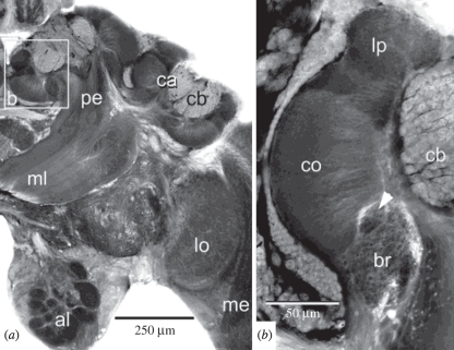
The mushroom bodies of the social aculeate Hymenoptera are typified by those of the honeybee, Apis mellifera (figure 1) [30–32]. In particular, the calyces are doubled, deeply cup-shaped and greatly expanded in size in relation to the lobes [33], a morphology we refer to here as ‘elaborate’. The calyces of social aculeates also receive visual input from the medulla and lobula of the optic lobes, in addition to olfactory and gustatory inputs that are more generally observed across the insects [31,34–37]. Morphologically distinct subcompartments in the calyces receive input from each of these sensory modalities, with the collar region specifically receiving optic lobe input [31].
Figure 1.
Mushroom body architecture in aculeate hymenopterans, as illustrated by the honeybee Apis mellifera (Apidae, Apoidea). (a) The mushroom body neuropil consisting of the calyces (ca), pedunculus (pe) and lobes (medial lobe (ml) visible in this plane of section). The calyces are elaborated into deeply invaginated cups, and the cell bodies of mushroom body intrinsic neurons, called Kenyon cells (cb) fill and surround the calyces. (b) High-magnification view of boxed area in (a) showing functional subdivisions of the calyx. Olfactory projection neurons target the lip (lp), visual projection neurons target the collar (co) and the basal ring (br) receives collaterals from both. Figures borrowed with permission from Ehmer & Gronenberg [37].
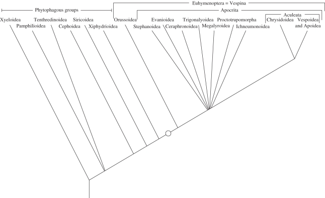
The Hymenoptera comprise one of the largest and most diverse insect orders. The ancestral species from which all Hymenoptera evolved probably had phytophagous larvae, as do extant members of the basal lineages [38] (figure 2). A prominent step in hymenopteran evolution was the transition from phytophagy to parasitism at the base of the Euhymenoptera, a group that contains the superfamily Orussoidea and the Apocrita [38,39]. Monophyly of Euhymenoptera is strongly supported by morphological, molecular and joint analyses [40–42]. Thus, the present account will not use the traditional classification of Hymenoptera as divided into Symphyta and Apocrita, but will instead refer to ‘phytophagous lineages’ versus Euhymenoptera.
Figure 2.
Cautious estimate of the phylogeny of Hymenoptera based on [41,56,120,121] and recent molecular analyses by the HymAToL project (not yet published). Only relationships that are supported independently by morphological and molecular analyses are shown. The present study examined brains of species representing all groups indicated in the tree with the exception of the phytophagous Pamphilioidea. Proctotrupomorpha includes Chalcidoidea, Proctotrupoidea, Cynipoidea, Platygastroidea, Diaprioidea and Mymarommatoidea. Circle indicates the appearance of large, elaborate mushroom bodies at the base of the Euhymenoptera. Eusocial species are found only within the Vespoidea and Apoidea.
Another prominent step in the evolution of the Hymenoptera was the transformation of the ovipositor into a stinger in the ancestor of Aculeata in the late Jurassic and early Cretaceous [43]. The Aculeata are split into Chrysidoidea and a clade comprising Vespoidea and Apoidea. Eusociality was another important evolutionary innovation and has probably evolved four times independently within Vespoidea and Apoidea [44].
In the first test of the evolutionary relationship between sociality and brain morphology in an invertebrate clade, we examined mushroom body morphologies of solitary species representing nearly all major hymenopteran lineages outside of the Aculeata, one basal solitary aculeate and one social aculeate (electronic supplementary material, table S1). We sought to identify three events in the evolution of hymenopteran mushroom bodies, and to determine whether any of these events was associated with the acquisition of sociality: (i) the expansion of mushroom body volume relative to the rest of the brain, (ii) the expansion of the calyces relative to the lobes, and (iii) the acquisition of visual inputs to the calyces from the optic lobes. Volumes of the mushroom bodies, their calyces and lobes were obtained from measurements of histological preparations, and when available, live-captured insects were used for fluorescent dextran tracing of optic lobe inputs to the mushroom bodies. These data were also compared with accounts of mushroom body morphologies of social and solitary aculeates from the published literature (electronic supplementary material, table S1).
2. Material and methods
(a). Insects
Insects were either live-captured in the Morgantown, WV area or obtained as ethanol- or alcoholic Bouin's-preserved specimens.
(b). Analysis of mushroom body morphology
Live-captured specimens were chilled on ice and brains dissected in physiological saline [45]. Brains were processed in one of two ways: (i) fixed in Carnoy's fixative for 2 h at room temperature and stored in 70 per cent ethanol at 4°C overnight, embedded in paraffin, sectioned at 10 µm on a rotary microtome and processed for Cason's staining [46]; or (ii) fixed in 4 per cent paraformaldehyde in phosphate-buffered saline overnight at 4°C, embedded in agarose, vibratome sectioned at 70 µm, and immunostained using the anti-DC0 antibody as described by Farris [47]. The anti-DC0 antibody (a gift of Dr Daniel Kalderon) robustly stains insect Kenyon cells [16,48]. For Cason's stained brains, mushroom body and protocerebral volumes were calculated from area measurements of traced brain regions from 10 µm paraffin sections using Zeiss AxioVision 4 software (Carl Zeiss AG, Oberkochen, Germany) as described by Farris & Roberts [49]. For anti-DC0 stained brains, sections were viewed on an Olympus Fluoview 1000 confocal microscope, and image stacks consisting of 10 µm optical sections of each section captured and saved as .avi files. Files were imported into ImageJ 1.43u software (http://rsbweb.nih.gov/ij/) for quantification of brain volumes using a point-counting grid (‘Grid’ plug-in). Points falling within each brain region of interest were counted for each optical section, and volumes were calculated from point numbers taking into account magnification, section thickness and grid size [50].
Rare species obtained as alcohol- or alcoholic Bouin's-fixed specimens were found to be unsuitable for paraffin histology and subsequent quantification owing to sectioning damage to the tissue. Better tissue condition for such specimens was obtained by post-fixing in 4 per cent paraformaldehyde followed by anti-DC0 immunostaining and brain region measurements as described above.
(c). Fluorescent dextran fills of sensory inputs to the calyces
Live insects were cold-anaesthetized, and brains removed rapidly in cold physiological saline. The tip of a pulled glass electrode was broken against a glass slide and then coated in a solution of 5 per cent Texas red- or fluorescein-conjugated 3000 MW dextran (Molecular Probes, Inc. (Invitrogen), Eugene, OR, USA). The dextran-coated electrode was applied to the optic or antennal lobes by hand. Brains were then incubated in the dark at room temperature in physiological saline for 4 h on a rapidly rotating orbital shaker. Brains were fixed in 4 per cent paraformaldehyde, embedded in agarose, sectioned and viewed on the confocal microscope. Most of the brains processed in this way were also of suitable condition for brain region measurements, so confocal stacks of 10 µm optical sections were captured and imported to ImageJ as described above.
(d). Statistical analyses
Volume measurements for the protocerebrum versus the mushroom bodies and the calyces versus the lobes were log-transformed, plotted and analysed using GraphPad Prism 5.0c software (GraphPad Software Inc., La Jolla, CA, USA). Linear regressions were fit for phytophagous species and Euhymenoptera, and slopes and intercepts compared between the two lines for the two groups using two-way analysis of covariance.
3. Results
(a). Mushroom body morphology in phytophagous versus parasitoid Hymenoptera
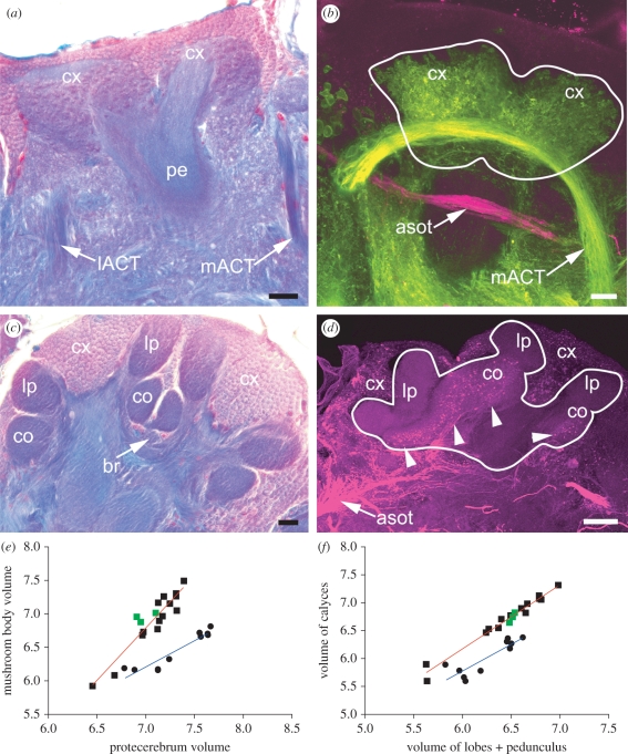
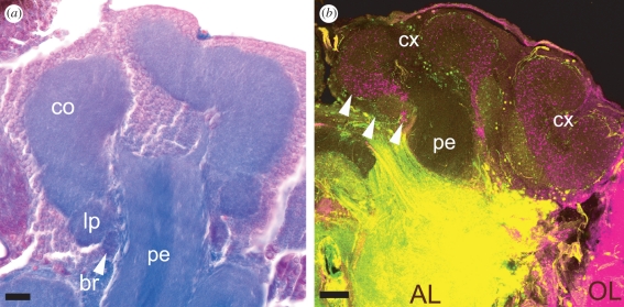
Comparisons of a phytophagous species (Dolerus sp., Tenthredinidae, Tenthredinoidea) and a euhymenopteran parasitoid species (Ophion sp., Ichneumonidae, Ichneumonoidea) reveal dramatic differences in mushroom body size, calyx morphology and afferent input (figure 3). Both the Tenthredinidae and the Ichneumonidae are composed entirely of solitary species [43]. The mushroom bodies of tenthredinids possessed simple, ovoid calyces lacking subcompartments (figure 3a). Fluorescent dextran fills of the antennal lobes and optic lobes of tenthredinids labelled antennal lobe projection neurons innervating the entire calyx, and outputs from the optic neuropils that passed ventral to the calyces without providing collaterals (figure 3b). By contrast, the mushroom bodies of ichneumonids were large and elaborate (figure 3c), with calyces partitioned into subcompartments corresponding to the lip, collar and basal ring regions of aculeate species. Dextran-labelled projection neurons from the optic lobes innervated the calyx collar, but not the lip (figure 3d).
Figure 3.
Comparisons of mushroom body morphology in phytophagous versus parasitoid Hymenoptera. (a) Mushroom bodies of the phytophagous Dolerus sp. (Tenthredinidae) showing simple, ovoid mushroom body calyces (cx) with no obvious compartmentalization into lip, collar or basal ring regions. (b) Dextran fills of the antennal lobes (green) reveal arborizations of olfactory projection neurons from the mACT throughout both calyces. Dextran fills of the optic lobes (magenta) label projection neuron axons in the anterior superior optic tract (asot), which passes beneath the mushroom bodies without providing collaterals to the calyces. (c) Mushroom bodies of the euhymenopteran parasitoid Ophion sp. (Ichneumonidae) have elaborate, deeply invaginated calyces divided into subcompartments like those observed in aculeate hymenopterans (lip (lp), collar (co) and basal ring (br), labelled in one calyx). (d) Dextran fills of the optic lobe of an ichneumonid reveal innervation of the calyx collar by optic lobe projection neurons (arrowheads). (e) Linear regression of log-transformed mushroom body volumes relative to the volume of the remaining protocerebrum in phytophagous (blue line) versus euhymenopteran (red line) species. Green points indicate measurements for the social aculeate Tetramorium caespitum (Formicidae, Vespoidea). (f) Linear regression of log-transformed calyx volumes versus lobe volumes (n = 10 for phytophagous species, n = 17 for parasitoid species). lACT, mACT; lateral and medial antennocerebral tracts. Scale bars (a–c) 20 µm; (d) 50 µm.
For all species in which brain regions were measured (electronic supplementary material, table S1), the relationship between mushroom body volume and the volume of the remaining protocerebrum (not including the mushroom bodies or optic lobes) was linear for both the phytophagous species (r2 = 0.88) and euhymenopteran species (r2 = 0.86), but the slopes of the plotted lines were significantly different (F = 15.07, p = 0.0008; figure 3e). This suggests that the allometric relationship between the mushroom bodies and the protocerebrum is different for the two groups of insects, such that mushroom body volume increases more with protocerebral volume in euhymenopterans. As a result, with the exception of the species with the smallest protocerebra (Megaspilus sp., and Stephanus serrator; indicative of small brain and thus body size; [33]), the mushroom bodies are larger relative to the protocerebrum in euhymenopterans. Despite their relatively small size, however, the mushroom bodies of both Megaspilus and Stephanus possessed invaginated and subcompartmentalized calyces typical of elaborate mushroom bodies (figure 4g). The relationship between calyx volume versus lobes + pedunculus volume was also linear in both phytophagous species (r2 = 0.76) and euhymenopteran species (r2 = 0.97), but the slopes were not significantly different (F = 1.58, p = 0.22; figure 3f). Instead, the y-intercepts for these measurements were significantly different (F = 93.83, p < 0.0001). This suggests a grade shift in the relationship between calyx volume and lobes + pedunculus volume [51,52], in which for a given volume of lobes + pedunculus, euhymenopterans have a significantly larger calyx, reflective of the expanded cup-shaped calyx morphology in these species.
Figure 4.
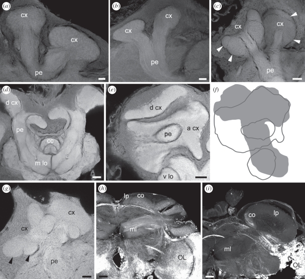
Mushroom body morphology in phytophagous and parasitoid Hymenoptera. Simple ovoid calyces (cx) without subcompartments were observed in the phytophagous (a) Tremex columba (Siricidae) and (b) Xiphydria maculata (Xiphydriidae). (c) The calyces of stem-boring sawflies such as Cephus spinipes (Cephidae) appeared to possess small subcompartments, although they were not otherwise elaborated like those of euhymenopterans (d–i). (d,e) The elaborate calyces of Orussus abietinus (Orussidae) were greatly enlarged, with one calyx oriented dorsally (d cx) and one anteriorly (a cx). (f) Outline of the Orussus mushroom bodies illustrating the great size of the calyces. (g) Subcompartments (arrowheads) in the elaborate calyces of the basal parasitoid Stephanus serrator (Stephanidae) are structurally similar to the lip and collar of the higher Euhymenoptera, suggesting that they are similarly subdivided by olfactory and visual inputs. (h) Optic lobe dextran fills reveal visual input to collar (co) subcompartments of the elaborate calyces of the parasitoid Gasteruption sp. (Gasteruptiidae, Evanoidea). Unlabelled dorsal compartments probably correspond to the lip (lp). (i) The mushroom bodies of Leucospis sp. (Leucospidae, Chalcidoidea) possess a single flask-shaped calyx that receives dextran-labelled optic lobe projection neurons to a small ventral collar (co). Again, the dorsal unfilled compartment probably represents an olfactory input-receiving lip. Medial lobe (m lo); optic lobe (OL); pedunculus (pe); vertical lobe (v lo). Scale bars (a–c,g) 20 µm; (d,e,h,i) 50 µm.
For both comparisons (mushroom body versus protocerebral volume and calyx versus lobes + pedunculus volume), three points representing measurements made for the social aculeate Tetramorium caespitum (Formicidae, Vespoidea) fell near the regression line calculated for all Euhymenoptera (green points in figure 3e,f). Removing these points and recalculating the regressions did not significantly change the slope or the intercept of the line for either comparison (r = 0.92 for mushroom bodies versus protocerebrum without Tetramorium data, F = 0.19, p = 0.67 for comparison of slopes; F = 0.55, p = 0.46 for comparison of intercepts; r = 0.97 for calyx versus lobes + pedunculus without Tetramorium data, F = 0.0007, p = 1.0 for comparison of slopes; F = 0.04, p = 0.84 for comparison of intercepts).
(b). Mushroom body morphology in phytophagous Hymenoptera
To explore the transition from small, simple mushroom bodies to large, elaborate mushroom bodies in more detail, we first examined species from among the basal phytophagous lineages (electronic supplementary material, table S1). Macroxyela ferruginea, of the basal-most superfamily Xyeloidea [41], possessed mushroom bodies with simple, ovoid calyces lacking lip- or collar-like subcompartments (data not shown). Mushroom bodies of species representing the more derived families of the phytophagous Hymenoptera [38], Tremex columba (Siricoidea, figure 4a) and Xiphydria maculata (Xiphydrioidea, figure 4b) were of similar morphology to those of Macroxyela. Species of the phytophagous Cephoidea, Cephus spinipes and Calameuta filiformis, had subcompartments in the calyces (figure 4c), although the mushroom bodies were otherwise similar in morphology to those of the other phytophagous species (not elaborate; they fell within the linear fits for the other phytophagous species for mushroom body volume versus protocerebral volume and calyx volume versus lobes + pedunculus volume, figure 3e,f). We were unable to identify the sources of afferent input to the cephid mushroom bodies from the tissue available for this study.
(c). Mushroom body morphology in parasitoid Euhymenoptera
Preparations of selected species spanning the parasitoid Euhymenoptera revealed the point of transition to large, elaborate mushroom bodies at the base of this clade. Wasps of the basal euhymenopteran superfamilies Orussoidea and Stephanoidea are ectoparasitoids of wood-boring insect larvae [38,39]. Mushroom bodies of the orussids Orussus abietinus (figure 4d–f) and Orussus occidentalis (data not shown) were dramatically expanded in size, with elaborate calyces that filled the dorsal and anterior brain (figure 4d–f). Although we could not determine with certainty whether there were subcompartments in the calyces of Orussus, they were distinctly larger when compared with those of the phytophagous hymenopterans, including the cephids. The mushroom bodies of Stephanus serrator (figure 4g) were also distinct from those of the phytophagous species, with elaborate calyces that were partitioned into subcompartments reminiscent of lip and collar regions. This suggests that the Stephanus calyces receive input from the optic lobes, although we were unable to confirm this in our alcohol-preserved specimens.
All of the remaining euhymenopteran species surveyed in this study were united with the social aculeates by possessing large, elaborate mushroom bodies with distinctly subcompartmentalized calyces (lip and collar; basal ring subcompartments were clearly identifiable only in the ichneumonids and aculeates). When fresh tissue was available, dextran fills to the optic lobes confirmed that the collar-like subcompartments of the calyces were innervated by optic lobe projection neurons.
Optic lobe dextran fills of the parasitoid Gasteruption sp. (Gasteruptiidae, Evaniodea) revealed large, elaborate mushroom bodies with clearly subcompartmentalized calyces that received input to the collar from optic lobe projection neurons (figure 4h). Smaller subcompartments dorsal to the collar were not innervated by optic lobe neurons, and are likely to correspond to the lip regions of aculeate Hymenoptera. The brains of three other apocritan parasitoids, Orthogonalys pulchella (Trigonalidae, Trigonalioidea), Megalyra sp. (Megalyridae, Megalyroidea) and Megaspilus sp. (Megaspilidae, Ceraphronoidea) possessed similar large and elaborate mushroom body morphologies (data not shown).
Species of the superfamily Chalcidoidea (Proctotrupomorpha) display diverse life histories and are among the smallest known insects [53,54], although the family Leucospidae contains species that are of relatively larger size (10–15 mm). Leucospis sp. mushroom bodies were large and elaborate, but possessed only a single flask-shaped calyx (figure 4i). Dextran fills to the optic lobes revealed innervation of a small, layered collar subcompartment in the ventral calyx, with the uninnervated majority of the calyx neuropil presumably corresponding to the lip. A similar mushroom body morphology was observed in Ropronia sp. (Roproniidae, data not shown). It is possible that the loss of a calyx in both Ropronia and Leucospis reflect the effects of miniaturization in the Chalcidoidea, which may result in the reduction and loss of morphological elements [55]. The single calyx may also be a synapomorphy for a clade containing both the Roproniidae and Leucospidae within the monophyletic Proctotrupomorpha [43].
(d). Mushroom body morphology in a solitary aculeate
The Chrysidoidea are solitary aculeate wasps considered to be the sister group to the remaining two aculeate superfamilies Vespoidea and Apoidea [56]. Many chrysidids are cleptoparasitic, laying their eggs within the nests or burrows of other aculeate species. Chrysidid mushroom bodies were large and elaborate, with extensively convoluted calyces clearly subdivided into lip, collar and basal ring regions (figure 5a). Optic and antennal lobe dextran fills revealed that their inputs to the calyces were inverted with respect to what is typically observed (compare figure 1 with figure 5b): optic lobe input filled a massively expanded dorsal calyx compartment (magenta), while antennal lobe input targeted a far smaller ventral compartment (green). Visual projection neurons from the optic lobes also provided collaterals to the basal ring neuropil.
Figure 5.
Mushroom body calyces of a solitary aculeate wasp (Chrysididae, Chrysidoidea). (a) Chrysidids have elaborate and deeply invaginated calyces (one visible here) with a clear lip, collar and basal ring (arrow). (b) Dextran fills of the antennal lobes (AL, green) and optic lobes (OL, magenta) show segregation of inputs into the calyx lip, collar and basal ring (arrows). The position of the collar and lip are inverted relative to what is observed in aculeates so that the greatly enlarged collar lies on top of the much smaller lip. Scale bars (a) 20 µm; (b) 50 µm.
4. Discussion
Eusociality is thought to have evolved at least four times independently in the aculeate Hymenoptera [43,44]. All aculeates investigated to date, ranging from solitary to presocial to eusocial, possess large, elaborate mushroom bodies with lip, collar and basal ring subcompartments in the calyx; in species in which calyx inputs have been traced, the collar receives visual input from the optic lobes (the only exception being secondarily blind species of ants in which both the optic lobes and calyx collar have been lost) (electronic supplementary material, table S1) (present account; [31,50,57–60]). In facultatively social and eusocial aculeates, intraspecific differences in mushroom body size have been correlated with social hierarchy [61–64] and foraging and other types of behavioural experience [50,65,66]; these differences may also be seen at the cellular level in terms of Kenyon cell dendrite morphology in the calyx [67–71]. Similarly, in a facultatively social species outside of the Hymenoptera, the gregarious locust Schistocerca gregaria; gregarious phase individuals have larger mushroom body calyces than do solitarious individuals [51]. However, cross-species comparisons of solitary and social species of aculeate Hymenoptera have thus far revealed only relatively minor modifications of calyx subcompartments associated with social organization [72]. Taken together, it is clear that the evolution of sociality within the Aculeata did not drive the acquisition of elaborate mushroom bodies; it is more parsimonious to assume that the common ancestor of the aculeates already possessed elaborate mushroom bodies prior to the subsequent evolution of sociality.
Jawlowski [73] noted that ichneumonid parasitoid wasps, which are evolutionarily basal to the Aculeata, have large and elaborate mushroom bodies, although species belonging to the basal phytophagous lineages do not [74]. These findings together with the present account provide substantial evidence that the evolution of sociality did not drive the initial expansion and elaboration of the mushroom bodies in the Hymenoptera. Rather, mushroom body elaboration, marked by an increase in size and the expansion and subcompartmentalization of the calyces, occurred prior to the evolution of the Aculeata and the social groups within. Specifically, our results suggest that this event occurred at the base of the Euhymenoptera, concurrent with the acquisition of a parasitoid behavioural ecology (figure 2). The fossil record supports an early Jurassic origin for the first euhymenopteran parasitoids, while the first social aculeates are found in the early Cretaceous [43]. If parasitoidism evolved concurrently with elaborate mushroom bodies as the evidence presented here suggests, then this mushroom body morphology predated the advent of sociality by approximately 90 Myr.
What selective pressures might underlie the acquisition of elaborate mushroom bodies in parasitoid wasps? In both vertebrates and invertebrates, the capacity for flexible behaviours, such as those associated with food acquisition, is correlated with overall brain expansion [21,27,47,49,75–77]. In addition, behavioural ecologies that rely heavily on particular behaviours drive size increases in the necessary brain regions; for example, food-caching birds that employ spatial learning to remember the locations of hidden food items have larger hippocampuses [78], while pelagic sharks that pursue agile prey have larger cerebellums [79]. In insects, the acquisition of a generalist feeding ecology in scarab beetles is associated with an elaborate mushroom body morphology much like that observed in the Euhymenoptera [49], including expansion and subcompartmentalization of the calyces with the acquisition of optic lobe inputs [47]. Why might feeding generalists require larger mushroom bodies with direct visual inputs? Generalists not only need to perceive and process a wider variety of sensory cues to detect palatable food sources, but they may also need to learn and remember characteristics of food sources and their locations. As sensory integration, learning and memory centres, the mushroom bodies would play a role in these functions, and the increased size of elaborate mushroom bodies might provide additional circuitry for associating and remembering multiple sensory cues in time and space.
Many adult parasitoid wasps may be considered feeding generalists, visiting flowers to collect pollen and nectar, or collecting honeydew from homopteran insects [80]. However, the adults of most phytophagous hymenopterans have a similar feeding ecology [81], so this cannot explain the abrupt enlargement and elaboration of the mushroom bodies at the base of the Euhymenoptera. Our results imply that some novel aspect of the concurrently acquired parasitoid behavioural ecology required enhanced processing by morphologically elaborate mushroom bodies relative to what is employed by basal, non-parasitoid species. One possibility is increased demand for learning and memory capabilities employed during host location; parasitoids are adept at learning both visual and olfactory cues in this context [82–87]. In particular, spatial learning plays an important role in host location in some species. For example, the parasitoid Hyposoter horticola (Ichneumonidae) employs spatial learning to monitor previously identified hosts over a span of several days [86,87]. Similarly, Argochrysis armilla (Chrysididae; [88]) and Dasymutilla coccineohirta (Mutillidae, Vespoidea; [89]), both kleptoparasitic solitary aculeates, employ spatial learning to monitor the burrows of hosts. Species in all three families possess large, elaborate mushroom bodies (present account and [90]). Although host-finding behaviours occur only in females, males also possess elaborate mushroom bodies, as observed for many of the species of parasitoids surveyed in the present account (electronic supplementary material, table S1). In social aculeates as well, both sexes possess elaborate mushroom bodies although those of males are smaller in size [91]. This may be because mushroom body development does not differ dramatically between the sexes [92], so males may possess elaborate mushroom bodies even though their behavioural repertoire is simpler than that of females. In some parasitoid species, however, males may employ associative learning to assist in locating females [93], while males of social aculeates such as the honeybee may take repeated mating flights that require them to learn the location of the nest [94]; such behaviours may also be facilitated by elaborate mushroom bodies.
Laboratory studies in the cockroach Periplaneta americana, an insect which also possesses large, elaborate mushroom bodies that receive visual input [95], have shown that these brain centres are necessary for spatial learning [4,5]. By contrast, spatial learning in the fruitfly Drosophila melanogaster is supported by neurons in the central complex rather than the mushroom bodies [96–98], and the mushroom bodies in this species are small and do not receive visual input to the calyces [99]. Spatial learning in Drosophila also appears to be somewhat limited, as laboratory assays have demonstrated memories only for near-field visual cues that decay within minutes to hours [98,100,101]. It is possible that Drosophila does not rely extensively on spatial learning for locating food sources, mates or oviposition sites (all of which occur on fallen and rotting fruits which are likely to be clustered together in space [102]). However, for insects that must navigate among multiple distant sites for feeding and reproduction, learning the locations of these sites for repeat visits may be of importance. In such species, the processing demands of spatial learning may have promoted the evolution of larger mushroom bodies with novel circuits for processing visual information and forming associative and spatial memories. We may therefore predict that insects outside of the Hymenoptera, with different behavioural ecologies, may be expected to possess elaborate mushroom bodies if they rely heavily on associative and particularly spatial learning. For example, many lineages of flies (Diptera) are parasitoids; species in one group, the Bombyliidae, have larger mushroom bodies relative to those of non-parasitoid flies [103]. Species of Heliconius butterflies (Lepidoptera) that trapline foraging sites (repeatedly visiting learned host plant locations in a specific order) and return to a specific roost location each night also have larger mushroom bodies than do species that do not share these spatial learning-reliant behaviours [104]; the butterfly Pieris rapae possesses elaborate mushroom bodies that receive visual input, and individuals with larger mushroom bodies perform better at associating visual cues with host plants [105]. Finally, predatory species such as dragonflies that patrol their habitat from a central perch location have large mushroom bodies that receive visual input [106,107]. Thus, the preponderance of evidence suggests that insects which rely heavily upon spatial learning have acquired this particular mushroom body morphology, although a mechanistic understanding of the role of elaborate mushroom bodies in spatial learning awaits further studies.
The demand for associative and spatial learning is still present in the behavioural ecologies of modern aculeates, albeit in other contexts. Spatial learning is a necessity for the many aculeate Hymenoptera that are central place foragers, both solitary and social, in which burrows or nests containing eggs or larvae are provisioned with food through repeated foraging trips. These insects excel at learning the locations of food sources and nest sites in relation to visual landmarks [108–115]. Thus, mushroom bodies that were adapted for spatial learning in the context of host location by parasitoids may have been pre-adapted for spatial learning in the context of foraging from a central nest site as is observed in the solitary aculeates. The place-centred foraging ecology, in turn, was fertile ground for group nest construction and sociality, giving rise to social ants, bees and wasps on multiple independent occasions [116]. A similar trajectory may perhaps also be observed in the Dictyoptera, in which cockroaches possess large, elaborate mushroom bodies and gave rise to the eusocial termites, which also have large and complex mushroom bodies [117]. As sociality emerged in the Hymenoptera, enlarged mushroom bodies with the capacity for processing complex visual features may have further facilitated social interactions, such as the ability to recognize individuals using visual cues [118,119]. Future studies making use of the extensive behavioural diversity in the Hymenoptera will probably continue to reveal relationships between learning and memory, social behaviour and the evolution of mushroom body structure and function.
Acknowledgements
We thank Dr Kevin Daly and three anonymous reviewers for editorial comments that greatly improved the quality of this manuscript. This research was funded partially by National Science Foundation award no. 929572 to S.M.F.
Footnotes
One contribution to a Special Feature ‘Information processing in miniature brains’.
References
- 1.McGuire S. E., Le P. T., Davis R. L. 2001. The role of Drosophila mushroom body signaling in olfactory memory. Science 293, 1330–1333 10.1126/science.1062622 (doi:10.1126/science.1062622) [DOI] [PubMed] [Google Scholar]
- 2.Perez-Orive J., Mazor O., Turner G. C., Cassenaer S., Wilson R. I., Laurent G. 2002. Oscillations and sparsening of odor representation in the mushroom body. Science 297, 359–365 10.1126/science.1070502 (doi:10.1126/science.1070502) [DOI] [PubMed] [Google Scholar]
- 3.Blum A. L., Li W., Cressy M., Dubnau J. 2009. Short- and long-term memory in Drosophila require cAMP signaling in distinct cell types. Curr. Biol. 19, 1341–1350 10.1016/j.cub.2009.07.016 (doi:10.1016/j.cub.2009.07.016) [DOI] [PMC free article] [PubMed] [Google Scholar]
- 4.Mizunami M., Weibrecht J. M., Strausfeld N. J. 1993. A new role for the insect mushroom bodies: place memory and motor control. In Biological neural networks in invertebrate neuroethology and robotics (eds Beer R. D., Ritzman R. E., McKenna T.), pp. 199–225 New York, NY: Academic Press [Google Scholar]
- 5.Mizunami M., Weibrecht J. M., Strausfeld N. J. 1998. Mushroom bodies of the cockroach: their participation in place memory. J. Comp. Neurol. 402, 520–537 (doi:10.1002/(SICI)1096-9861(19981228)402:4<520::AID-CNE6>3.0.CO;2-K) [DOI] [PubMed] [Google Scholar]
- 6.Schildberger K. 1984. Multimodal interneurons in the cricket brain: properties of identified extrinsic mushroom body cells. J. Comp. Physiol. 154, 71–79 10.1007/BF00605392 (doi:10.1007/BF00605392) [DOI] [Google Scholar]
- 7.Li Y.-S., Strausfeld N. J. 1999. Multimodal efferent and recurrent neurons in the medial lobes of cockroach mushroom bodies. J. Comp. Neurol. 409, 647–663 (doi:10.1002/(SICI)1096-9861(19990712)409:4<647::AID-CNE9>3.0.CO;2-3) [DOI] [PubMed] [Google Scholar]
- 8.Brembs B., Wiener J. 2006. Context and occasion setting in Drosophila visual learning. Learn. Mem. 13, 618–628 10.1101/lm.318606 (doi:10.1101/lm.318606) [DOI] [PMC free article] [PubMed] [Google Scholar]
- 9.Peng Y., Xi W., Zhang K., Guo A. 2007. Experience improves feature extraction in Drosophila. J. Neurosci. 27, 5139–5145 10.1523/JNEUROSCI.0472-07.2007 (doi:10.1523/JNEUROSCI.0472-07.2007) [DOI] [PMC free article] [PubMed] [Google Scholar]
- 10.Xi W., Peng Y., Guo J., Ye Y., Zhang K., Yu F., Guo A. 2008. Mushroom bodies modulate salience-based fixation behavior in Drosophila. Eur. J. Neurosci. 27, 1441–1451 10.1111/j.1460-9568.2008.06114.x (doi:10.1111/j.1460-9568.2008.06114.x) [DOI] [PubMed] [Google Scholar]
- 11.Brembs B. 2009. Mushroom bodies regulate habit formation in Drosophila. Curr. Biol. 19, 1–5 [DOI] [PubMed] [Google Scholar]
- 12.Van Swinderen B., Brembs B. 2010. Attention-like deficit and hyperactivity in a Drosophila memory mutant. J. Neurosci. 30, 1003–1014 10.1523/JNEUROSCI.4516-09.2010 (doi:10.1523/JNEUROSCI.4516-09.2010) [DOI] [PMC free article] [PubMed] [Google Scholar]
- 13.Kenyon F. C. 1896. The brain of the bee. A preliminary contribution to the morphology of the nervous system of the Arthropoda. J. Comp. Neurol. 6, 133–210 10.1002/cne.910060302 (doi:10.1002/cne.910060302) [DOI] [Google Scholar]
- 14.Strausfeld N. J. 1976. Atlas of an insect brain. Berlin, Germany: Springer [Google Scholar]
- 15.Strausfeld N. J., Hansen L., Li Y.-S., Gomez R. S., Ito K. 1998. Evolution, discovery and interpretations of arthropod mushroom bodies. Learn. Mem. 5, 11–37 [PMC free article] [PubMed] [Google Scholar]
- 16.Farris S. M. 2005. Evolution of insect mushroom bodies: old clues, new insights. Arthropod Struct. Dev. 34, 211–234 10.1016/j.asd.2005.01.008 (doi:10.1016/j.asd.2005.01.008) [DOI] [Google Scholar]
- 17.Dujardin F. 1850. Mémoire sur le système nerveux des insectes. Ann. Sci. Natl Zool. 14, 195–206 [Google Scholar]
- 18.von Alten H. 1910. Zur Phylogenie des Hymenopterengehirns. Jenaische Zeitschrift für Naturwissenschaft 46, 511–590 [Google Scholar]
- 19.Howse P. E. 1975. Brain structure and behavior in insects. Annu. Rev. Entomol. 20, 359–379 10.1146/annurev.en.20.010175.002043 (doi:10.1146/annurev.en.20.010175.002043) [DOI] [PubMed] [Google Scholar]
- 20.Gronenberg W., Riveros A. J. 2009. Social brains and behavior: past and present. In Organization of insect societies (eds Gadau J., Fewell J.), pp. 377–401 Boston, MA: Harvard University Press [Google Scholar]
- 21.Barton R. A. 1996. Neocortex size and behavioural ecology in primates. Proc. R. Soc. Lond. B 263, 173–177 10.1098/rspb.1996.0028 (doi:10.1098/rspb.1996.0028) [DOI] [PubMed] [Google Scholar]
- 22.Burish M. J., Kueh H. Y., Wang H. 2004. Brain architecture and social complexity in modern and ancient birds. Brain Behav. Evol. 63, 107–124 10.1159/000075674 (doi:10.1159/000075674) [DOI] [PubMed] [Google Scholar]
- 23.Byrne R. W., Bates L. A. 2007. Sociality, evolution and cognition. Curr. Biol. 17, R714–R723 10.1016/j.cub.2007.05.069 (doi:10.1016/j.cub.2007.05.069) [DOI] [PubMed] [Google Scholar]
- 24.Dunbar R. I. M., Shultz S. 2007. Evolution in the social brain. Science 317, 1344–1347 10.1126/science.1145463 (doi:10.1126/science.1145463) [DOI] [PubMed] [Google Scholar]
- 25.Pérez-Barbería F. J., Shultz S., Dunbar R. I. M. 2007. Evidence for coevolution of sociality and relative brain size in three orders of mammals. Evolution 61, 2811–2821 10.1111/j.1558-5646.2007.00229.x (doi:10.1111/j.1558-5646.2007.00229.x) [DOI] [PubMed] [Google Scholar]
- 26.Schulz S., Dunbar R. I. M. 2006. Both social and ecological factors predict ungulate brain size. Proc. R. Soc. B 273, 207–215 10.1098/rspb.2005.3283 (doi:10.1098/rspb.2005.3283) [DOI] [PMC free article] [PubMed] [Google Scholar]
- 27.Schulz S., Dunbar R. I. M. 2007. The evolution of the social brain: anthropoid primates contrast with other vertebrates. Proc. R. Soc. B 274, 2429–2436 10.1098/rspb.2007.0693 (doi:10.1098/rspb.2007.0693) [DOI] [PMC free article] [PubMed] [Google Scholar]
- 28.Finarelli J. A., Flynn J. J. 2009. Brain-size evolution and sociality in Carnivora. Proc. Natl Acad. Sci. USA 106, 9345–9349 10.1073/pnas.0901780106 (doi:10.1073/pnas.0901780106) [DOI] [PMC free article] [PubMed] [Google Scholar]
- 29.MacLean E. L., Barrickman N. L., Johnson E. M., Wall C. E. 2009. Sociality, ecology, and relative brain size in lemurs. J. Hum. Evol. 56, 471–478 10.1016/j.jhevol.2008.12.005 (doi:10.1016/j.jhevol.2008.12.005) [DOI] [PubMed] [Google Scholar]
- 30.Mobbs P. G. 1982. The brain of the honeybee Apis mellifera L. The connections and spatial organization of the mushroom bodies. Phil. Trans. R. Soc. Lond. B 298, 309–354 10.1098/rstb.1982.0086 (doi:10.1098/rstb.1982.0086) [DOI] [Google Scholar]
- 31.Gronenberg W. 2001. Subdivisions of hymenopteran mushroom body calyces by their afferent supply. J. Comp. Neurol. 436, 474–489 [DOI] [PubMed] [Google Scholar]
- 32.Strausfeld N. J. 2002. Organization of the honey bee mushroom body: representation of the calyx within the vertical and gamma lobes. J. Comp. Neurol. 450, 4–33 10.1002/cne.10285 (doi:10.1002/cne.10285) [DOI] [PubMed] [Google Scholar]
- 33.Mares S., Ash L., Gronenberg W. 2005. Brain allometry in bumblebee and honey bee workers. Brain Behav. Evol. 66, 50–61 10.1159/000085047 (doi:10.1159/000085047) [DOI] [PubMed] [Google Scholar]
- 34.Schröter U., Menzel R. 2003. A new ascending sensory tract to the calyces of the honeybee mushroom body, the subesophageal-calycal tract. J. Comp. Neurol. 465, 168–178 10.1002/cne.10843 (doi:10.1002/cne.10843) [DOI] [PubMed] [Google Scholar]
- 35.Farris S. M. 2008. Tritocerebral tract input to the insect mushroom bodies. Arthropod Struct. Dev. 37, 492–503 10.1016/j.asd.2008.05.005 (doi:10.1016/j.asd.2008.05.005) [DOI] [PubMed] [Google Scholar]
- 36.Paulk A. C., Gronenberg W. 2008. Higher order visual input to the mushroom bodies in the bee, Bombus impatiens. Arthropod Struct. Dev. 37, 443–458 10.1016/j.asd.2008.03.002 (doi:10.1016/j.asd.2008.03.002) [DOI] [PMC free article] [PubMed] [Google Scholar]
- 37.Ehmer B., Gronenberg W. 2002. Segregation of visual input to the mushroom bodies in the honeybee (Apis mellifera). J. Comp. Neurol. 451, 362–373 10.1002/cne.10355 (doi:10.1002/cne.10355) [DOI] [PubMed] [Google Scholar]
- 38.Whitfield J. B. 2003. Phylogenetic insights into the evolution of parasitism in Hymenoptera. Adv. Parasitol. 54, 69–100 10.1016/S0065-308X(03)54002-7 (doi:10.1016/S0065-308X(03)54002-7) [DOI] [PubMed] [Google Scholar]
- 39.Whitfield J. B. 1998. Phylogeny and evolution of host-parasitoid interactions in Hymenoptera. Annu. Rev. Entomol. 43, 129–151 10.1146/annurev.ento.43.1.129 (doi:10.1146/annurev.ento.43.1.129) [DOI] [PubMed] [Google Scholar]
- 40.Vilhelmsen L., Isidoro N., Romani R., Basibuyuk H. H., Quicke D. L. J. 2001. Host location and oviposition in a basal group of parasitic wasps: the subgenual organ, ovipositor apparatus and associated structures in the Orussidae (Hymenoptera, Insecta). Zoomorphology 121, 63–84 10.1007/s004350100046 (doi:10.1007/s004350100046) [DOI] [Google Scholar]
- 41.Schulmeister S. 2003. Simultaneous analysis of basal Hymenoptera (Insecta): introducing robust-choice sensitivity analysis. Biol. J. Linn. Soc. 79, 245–275 10.1046/j.1095-8312.2003.00233.x (doi:10.1046/j.1095-8312.2003.00233.x) [DOI] [Google Scholar]
- 42.Schulmeister S. 2003. Review of morphological evidence on the phylogeny of basal Hymenoptera (Insecta), with a discussion of the ordering of characters. Biol. J. Linn. Soc. 79, 209–243 10.1046/j.1095-8312.2003.00232.x (doi:10.1046/j.1095-8312.2003.00232.x) [DOI] [Google Scholar]
- 43.Grimaldi D. A., Engel M. S. 2005. Evolution of the insects. New York, NY: Cambridge University Press [Google Scholar]
- 44.Hunt J. H. 1999. Trait mapping and salience in the evolution of eusocial vespid wasps. Evolution 53, 225–237 10.2307/2640935 (doi:10.2307/2640935) [DOI] [PubMed] [Google Scholar]
- 45.O'Shea M., Adams M. 1981. Pentapeptide (proctolin) associated with an identified neuron. Science 213, 567–569 10.1126/science.6113690 (doi:10.1126/science.6113690) [DOI] [PubMed] [Google Scholar]
- 46.Kiernan J. 1990. Histological and histochemical methods: theory and practice, 2nd edn. Oxford, UK: Pergamon Press [Google Scholar]
- 47.Farris S. M. 2008. Structural, functional and developmental convergence of the insect mushroom bodies with higher brain centers of vertebrates. Brain Behav. Evol. 72, 1–15 10.1159/000139457 (doi:10.1159/000139457) [DOI] [PubMed] [Google Scholar]
- 48.Skoulakis E. M. C., Kalderon D., Davis R. L. 1993. Preferential expression in mushroom bodies of the catalytic subunit of protein kinase A and its role in learning and memory. Neuron 11, 197–208 10.1016/0896-6273(93)90178-T (doi:10.1016/0896-6273(93)90178-T) [DOI] [PubMed] [Google Scholar]
- 49.Farris S. M., Roberts N. S. 2005. Coevolution of generalist feeding ecologies and gyrencephalic mushroom bodies in insects. Proc. Natl Acad. Sci. USA 102, 17 394–17 399 10.1073/pnas.0508430102 (doi:10.1073/pnas.0508430102) [DOI] [PMC free article] [PubMed] [Google Scholar]
- 50.Withers G. S., Day N. F., Talbot E. F., Dobson H. E. M., Wallace C. S. 2008. Experience-dependent plasticity in the mushroom bodies of the solitary bee Osmia lignaria (Megachilidae). Dev. Neurobiol. 68, 73–82 10.1002/dneu.20574 (doi:10.1002/dneu.20574) [DOI] [PubMed] [Google Scholar]
- 51.Ott S. R., Rogers S. M. 2010. Gregarious phase locusts have substantially larger brains with altered proportions compared with the solitarious phase. Proc. R. Sci. B 277, 3087–3096 10.1098/rspb.2010.0694 (doi:10.1098/rspb.2010.0694) [DOI] [PMC free article] [PubMed] [Google Scholar]
- 52.Striedter G. F. 2005. Principles of brain evolution. Sunderland, MA: Sinauer Associates Inc [Google Scholar]
- 53.Gibson G. A. P., Herarty J. M., Woolley J. B. 1999. Phylogenetics and classification of Chalcidoidea and Mymarommatoidea: a review of current concepts (Hymenoptera, Apocrita). Zool. Scripta 28, 87–124 10.1046/j.1463-6409.1999.00016.x (doi:10.1046/j.1463-6409.1999.00016.x) [DOI] [Google Scholar]
- 54.Triplehorn C. A., Johnson N. F. 2005. Borror and DeLong's introduction to the study of insects, 7th edn. Belmont, CA: Thomson Brooks/Cole [Google Scholar]
- 55.Yeh J. 2002. The effect of miniaturized body size on skeletal morphology in frogs. Evolution 56, 628–641 [DOI] [PubMed] [Google Scholar]
- 56.Brothers D. J., Carpenter J. M. 1993. Phylogeny of the Aculeata: Chrysidoidea and Vespoidea (Hymenoptera). J. Hymenoptera Res. 2, 227–304 [Google Scholar]
- 57.Jawlowski H. 1959. The structure of corpora pedunculata in Aculeata (Hymenoptera). Folia Biol. 7, 61–70 [Google Scholar]
- 58.Gronenberg W., Hölldobler B. 1999. Morphologic representation of visual and antennal information in the ant brain. J. Comp. Neurol. 412, 229–240 (doi:10.1002/(SICI)1096-9861(19990920)412:2<229::AID-CNE4>3.0.CO;2-E) [DOI] [PubMed] [Google Scholar]
- 59.Ehmer B., Hoy R. R. 2000. Mushroom bodies of vespid wasps. J. Comp. Neurol. 416, 93–100 (doi:10.1002/(SICI)1096-9861(20000103)416:1<93::AID-CNE7>3.0.CO;2-F) [DOI] [PubMed] [Google Scholar]
- 60.Lòpez-Riquelme G. O., Gronenberg W. 2004. Multisensory convergence in the mushroom bodies of ants and bees. Acta. Biol. Hung. 55, 31–37 [DOI] [PubMed] [Google Scholar]
- 61.Molina Y., O'Donnell S. 2007. Mushroom body volume is related to social aggression and ovary development in the paper wasp Polistes instabilis. Brain Behav. Evol. 70, 137–144 10.1159/000102975 (doi:10.1159/000102975) [DOI] [PubMed] [Google Scholar]
- 62.O'Donnell S., Donlan N., Jones T. 2007. Developmental and dominance-associated differences in mushroom body structure in the paper wasp Misocyttarus mastigophorus. Dev. Neurobiol. 67, 39–46 10.1002/dneu.20324 (doi:10.1002/dneu.20324) [DOI] [PubMed] [Google Scholar]
- 63.Molina Y., O'Donnell S. 2008. Age, sex, and dominance-related mushroom body plasticity in the paperwasp Mischocyttarus mastigophorus. Dev. Neurobiol. 68, 950–959 10.1002/dneu.20633 (doi:10.1002/dneu.20633) [DOI] [PubMed] [Google Scholar]
- 64.Smith A. R., Seid M. A., Jiménez L. C., Wcislo W. T. 2010. Socially induced brain development in a facultatively eusocial sweat bee Megalopta genalis (Halictidae). Proc. R. Soc. B 277, 2157–2163 10.1098/rspb.2010.0269 (doi:10.1098/rspb.2010.0269) [DOI] [PMC free article] [PubMed] [Google Scholar]
- 65.Withers G. S., Fahrbach S. E., Robinson G. E. 1993. Selective neuroanatomical plasticity and division of labour in the honeybee. Nature 364, 238–240 10.1038/364238a0 (doi:10.1038/364238a0) [DOI] [PubMed] [Google Scholar]
- 66.Durst C., Eichmuller S., Menzel R. 1994. Development and experience lead to increased volume of subcompartments of the honeybee mushroom body. Behav. Neural Biol. 62, 259–263 10.1016/S0163-1047(05)80025-1 (doi:10.1016/S0163-1047(05)80025-1) [DOI] [PubMed] [Google Scholar]
- 67.Groh C., Rössler W. 2008. Caste-specific postembryonic development of primary and secondary olfactory centers in the female honeybee brain. Arthropod Struct. Dev. 37, 459–468 10.1016/j.asd.2008.04.001 (doi:10.1016/j.asd.2008.04.001) [DOI] [PubMed] [Google Scholar]
- 68.Krofczik S., Khojasteh U., Hempel de Ibarra N., Menzel R. 2008. Adaptation of microglomerular complexes in the honeybee mushroom body lip to manipulations of behavioral maturation and sensory experience. Dev. Neurobiol. 68, 1007–1017 10.1002/dneu.20640 (doi:10.1002/dneu.20640) [DOI] [PubMed] [Google Scholar]
- 69.Seid M. A., Wehner R. 2009. Delayed axonal pruning in the ant brain: a study of developmental trajectories. Dev. Neurobiol. 69, 350–364 10.1002/dneu.20709 (doi:10.1002/dneu.20709) [DOI] [PubMed] [Google Scholar]
- 70.Hourcade B., Muenz T. S., Sandoz J.-C., Rössler W., Devaud M. 2010. Long-term synaptic reorganization in the mushroom bodies: a memory trace in the insect brain? J. Neurosci. 30, 6461–6465 10.1523/JNEUROSCI.0841-10.2010 (doi:10.1523/JNEUROSCI.0841-10.2010) [DOI] [PMC free article] [PubMed] [Google Scholar]
- 71.Stieb S. M., Muenz T. S., Wehner R., Rössler W. 2010. Visual experience and age affect synaptic organization in the mushroom bodies of the desert ant Cataglyphis fortis. Dev. Neurobiol. 70, 408–423 10.1002/dneu.20785 (doi:10.1002/dneu.20785) [DOI] [PubMed] [Google Scholar]
- 72.Molina Y., Harris R. M., O'Donnell S. 2009. Brain organization mirrors caste differences, colony founding and nest architecture in paper wasps (Hymenoptera: Vespidae). Proc. R. Soc. B 276, 3345–3351 10.1098/rspb.2009.0817 (doi:10.1098/rspb.2009.0817) [DOI] [PMC free article] [PubMed] [Google Scholar]
- 73.Jawlowski H. 1959. On the brain structure of the Ichneumonidae. Bulletin de l'Academie Polonaise des Sciences Cl. II. Serie des Sciences Biologiques 8, 123–125 [Google Scholar]
- 74.Jawlowski H. 1960. On the brain structure of the Symphyta (Hymenoptera). Bulletin de l'Academie Polonaise des Sciences. Serie des Sciences Biologiques 8, 265–268 [Google Scholar]
- 75.Lefebvre L., Reader S. M., Sol D. 2004. Brains, innovations and evolution in birds and primates. Brain Behav. Evol. 63, 233–246 10.1159/000076784 (doi:10.1159/000076784) [DOI] [PubMed] [Google Scholar]
- 76.Sol D., Duncan R. P., Blackburn T. M., Cassey P., Lefebvre L. 2005. Big brains, enhanced cognition, and response of birds to novel environments. Proc. Natl Acad. Sci. USA 102, 5460–5465 10.1073/pnas.0408145102 (doi:10.1073/pnas.0408145102) [DOI] [PMC free article] [PubMed] [Google Scholar]
- 77.Sol D., Lefebvre L., Rodriguez-Teijeiro J. D. 2005. Brain size, innovative propensity and migratory behavior in temperate Palearctic birds. Proc. R. Soc. B 272, 1433–1441 10.1098/rspb.2005.3099 (doi:10.1098/rspb.2005.3099) [DOI] [PMC free article] [PubMed] [Google Scholar]
- 78.Lucas J. R., Brodin A., de Kort S. R., Clayton N. S. 2004. Does hippocampal size correlate with the degree of caching specialization? Proc. R. Soc. Lond. B 271, 2423–2429 10.1098/rspb.2004.2912 (doi:10.1098/rspb.2004.2912) [DOI] [PMC free article] [PubMed] [Google Scholar]
- 79.Yopak K. E., Lisney T. J., Collin S. P., Montgomery J. C. 2007. Variation in brain organization and cerebellar foliation in chondrichthyans: sharks and holocephalans. Brain Behav. Evol. 69, 280–300 10.1159/000100037 (doi:10.1159/000100037) [DOI] [PubMed] [Google Scholar]
- 80.Jervis M. A., Kidd N. A. C., Walton M. 1992. A review of methods for determining dietary range in adult parasitoids. Entomophaga 37, 565–574 10.1007/BF02372326 (doi:10.1007/BF02372326) [DOI] [Google Scholar]
- 81.Willemstein S. C. 1987. An evolutionary basis for pollination ecology . Leiden, The Netherlands: E.J. Brill [Google Scholar]
- 82.Papaj D. R., Vet L. E. M. 1990. Odor learning and foraging success in the parasitoid Leptopilina heterotoma. J. Chem. Ecol. 16, 3137–3150 10.1007/BF00979616 (doi:10.1007/BF00979616) [DOI] [PubMed] [Google Scholar]
- 83.Sheehan W., Wäckers F. L., Lewis W. J. 1993. Discrimination of previously searched, host-free sites by Microplitis croceipes (Hymenoptera: Braconidae). J. Insect Behav. 6, 323–331 10.1007/BF01048113 (doi:10.1007/BF01048113) [DOI] [Google Scholar]
- 84.Turlings T. C. J., Wäckers R. L., Vet L. E. M., Lewis W. J., Tumlinson J. H. 1993. Learning of host-finding cues by hymenopterous parasitoids. In Insect learning: ecological and evolutionary perspectives (eds Papaj D. R., Lewis A.), pp. 51–78 New York, NY: Chapman & Hall [Google Scholar]
- 85.Steidle J. L. M. 1998. Learning pays off: influence of experience on host finding and parasitism in Lariophagus distinguendus. Ecol. Entomol. 23, 451–456 10.1046/j.1365-2311.1998.00144.x (doi:10.1046/j.1365-2311.1998.00144.x) [DOI] [Google Scholar]
- 86.Van Nouhuys S., Ehrnsten J. 2004. Wasp behavior leads to uniform parasitism of a host available for only a few hours per year. Behav. Ecol. 15, 661–665 10.1093/beheco/arh059 (doi:10.1093/beheco/arh059) [DOI] [Google Scholar]
- 87.Van Nouhuys S., Kaartinen R. 2008. A parasitoid wasp uses landmarks while monitoring potential resources. Proc. R. Soc. B 275, 377–385 10.1098/rspb.2007.1446 (doi:10.1098/rspb.2007.1446) [DOI] [PMC free article] [PubMed] [Google Scholar]
- 88.Rosenheim J. A. 1987. Host location and exploitation by the cleptoparasitic wasp Argochrysis armilla: the role of learning (Hymenoptera: Chrysididae). Behav. Ecol. Sociobiol. 21, 401–406 10.1007/BF00299935 (doi:10.1007/BF00299935) [DOI] [Google Scholar]
- 89.VanderSal N. D. 2008. Rapid spatial learning in a velvet ant (Dasymutilla coccineohirta). Anim. Cogn. 11, 563–567 10.1007/s10071-008-0145-4 (doi:10.1007/s10071-008-0145-4) [DOI] [PubMed] [Google Scholar]
- 90.Strausfeld N. J. 2001. Insect brain. In Brain, evolution and cognition (eds Roth G., Wulliman M. F.), pp. 367–400 New York, NY: John Wiley and Sons [Google Scholar]
- 91.Ehmer B., Gronenberg W. 2004. Mushroom body volumes and visual interneurons in ants: comparison between sexes and castes. J. Comp. Neurol. 469, 198–213 10.1002/cne.11014 (doi:10.1002/cne.11014) [DOI] [PubMed] [Google Scholar]
- 92.Roat T. C., da Cruz Landim C. 2008. Temporal and morphological differences in post-embryonic differentiation of the mushroom bodies in the brain of workers, queens, and drones of Apis mellifera (Hymenoptera, Apidae). Micron 39, 1171–1178 10.1016/j.micron.2008.05.004 (doi:10.1016/j.micron.2008.05.004) [DOI] [PubMed] [Google Scholar]
- 93.Godfray H. C. J. 1994. Parasitoids: behavioral and evolutionary ecology. Princeton, NJ: Princeton University Press [Google Scholar]
- 94.Fahrboch S. E., Giray T., Farris S. M., Robinson G. E. 1997. Expansion of the neuropil of the mushroom bodies in male honey bees is coincident with initiation of flight. Neurosci. Lett. 236, 135–138 10.1016/S0304-3940(97)00772-6 (doi:10.1016/S0304-3940(97)00772-6) [DOI] [PubMed] [Google Scholar]
- 95.Li Y.-S., Strausfeld N. J. 1997. Morphology and sensory modality of mushroom body extrinsic neurons in the brain of the cockroach, Periplaneta americana. J. Comp. Neurol. 387, 631–650 (doi:10.1002/(SICI)1096-9861(19971103)387:4<631::AID-CNE9>3.0.CO;2-3) [DOI] [PubMed] [Google Scholar]
- 96.Wolf R., Wittig T., Liu L., Wustmann G., Eyding D., Heisenberg M. 1998. Drosophila mushroom bodies are dispensable for visual, tactile, and motor learning. Learn. Mem. 5, 166–178 [PMC free article] [PubMed] [Google Scholar]
- 97.Liu G., Seiler H., Wen A., Zars T., Ito K., Wolf R., Heisenberg M., Liu L. 2006. Distinct memory traces for two visual features in the Drosophila brain. Nature 439, 551–556 10.1038/nature04381 (doi:10.1038/nature04381) [DOI] [PubMed] [Google Scholar]
- 98.Neuser K., Triphan T., Mronz M., Poeck B., Strauss R. 2008. Analysis of a spatial orientation memory in Drosophila. Nature 453, 1244–1247 10.1038/nature07003 (doi:10.1038/nature07003) [DOI] [PubMed] [Google Scholar]
- 99.Strausfeld N. J., Sinakevitch I., Vilinsky I. 2003. The mushroom bodies of Drosophila melanogaster: an immunocytological and Golgi study of Kenyon cell organization in the calyces and lobes. Microsc. Res. Tech. 62, 151–169 10.1002/jemt.10368 (doi:10.1002/jemt.10368) [DOI] [PubMed] [Google Scholar]
- 100.Putz G., Heisenberg M. 2002. Memories in Drosophila heat-box learning. Learn. Mem. 9, 349–359 10.1101/lm.50402 (doi:10.1101/lm.50402) [DOI] [PMC free article] [PubMed] [Google Scholar]
- 101.Zars T. 2009. Spatial orientation in Drosophila. J. Neurogenet. 23, 104–110 10.1080/01677060802441364 (doi:10.1080/01677060802441364) [DOI] [PubMed] [Google Scholar]
- 102.Markow T. A. 1988. Reproductive behavior of Drosophila melanogaster and D. nigrospiracula in the field and in the laboratory. J. Comp. Psychol. 102, 169–173 10.1037/0735-7036.102.2.169 (doi:10.1037/0735-7036.102.2.169) [DOI] [PubMed] [Google Scholar]
- 103.Panov A. A. 2009. General structure of the mushroom body calyx in Brachycera Orthorrapha flies (Diptera). Biol. Bull. 36, 267–276 [PubMed] [Google Scholar]
- 104.Sivinski J. 1989. Mushroom body development in nymphalid butterflies: a correlate of learning? J. Insect. Behav. 2, 277–283 10.1007/BF01053299 (doi:10.1007/BF01053299) [DOI] [Google Scholar]
- 105.Snell-Rood E. C., Papaj D. R., Gronenberg W. 2009. Brain size: a global or induced cost of learning? Brain. Behav. Evol. 72, 111–128 [DOI] [PubMed] [Google Scholar]
- 106.Svidersky V. L., Plotnikova S. I. 2004. On structural–functional organization of dragonfly mushroom bodies and some general considerations about purpose of these formations. J. Evol. Biochem. Physiol. 40, 608–624 10.1007/s10893-005-0018-2 (doi:10.1007/s10893-005-0018-2) [DOI] [Google Scholar]
- 107.Strausfeld N. J., Sinakevitch I., Brown S. M., Farris S. M. 2009. Ground plan of the insect mushroom body: functional and evolutionary implications. J. Comp. Neurol. 513, 265–291 10.1002/cne.21948 (doi:10.1002/cne.21948) [DOI] [PMC free article] [PubMed] [Google Scholar]
- 108.Tinbergen N. 1972. On the orientation of the digger wasp Philanthus trangulum Fabr. I. In The animal in its world, vol. 1 (ed. Tinbergen N.), pp. 103–127 Cambridge, MA: Harvard University Press [Google Scholar]
- 109.Capaldi E. A., Dyer F. C. 1999. The role of orientation flights on homing performance in honeybees. J. Exp. Biol. 202, 1655–1666 [DOI] [PubMed] [Google Scholar]
- 110.Zhang S., Lehrer M., Srinivasan M. V. 1999. Honeybee memory: navigation by associative grouping and recall of visual stimuli. Neurobiol. Learn. Mem. 72, 180–201 10.1006/nlme.1998.3901 (doi:10.1006/nlme.1998.3901) [DOI] [PubMed] [Google Scholar]
- 111.Collett T. S., Collett M., Wehner R. 2001. The guidance of desert ants by extended landmarks. J. Exp. Biol. 204, 1635–1639 [DOI] [PubMed] [Google Scholar]
- 112.Fukushi T., Wehner R. 2004. Navigation in wood ants Formica japonica: context dependent use of landmarks. J. Exp. Biol. 207, 3431–3439 10.1242/jeb.01159 (doi:10.1242/jeb.01159) [DOI] [PubMed] [Google Scholar]
- 113.Warrant E. J., Kelber A., Gislén A., Greiner B., Ribi W., Wcislo W. T. 2004. Nocturnal vision and landmark orientation in a tropical halictid bee. Curr. Biol. 14, 1309–1318 10.1016/j.cub.2004.07.057 (doi:10.1016/j.cub.2004.07.057) [DOI] [PubMed] [Google Scholar]
- 114.Menzel R., et al. 2005. Honey bees navigate according to a map-like spatial memory. Proc. Natl Acad. Sci. USA 102, 3040–3045 10.1073/pnas.0408550102 (doi:10.1073/pnas.0408550102) [DOI] [PMC free article] [PubMed] [Google Scholar]
- 115.Saleh N., Chittka L. 2007. Traplining in bumblebees (Bombus impatiens): a foraging strategy's ontogeny and the importance of spatial reference memory in short-range foraging. Oecologia 151, 719–730 10.1007/s00442-006-0607-9 (doi:10.1007/s00442-006-0607-9) [DOI] [PubMed] [Google Scholar]
- 116.Wilson E. O., Hölldobler B. 2005. Eusociality: origins and consequences. Proc. Natl Acad. Sci. USA 102, 13 367–13 371 10.1073/pnas.0505858102 (doi:10.1073/pnas.0505858102) [DOI] [PMC free article] [PubMed] [Google Scholar]
- 117.Farris S. M., Strausfeld N. J. 2003. A unique mushroom body substructure common to both basal cockroaches and to termites. J. Comp. Neurol. 456, 305–320 10.1002/cne.10517 (doi:10.1002/cne.10517) [DOI] [PubMed] [Google Scholar]
- 118.Tibbetts E. A. 2002. Visual signals of individual identity in the wasp Polistes fuscatus. Proc. R. Soc. Lond. B 269, 1423–1428 10.1098/rspb.2002.2031 (doi:10.1098/rspb.2002.2031) [DOI] [PMC free article] [PubMed] [Google Scholar]
- 119.Sheehan M. J., Tibbetts E. A. 2008. Robust long-term social memories in a paper wasp. Curr. Biol. 18, 851–852 [DOI] [PubMed] [Google Scholar]
- 120.Dowton M., Austin A. D. 1994. Molecular phylogeny of the insect order Hymenoptera: apocritan relationships. Proc. Natl Acad. Sci. USA 91, 9911–9915 10.1073/pnas.91.21.9911 (doi:10.1073/pnas.91.21.9911) [DOI] [PMC free article] [PubMed] [Google Scholar]
- 121.Vilhelmsen L. 2001. Phylogeny and classification of the extant basal lineages of the Hymenoptera. Zool. J. Linn. Soc. 131, 393–442 10.1111/j.1096-3642.2001.tb01320.x (doi:10.1111/j.1096-3642.2001.tb01320.x) [DOI] [Google Scholar]


