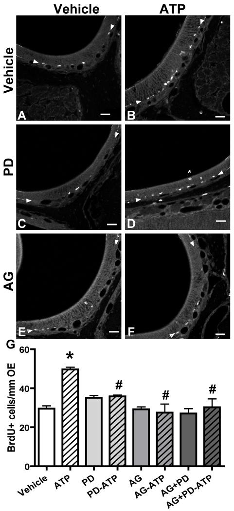
Figure 7. FGF and EGF receptors mediate ATP-induced increase in BrdU incorporation in adult mouse OE.
Mice were instilled with vehicle (A, C, E) or ATP (B, D, F) followed by vehicle (A, B) FGF receptor inhibitor PD173074 (PD; C, D), TGFα receptor inhibitor AG1478 (AG; E, F) or both (G). Representative images of a single confocal plane depicting BrdU-IR from ectoturbinate 2 are shown. Dotted lines demark the OE. Arrowheads indicate the basal cell layer. Scale bar = 50μm. (G) Quantification of BrdU+ cells in endoturbinate II and ectoturbinate 2. * p<0.001 v. vehicle. #, p<0.001 v. ATP.

