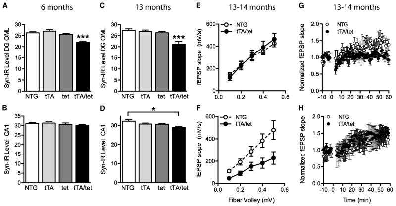
Figure 6.
Synaptic deficits in EC-APP mice. (A–D) Percent area of sections occupied by synaptophysin-immunoreactive structures in the outer molecular layer of the DG and stratum radiatum of CA1 in mice at 6 (A,B) and 13 (C,D) months of age. (A,C) At both ages, synaptophysin levels were lower in the DG of EC-APP (tTA/tet) mice than in all control groups. (B,D) Genotype affected synaptophysin levels in CA1 at 13, but not 6, months. n=6–9 mice/group. *p<0.05, ***p<0.0005 vs. all other groups, or as indicated by the bracket (Tukey post-hoc test). Values are mean ± SEM. (E–H) Electrophysiological recordings were obtained in acute hippocampal slices from 13–14-month-old EC-APP and NTG mice. The input-output relationship in EC-APP mice was normal, compared with NTG mice, at the perforant path-granule cell synapse in the DG (E; n=6–7 slices from 3 mice/genotype), but impaired at the Schaffer collateral-CA1 synapse (F; p<0.05 by 2-way repeated-measures ANOVA, n=9–10 slices from 3–5 mice/genotype). LTP was impaired at the perforant path-granule cell synapse in the DG of EC-APP mice relative to controls (G; p<0.05 by repeated-measures ANOVA from minutes 50–60, n=3–4 slices from 3 mice/genotype), but not at the Schaffer collateral-CA1 synapse (H; n=3–4 slices from 2–3 mice/genotype).

