

Abstract
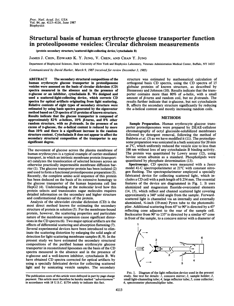
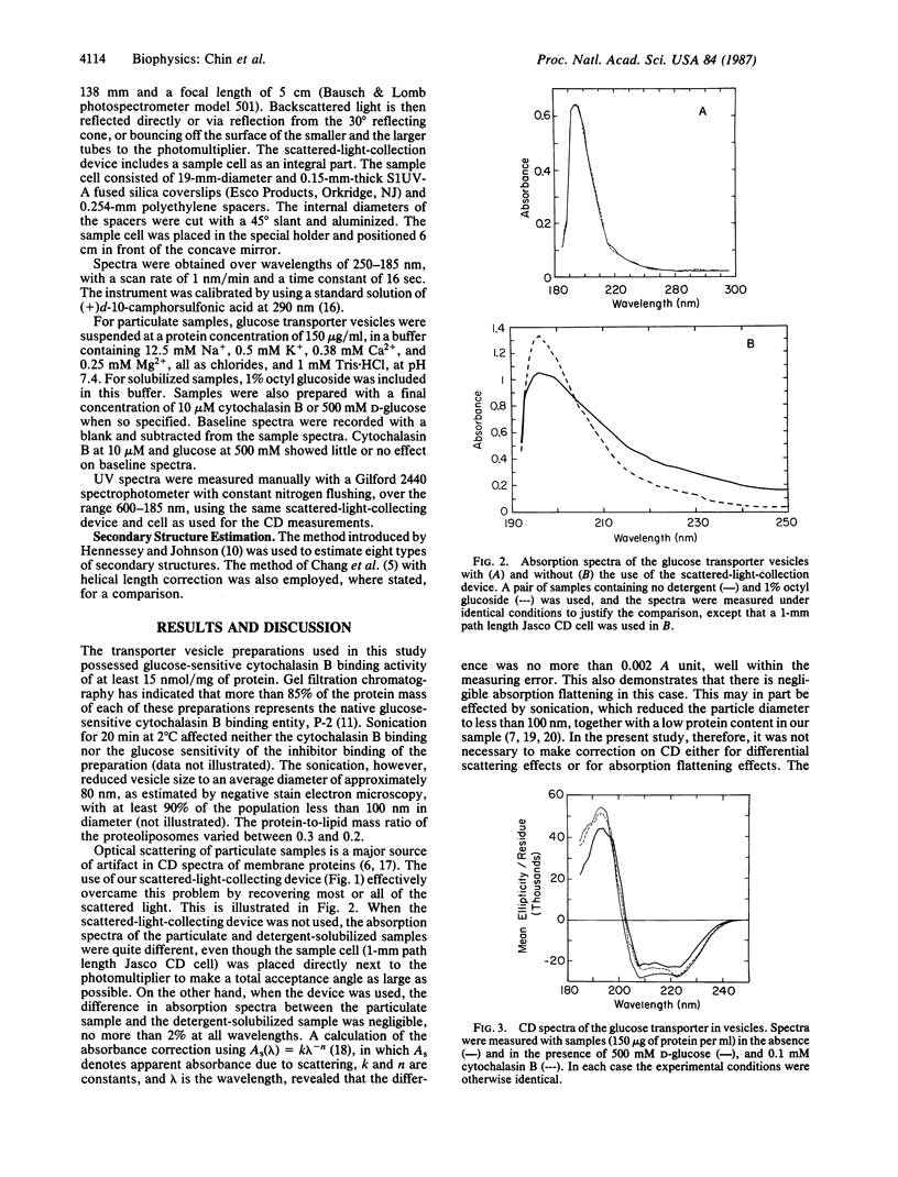
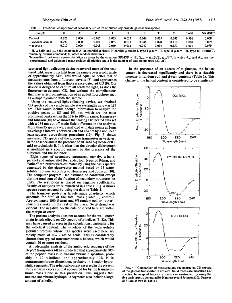
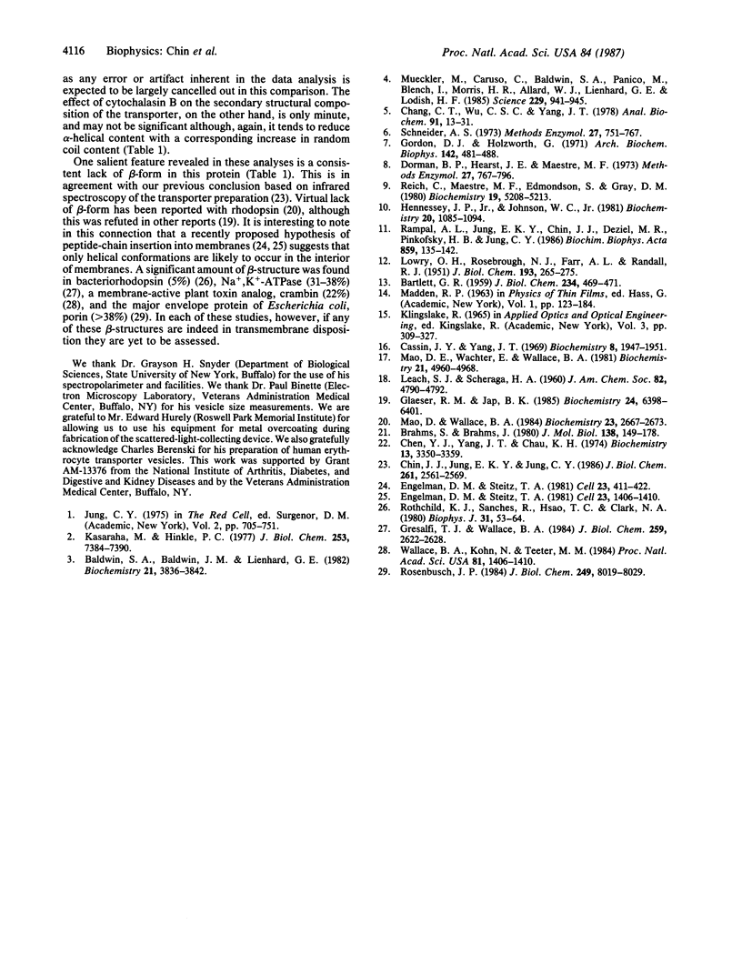
The secondary structural compositions of the human erythrocyte glucose transporter in proteoliposome vesicles were assessed on the basis of circular dichroism (CD) spectra measured in the absence and in the presence of D-glucose or an inhibitor, cytochalasin B. We designed and used a scattered-light-collecting device, which corrects CD spectra for optical artifacts originating from light scattering. Relative contents of eight types of secondary structure were estimated by using basis spectra generated by the eigenvector method based on CD spectra of 15 proteins of known structure. Results indicate that the glucose transporter is composed of approximately 82% alpha-helices, 10% beta-turns, and 8% other random structure, with no beta-strands. In the presence of an excess of D-glucose, the alpha-helical content is reduced by more than 10% and there is a significant increase in the random structure content. Cytochalasin B does not appear to affect the secondary structural composition of the transporter to any significant degree.
Full text
PDF
Selected References
These references are in PubMed. This may not be the complete list of references from this article.
- BARTLETT G. R. Colorimetric assay methods for free and phosphorylated glyceric acids. J Biol Chem. 1959 Mar;234(3):469–471. [PubMed] [Google Scholar]
- Baldwin S. A., Baldwin J. M., Lienhard G. E. Monosaccharide transporter of the human erythrocyte. Characterization of an improved preparation. Biochemistry. 1982 Aug 3;21(16):3836–3842. doi: 10.1021/bi00259a018. [DOI] [PubMed] [Google Scholar]
- Brahms S., Brahms J. Determination of protein secondary structure in solution by vacuum ultraviolet circular dichroism. J Mol Biol. 1980 Apr;138(2):149–178. doi: 10.1016/0022-2836(80)90282-x. [DOI] [PubMed] [Google Scholar]
- Cassim J. Y., Yang J. T. A computerized calibration of the circular dichrometer. Biochemistry. 1969 May;8(5):1947–1951. doi: 10.1021/bi00833a026. [DOI] [PubMed] [Google Scholar]
- Chang C. T., Wu C. S., Yang J. T. Circular dichroic analysis of protein conformation: inclusion of the beta-turns. Anal Biochem. 1978 Nov;91(1):13–31. doi: 10.1016/0003-2697(78)90812-6. [DOI] [PubMed] [Google Scholar]
- Chen Y. H., Yang J. T., Chau K. H. Determination of the helix and beta form of proteins in aqueous solution by circular dichroism. Biochemistry. 1974 Jul 30;13(16):3350–3359. doi: 10.1021/bi00713a027. [DOI] [PubMed] [Google Scholar]
- Dorman B. P., Hearst J. E., Maestre M. F. UV absorption and circular dichroism measurements on light scattering biological specimens; fluorescent cell and related large-angle light detection techniques. Methods Enzymol. 1973;27:767–96?. doi: 10.1016/s0076-6879(73)27033-7. [DOI] [PubMed] [Google Scholar]
- Engelman D. M., Steitz T. A. The spontaneous insertion of proteins into and across membranes: the helical hairpin hypothesis. Cell. 1981 Feb;23(2):411–422. doi: 10.1016/0092-8674(81)90136-7. [DOI] [PubMed] [Google Scholar]
- Glaeser R. M., Jap B. K. Absorption flattening in the circular dichroism spectra of small membrane fragments. Biochemistry. 1985 Nov 5;24(23):6398–6401. doi: 10.1021/bi00344a012. [DOI] [PubMed] [Google Scholar]
- Gordon D. J., Holzwarth G. Artifacts in the measured optic activity of membrane suspensions. Arch Biochem Biophys. 1971 Feb;142(2):481–488. doi: 10.1016/0003-9861(71)90511-x. [DOI] [PubMed] [Google Scholar]
- Gresalfi T. J., Wallace B. A. Secondary structural composition of the Na/K-ATPase E1 and E2 conformers. J Biol Chem. 1984 Feb 25;259(4):2622–2628. [PubMed] [Google Scholar]
- Hennessey J. P., Jr, Johnson W. C., Jr Information content in the circular dichroism of proteins. Biochemistry. 1981 Mar 3;20(5):1085–1094. doi: 10.1021/bi00508a007. [DOI] [PubMed] [Google Scholar]
- Kasahara M., Hinkle P. C. Reconstitution and purification of the D-glucose transporter from human erythrocytes. J Biol Chem. 1977 Oct 25;252(20):7384–7390. [PubMed] [Google Scholar]
- LOWRY O. H., ROSEBROUGH N. J., FARR A. L., RANDALL R. J. Protein measurement with the Folin phenol reagent. J Biol Chem. 1951 Nov;193(1):265–275. [PubMed] [Google Scholar]
- Mao D., Wachter E., Wallace B. A. Folding of the mitochondrial proton adenosinetriphosphatase proteolipid channel in phospholipid vesicles. Biochemistry. 1982 Sep 28;21(20):4960–4968. doi: 10.1021/bi00263a020. [DOI] [PubMed] [Google Scholar]
- Mao D., Wallace B. A. Differential light scattering and absorption flattening optical effects are minimal in the circular dichroism spectra of small unilamellar vesicles. Biochemistry. 1984 Jun 5;23(12):2667–2673. doi: 10.1021/bi00307a020. [DOI] [PubMed] [Google Scholar]
- Mueckler M., Caruso C., Baldwin S. A., Panico M., Blench I., Morris H. R., Allard W. J., Lienhard G. E., Lodish H. F. Sequence and structure of a human glucose transporter. Science. 1985 Sep 6;229(4717):941–945. doi: 10.1126/science.3839598. [DOI] [PubMed] [Google Scholar]
- Rampal A. L., Jung E. K., Chin J. J., Deziel M. R., Pinkofsky H. B., Jung C. Y. Further characterization and chemical purity assessment of the human erythrocyte glucose transporter preparation. Biochim Biophys Acta. 1986 Jul 24;859(2):135–142. doi: 10.1016/0005-2736(86)90208-7. [DOI] [PubMed] [Google Scholar]
- Reich C., Maestre M. F., Edmondson S., Gray D. M. Circular dichroism and fluorescence-detected circular dichroism of deoxyribonucleic acid and poly[d(A-C).d(G-T)] in ethanolic solutions: a new method for estimating circular intensity differential scattering. Biochemistry. 1980 Nov 11;19(23):5208–5213. doi: 10.1021/bi00564a009. [DOI] [PubMed] [Google Scholar]
- Rosenbusch J. P. Characterization of the major envelope protein from Escherichia coli. Regular arrangement on the peptidoglycan and unusual dodecyl sulfate binding. J Biol Chem. 1974 Dec 25;249(24):8019–8029. [PubMed] [Google Scholar]
- Rothschild K. J., Sanches R., Hsiao T. L., Clark N. A. A spectroscopic study of rhodopsin alpha-helix orientation. Biophys J. 1980 Jul;31(1):53–64. doi: 10.1016/S0006-3495(80)85040-5. [DOI] [PMC free article] [PubMed] [Google Scholar]
- Schneider A. S. Analysis of optical activity spectra of turbid biological suspensions. Methods Enzymol. 1973;27:751–767. doi: 10.1016/s0076-6879(73)27032-5. [DOI] [PubMed] [Google Scholar]
- Wallace B. A., Kohl N., Teeter M. M. Crambin in phospholipid vesicles: Circular dichroism analysis of crystal structure relevance. Proc Natl Acad Sci U S A. 1984 Mar;81(5):1406–1410. doi: 10.1073/pnas.81.5.1406. [DOI] [PMC free article] [PubMed] [Google Scholar]