Abstract
Assignment of the protonation state of the residue Glu-181 is important to our understanding of the primary event, activation processes and wavelength selection in rhodopsin. Despite extensive study, there is no general agreement on the protonation state of this residue in the literature. Electronic assignment is complicated by the location of Glu-181 near the nodal point in the electrostatic charge shift that accompanies excitation of the chromophore into the low-lying, strongly allowed ππ* state. Thus, the charge on this residue is effectively hidden from electronic spectroscopy. This situation is resolved in bathorhodopsin, because photoisomerization of the chromophore places Glu-181 well within the region of negative charge shift following excitation. We demonstrate that Glu-181 is negatively charged in bathorhodopsin based on the shift in the batho absorption maxima at 10K [λmax band (native)= 544±2 nm, λmax band (E181Q)= 556±3 nm] and the decrease in the λmax band oscillator strength (0.069±0.004) of E181Q relative to the native protein. Because the primary event in rhodopsin does not include a proton translocation or disruption of the hydrogen-bonding network within the binding pocket, we may conclude that the Glu-181 residue in rhodopsin is also charged.
Rhodopsin is a membrane bound photoreceptor protein responsible for scotopic (dim light) vision in humans and animals with image resolving eyes. Rhodopsin is the first G protein coupled receptor (GPCR) for which a crystal structure was obtained.1,2 The protein consists of seven transmembrane α-helices and an 11-cis retinal chromophore covalently bound via a protonated Schiff based linkage to Lys-296. The primary photochemical event generates the intermediate bathorhodopsin (batho), which is stable at low temperatures and contains an all-trans chromophore.3 Thermal decay of batho generates a series of less energetic intermediates (BSI, Lumi, Meta I and Meta II). The Meta II intermediate is responsible for activating the heterotrimeric G-protein, transducin, which in turn initiates the visual signal cascade.4,5 Because GPCRs comprise the largest protein family in the human genome, a greater understanding of the activation pathway is important to drug discovery and development.6 Elucidation of the photoactivation mechanism of rhodopsin should yield insight into the activation pathway of all class A GPCRs.
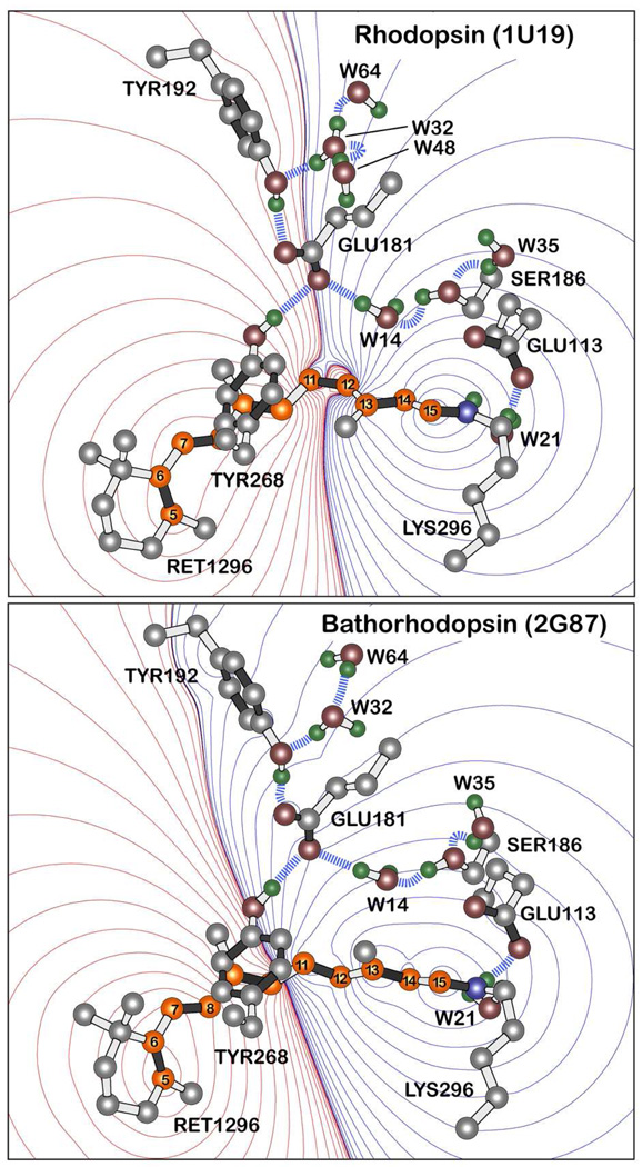
Recently, a new mechanism of rhodopsin activation has been proposed based on the observation of a counterion-switch during the photobleaching sequence.7 Subsequent studies of cone pigments indicate that a counterion switch also occurs in the blue and ultraviolet cone pigments.8,9 These studies support the concept that a counterion switch may be a generic requisite for GPCR activation.7,10 The basic elements of the counterion switch can be understood by reference to Figure 1. Glu-113, the primary counterion in the dark state, forms a water mediated salt bridge with the imine linkage of the protonated Schiff base of the 11-cis retinal chromophore. In the original counterion-switch model, Glu-181 is protonated, and a hydrogen bonding network connects this residue to Ser-186 which is in close proximity, or hydrogen bonded, to Glu-113. During the Lumi to Meta I transition, Glu-181 transfers its proton to the hydrogen bonding network which directly, or indirectly, donates the proton to Glu-113. Glu-181 now becomes the primary counterion, and creates a large electrostatic shift within the protein that plays a role in activating the protein and expelling the chromophore.
Figure 1.
Charge shifts upon excitation of the chromophore in rhodopsin (top) and bathorhodopsin (bottom) into the lowest-lying strongly allowed 1Bu+-like excited singlet states based on SAC-CISD calculations (see text). Red contours indicate regions of increased positive charge and blue contours regions of increased negative charge. Note that the carboxylate oxygen atoms of the Glu-181 residue in rhodopsin lie along the nodal line whereas in bathorhodopsin, these two atoms lie within the region of net negative charge. The contours are drawn at the following first order electrostatic energies: 0 (black), ±0.282, ±2.26, ±7.63, ±18, ±35.3, ±61, ±96.9, ±144, ±206, ±282, ±376, ±488, ±621, ±775 kJ/mol. Key hydrogen bonds are indicated with blue dashed lines, and the polyene atoms of the retinal chromophore are shown in orange and numbered following convention. The heavy atom coordinates of the binding sites were taken from the 1U192 and 2G873 crystal structures of rhodopsin and bathorhodopsin, respectively. Waters are labeled using the PDB numbers minus 2000. Only polar hydrogen atoms are shown, but all hydrogen atoms were included in the calculations and were optimized along with the chromophore by using B3LYP/6-31G(d) procedures.
The question we address in this study is whether Glu-181 is neutral or charged in the bathorhodopsin photointermediate of rhodopsin. If it is neutral, the counterion-switch mechanism as originally envisioned remains viable. If charged, a revision is required (see below). A significant number of experimental7,11–19 and theoretical20–26 studies have examined this issue, not only because of the potential role of this residue in the counterion switch, but participation of Glu-181 in wavelength regulation and photoisomerization. The previous studies on the protonation state of Glu-181 are described and tabulated in the supplementary section.
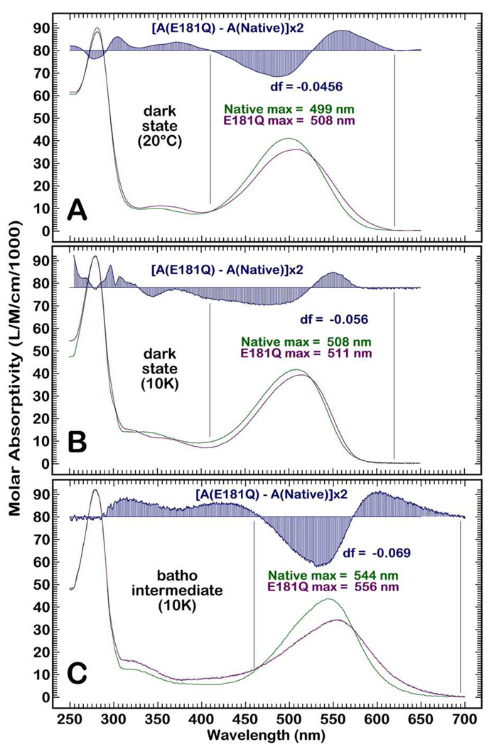
Because the Schiff base chromophore is protonated and undergoes a large charge shift upon excitation27,28, one might anticipate that electronic spectroscopy would be the most sensitive technique for protonation state assignment. In the case of rhodopsin, this assumption is not correct. As shown in Figure 1, Glu-181 is located at a nodal point in the charge shift contours associated with excitation into the low-lying strongly allowed state. The substitution of Glu-181 with Glutamine (E181Q) has a modest impact on the spectra of rhodopsin (Figures 2A & 2B), too small to provide a definitive assignment of the protonation state.22 The situation is different for the primary photoproduct, bathorhodopsin. In this photoproduct, photoisomerization has generated a distorted all-trans chromophore3 and the charge shift contours change significantly. The Glu-181 residue is now located well within the negative region of the contours and the impact on the electronic spectrum calculated with more reliability (Figure 1). Substitution of Glu-181 with Gln-181 now has a larger impact on both the absorption maximum and the oscillator strength of the λmax band of bathorhodopsin (Figure 2C). We now demonstrate that these changes are only consistent with a negatively charged Glu-181.
Figure 2.
Absorption spectra of rhodopsin at 20°C (A), at 10K (B) and the batho intermediates at 10K (C) for the native protein (green) and the E181Q mutant (purple). The difference spectra [A(E181Q) – A(native)] multiplied by two are shown in blue above the spectra. The absorption maxima are listed above the spectra, and the change in the oscillator strength of the main absorption band, df, is shown in blue, where a negative number indicates a lower oscillator strength for this band in E181Q. The regions of integration are marked by using vertical blue lines. The absorption maxima are accurate to ±1 nm and the oscillator strengths differences are accurate to ±0.005.
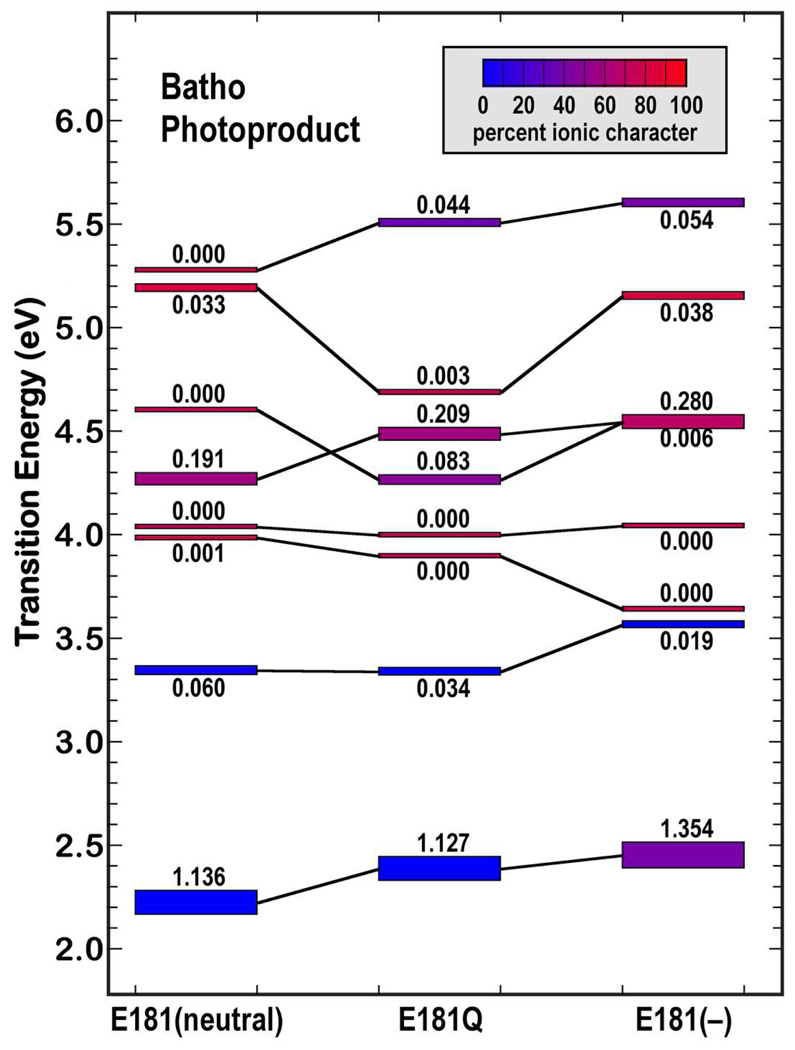
Comparison of the experimental results, shown in Fig. 2, with theoretical results provides a valuable perspective on this assignment. The theoretical methods are described in detail in the supporting information. Briefly, the B coordinates of the 2G87 crystal geometry of bathorhodopsin were selected, following the recommendations of Schreiber et al.29 The chromophore and the hydrogen atoms were minimized using B3LYP/6-31G(d) methods, while all other binding site heavy atoms were held at the crystal coordinates. The resulting chromophore geometry was nearly identical (RMS deviations less than ±0.012Å) to that generated by the DFTTB methods of Schreiber et al.29 We carried out MNDO-PSDCI and SAC-CI calculations on the chromophore binding site of bathorhodopsin. All residues within 5.6Å of the chromophore were included in the MNDO-PSDCI calculations. The results are presented in the supporting information (SI). The residues and water molecules shown in Fig. 1 were included in the SAC-CI calculations along with Glu-122, His-211 in selected calculations. The key goal of these calculations was to determine the changes in the electronic properties in the low-lying excited singlet states when the charge of Glu-181 was modified (protonated or unprotonated) or the residue replaced, as in the E181Q mutation. By including the entire hydrogen bonding network in the SAC-CI calculation, this goal is achieved. The results for the highest accuracy (level three) SAC-CI calculation are shown in Fig. 3. These calculations assumed neutral Glu-122 and His-211 in keeping with experimental observation.30,31 Other calculations explored Glu-122(−) and His-211(+), which combine to generate minor red shifts in the low-lying transitions (see SI).
Figure 3.
Level ordering of the low-lying excited singlet states of bathorhodopsin based on SAC-CI molecular orbital theory for three cases: Glu-181 neutral (left), E181Q (middle) and Glu-181 negatively charged (right). The calculations included the 190 highest energy occupied molecular orbitals and the 190 lowest energy unoccupied molecular orbitals, with single and double excitation configuration interaction based on level three (maximum CISD) selection (36,100 singles and roughly 600,000 doubles). The covalent versus ionic character of the state is indicated by the color of the state marker and varies from blue (covalent) to red (ionic) based on the scale shown at top left. The oscillator strength of the electronic transition from the ground state is written directly above or below the state marker.
Replacing Glu-181 with a Glutamine residue (E181Q) generates a red shift (12nm) in the absorption maximum of the λmax band (Fig 2). If we assume Glu-181 is charged, the calculations (Fig. 3) reproduce the red shift within 2nm (14nm). If we assume Glu-181 is neutral, the calculations predict a large blue shift in the excitation energy (−38nm). The oscillator strength of the λmax band is observed to decrease by 0.07 in going from the native batho to E181Q batho (Fig 2). The Glu-181(−) calculations predict a decrease in oscillator strength, but overestimate the magnitude by three-fold. The Glu-181(neutral) calculations underestimate the magnitude by eight-fold. Neither calculation is very successful, but the Glu-181(−) calculation is closer to experiment. More revealing, the increase in the oscillator strength of the higher energy band at ~3.3 eV (~380 nm) in E181Q is well described assuming Glu-181(−) and poorly described assuming Glu-181(neutral). In particular, the calculation on Glu-181(neutral) predicts the ~380nm band should be more intense in the native protein, which is exactly the opposite of experiment. All the calculations were carried out by using active region restricted CISD because a full CISD calculation is intractable. We carried out a series of level three calculations using filled-to-unfilled MOs of 20×20, 40×40, 80×80, 120×120, 160×160 and 190×190 (the latter is shown in Fig 3). We can extrapolate the results to a full valence CISD. The extrapolated λmax for Glu-181(−) is 557±14 nm and for Glu-181(neutral) the extrapolated value is 599±15 nm. The former range is consistent with the observed value of 544nm, whereas the latter range is more than 40 nm red shifted from the experimental value. All of the above results support Glu-181(−) and most of the results strongly argue against Glu-181(neutral) because the calculated trends are in the opposite direction of those observed. We conclude with confidence that Glu-181 is negatively charged in bathorhodopsin.
What remains is to explain why this assignment indicates that Glu-181 is also negatively charged in the dark state of rhodopsin. There is general agreement that the primary photochemical event is associated with the cis-trans photoisomerization along the 11–12 torsional coordinate of the retinal chromophore.32–34 Early picosecond studies proposed that a proton transfer from the protonated Schiff base (PSB) occurred during Batho formation.35 The notion, however, was subsequently shown to be incorrect by Raman experiments, which revealed a PSB in the structure of Batho36–38, and ultrafast time-resolved spectroscopy, which revealed little to no deuteration effect on the isomerization dynamics at room temperature39. Subsequent experimental and theoretical studies also indicate a fully protonated Schiff base in bathorhodopsin as well as little to no disruption of the hydrogen-bonding network within the binding pocket during the photoisomerization of the retinal chromophore.40–44 Thus we conclude that deprotonation of Glu-181 is not feasible during the transition to the batho photoproduct and as a result Glu-181 must also be charged in the dark-adapted form of the protein. We note further that the spectra of Fig. 2 and SAC-CI calculations on rhodopsin (Table S2) also support a negatively charged Glu-181, but with a lower level of confidence (see Supporting Information).
A negatively charged Glu-181 may provide mechanistic advantages by creating a more stable hydrogen bonding network and assuring a high pKa of the chromophore PSB in the dark state.18 A high pKa of the chromophore decreases the probability of finding deprotonated chromophores within the ensemble of rhodopsin molecules, and minimizes potential dark noise associated with dark isomerization.45 Although a negatively charged Glu-181 requires a modification of the counterion switch model, it does not force retraction. A revised model of the counterion switch mechanism, proposed by Lüdeke et al., involves the rearrangement of the hydrogen-bonding network and PSB rather than a proton transfer from Glu-181 to Glu-113. The proposed complex counterion model allows both Glu-113 and Glu-181 to be negatively charged in the dark state and serve as counterion to the PSB, with Glu-113 contributing primarily in the dark state and Glu-181 becoming the primary counterion in Meta I.18 The notion of a counterion switch involving a complex counterion is consistent with our results.18,46
In closing we note that a key conclusion of our previous two-photon study of rhodopsin was incorrect.11 Failure to consider the possibility that one of the counterions could be hidden in a null point in the charge shift field led to the incorrect conclusion that the binding site is neutral. Conversely, it is interesting to note that the external point charge model of Honig and coworkers published in 1979 turns out to be surprisingly accurate.13 The success of the latter study demonstrates the power of combining theory and chromophore analogs in the study of the electrostatic properties of protein binding sites.
Supplementary Material
Acknowledgment
This research was supported in parts by grants from the National Institutes of Health to R.R.B. (GM-34548) and B.E.K. (EY-11256 and EY-12975) and the National Science Foundation to R.R.B. (EMT-0829916). Graduate student support and low-temperature spectroscopy facilities were provided by The Harold S. Schwenk Sr. Distinguished Chair in Chemistry.
Footnotes
Supporting Information Available: A summary table of Glu-181 protonation state literature assignments as well as discussion of sample preparation, cryogenic electronic spectroscopy, computational methods, MNDO-PSDCI calculations and additional SAC-CI results. This information is available free of charge via the Internet at http://pubs.acs.org.
References
- 1.Palczewski K, Kumasaka T, Hori T, Behnke CA, Motoshima H, Fox BA, Le Trong I, Teller DC, Okada T, Stenkamp RE, Yamamoto M, Miyano M. Science. 2000;289:739–745. doi: 10.1126/science.289.5480.739. [DOI] [PubMed] [Google Scholar]
- 2.Okada T, Sugihara M, Bondar A, Elstner M, Entel P, Buss V. J. Mol. Biol. 2004;342:571–583. doi: 10.1016/j.jmb.2004.07.044. [DOI] [PubMed] [Google Scholar]
- 3.Nakamichi H, Okada T. Angew. Chem. Int. Ed. 2006;45:4270–4273. doi: 10.1002/anie.200600595. [DOI] [PubMed] [Google Scholar]
- 4.Hofmann KP, Jäger S, Ernst OP. Isr. J. Chem. 1995;35:339–355. [Google Scholar]
- 5.Fung BKK, Stryer L. Proc. Natl. Acad. Sci. USA. 1980;77:2500–2504. doi: 10.1073/pnas.77.5.2500. [DOI] [PMC free article] [PubMed] [Google Scholar]
- 6.Fanelli F, De Benedetti PG. Chem. Rev. 2005;105:3297–3351. doi: 10.1021/cr000095n. [DOI] [PubMed] [Google Scholar]
- 7.Yan ECY, Kasmi MA, Ganim Z, Hou JM, Pan D, Chang BSW, Sakmar TP, Mathies RA. Proc. Natl. Acad. Sci. USA. 2003;100:9262–9267. doi: 10.1073/pnas.1531970100. [DOI] [PMC free article] [PubMed] [Google Scholar]
- 8.Kusnetzow AK, Dukkipati A, Babu KR, Ramos L, Knox BE, Birge RR. Proc Natl Acad Sci USA. 2004;101:941–946. doi: 10.1073/pnas.0305206101. [DOI] [PMC free article] [PubMed] [Google Scholar]
- 9.Ramos LS, Chen MH, Knox BE, Birge RR. Biochemistry. 2007;46:5330–5340. doi: 10.1021/bi700138g. [DOI] [PubMed] [Google Scholar]
- 10.Birge RR, Knox BE. Proc. Natl. Acad. Sci. USA. 2003;100:9105–9107. doi: 10.1073/pnas.1733801100. [DOI] [PMC free article] [PubMed] [Google Scholar]
- 11.Birge RR, Murray LP, Pierce BM, Akita H, Balogh-Nair V, Findsen LA, Nakanishi K. Proc. Natl. Acad. Sci. USA. 1985;82:4117–4121. doi: 10.1073/pnas.82.12.4117. [DOI] [PMC free article] [PubMed] [Google Scholar]
- 12.Yan ECY, Ganim Z, Kazmi MA, Chang BSW, Sakmar TP, Mathies RA. Biochemistry. 2004;43:10867–10876. doi: 10.1021/bi0400148. [DOI] [PMC free article] [PubMed] [Google Scholar]
- 13.Honig B, Dinur U, Nakanishi K, Balogh-Nair V, Gawinowicz MA, Arnaboldi M, Motto MG. J. Am. Chem. Soc. 1979;101:7084–7086. [Google Scholar]
- 14.Mollevanger LC, Kentgens AP, Pardoen JA, Courtin JM, Veeman WS, Lugtenburg J, de Grip WJ. Eur. J. Biochem. 1987;163:9–14. doi: 10.1111/j.1432-1033.1987.tb10729.x. [DOI] [PubMed] [Google Scholar]
- 15.Smith SO, Palings I, Miley ME, Courtin JM, de Groot H, Lugtenburg J, Mathies RA, Griffin RG. Biochemistry. 1990;29:8158–8164. doi: 10.1021/bi00487a025. [DOI] [PubMed] [Google Scholar]
- 16.Han M, Smith SO. Biochemistry. 1995;34:1425–1432. doi: 10.1021/bi00004a037. [DOI] [PubMed] [Google Scholar]
- 17.Nagata T, Terakita A, Kandori H, Shichida Y, Maeda A. Biochemistry. 1998;37:17216–17222. doi: 10.1021/bi9810149. [DOI] [PubMed] [Google Scholar]
- 18.Lüdeke S, Beck M, Yan ECY, Sakmar TP, Siebert F, Vogel R. J. Mol. Biol. 2005;353:345–356. doi: 10.1016/j.jmb.2005.08.039. [DOI] [PubMed] [Google Scholar]
- 19.Lewis JW, Szundi I, Kazmi MA, Sakmar TP, Kliger DS. Biochemistry. 2004;43:12614–12621. doi: 10.1021/bi049581l. [DOI] [PubMed] [Google Scholar]
- 20.Röhrig UF, Guidoni L, Rothlisberger U. Biochemistry. 2002;41:10799–10809. doi: 10.1021/bi026011h. [DOI] [PubMed] [Google Scholar]
- 21.Martínez-Mayorga K, Pitman MC, Grossfield A, Feller SE, Brown MF. J. Am. Chem. Soc. 2006;128:16502–16503. doi: 10.1021/ja0671971. [DOI] [PubMed] [Google Scholar]
- 22.Hall KF, Vreven T, Frisch MJ, Bearpark MJ. J. Mol. Biol. 2008;383:106–121. doi: 10.1016/j.jmb.2008.08.007. [DOI] [PubMed] [Google Scholar]
- 23.Sekharan S, Buss V. J. Am. Chem. Soc. 2008;130:17220–17221. doi: 10.1021/ja805992d. [DOI] [PubMed] [Google Scholar]
- 24.Frähmcke JS, Wanko M, Phatak P, Mroginski MA, Elstner M. J. Phys. Chem. B. 2010;114:11338–11352. doi: 10.1021/jp104537w. [DOI] [PubMed] [Google Scholar]
- 25.Grossfield A, Pitman MC, Feller SE, Soubias O, Gawrisch K. J. Mol. Biol. 2008;381:478–486. doi: 10.1016/j.jmb.2008.05.036. [DOI] [PMC free article] [PubMed] [Google Scholar]
- 26.Tomasello G, Olaso-González G, Altoè P, Stenta M, Serrano-Andrés L, Merchán M, Orlandi G, Bottoni A, Garavelli M. J. Am. Chem. Soc. 2009;131:5172–5186. doi: 10.1021/ja808424b. [DOI] [PubMed] [Google Scholar]
- 27.Mathies R, Stryer L. Proc. Natl. Acad. Sci. USA. 1976;73:2169–2173. doi: 10.1073/pnas.73.7.2169. [DOI] [PMC free article] [PubMed] [Google Scholar]
- 28.Birge RR. Ann. Rev. Biophys. Bioeng. 1981;10:315–354. doi: 10.1146/annurev.bb.10.060181.001531. [DOI] [PubMed] [Google Scholar]
- 29.Schreiber M, Sugihara M, Okada T, Buss V. Angew. Chem. Int. Ed. 2006;45:4274–4277. doi: 10.1002/anie.200600585. [DOI] [PubMed] [Google Scholar]
- 30.Patel AB, Crocker E, Reeves PJ, Getmanova EV, Eilers M, Khorana HG, Smith SO. J. Mol. Biol. 2005;347:803–812. doi: 10.1016/j.jmb.2005.01.069. [DOI] [PubMed] [Google Scholar]
- 31.Fahmy K, Jäger F, Beck M, Zvyaga TA, Sakmar TP, Siebert F. Proc. Natl. Acad. Sci. USA. 1993;90:10206–10210. doi: 10.1073/pnas.90.21.10206. [DOI] [PMC free article] [PubMed] [Google Scholar]
- 32.Ottolenghi M. Adv. Photochem. 1980;12:97–200. [Google Scholar]
- 33.Birge RR, Hubbard LM. Biophys.J. 1981;34:517–534. doi: 10.1016/S0006-3495(81)84865-5. [DOI] [PMC free article] [PubMed] [Google Scholar]
- 34.Doukas AG, Junnarkar MR, Alfano RR, Callender RH, Balogh-Nair V. Biophys. J. 1985;47:795–798. doi: 10.1016/S0006-3495(85)83983-7. [DOI] [PMC free article] [PubMed] [Google Scholar]
- 35.Peters KS, Applebury ML, Rentzepis PM. Proc. Natl. Acad. Sci. USA. 1977;74:3119–3123. doi: 10.1073/pnas.74.8.3119. [DOI] [PMC free article] [PubMed] [Google Scholar]
- 36.Eyring G, Mathies RA. Proc. Natl. Acad. Sci. USA. 1979;76:33–37. doi: 10.1073/pnas.76.1.33. [DOI] [PMC free article] [PubMed] [Google Scholar]
- 37.Aton B, Doukas A, Narva D, Callender R, Dinur U, Honig B. Biophys. J. 1980;29:79–94. doi: 10.1016/S0006-3495(80)85119-8. [DOI] [PMC free article] [PubMed] [Google Scholar]
- 38.Narva D, Callender RH. Photochem. Photobiol. 1980;32:273–276. doi: 10.1111/j.1751-1097.1980.tb04021.x. [DOI] [PubMed] [Google Scholar]
- 39.Yan M, Manor D, Weng G, Chao H, Rothberg L, Jedju TM, Alfano RR, Callender RH. Proc. Natl. Acad. Sci. USA. 1991;88:9809–9812. doi: 10.1073/pnas.88.21.9809. [DOI] [PMC free article] [PubMed] [Google Scholar]
- 40.Bagley K, Balogh-Nair V, Croteau AA, Dollinger G, Ebrey TG, Eisenstein L, Hong MK, Nakanishi K, Vittitow J. Biochemistry. 1985;24:6055–6071. doi: 10.1021/bi00343a006. [DOI] [PubMed] [Google Scholar]
- 41.Deng H, Callender RH. Biochemistry. 1987;26:7418–7426. doi: 10.1021/bi00397a033. [DOI] [PubMed] [Google Scholar]
- 42.Gilson HSR, Honig BH, Croteau A, Zarrilli G, Nakanishi K. Biophys. J. 1988;53:261–269. doi: 10.1016/S0006-3495(88)83087-X. [DOI] [PMC free article] [PubMed] [Google Scholar]
- 43.Yan ECY, Epps J, Lewis JW, Szundi I, Bhagat A, Sakmar TP, Kliger DS. J. Phys. Chem. C. 2007;111:8843–8848. [Google Scholar]
- 44.Bondar A-N, Sugihara M. Rev. Chim. 2008;59:1260–1262. [Google Scholar]
- 45.Barlow RB, Birge RR, Kaplan E, Tallent JR. Nature. 1993;366:64–66. doi: 10.1038/366064a0. [DOI] [PubMed] [Google Scholar]
- 46.Standfuss J, Zaitseva E, Mahalingam M, Vogel R. J. Mol. Biol. 2008;380:145–157. doi: 10.1016/j.jmb.2008.04.055. [DOI] [PMC free article] [PubMed] [Google Scholar]
Associated Data
This section collects any data citations, data availability statements, or supplementary materials included in this article.


