Fig. 2.
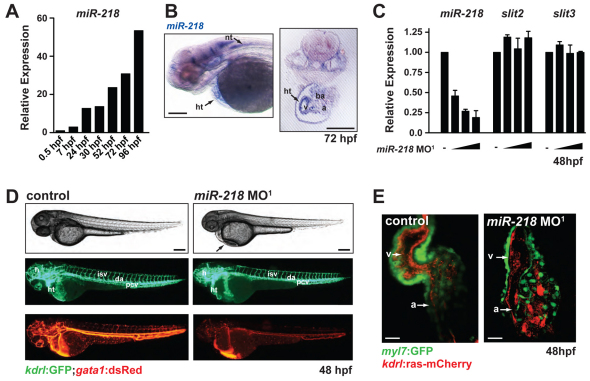
miR-218 regulates heart formation and function. (A) Expression of miR-218 (relative to 0.5 hpf timepoint) was monitored by real-time qRT-PCR during zebrafish development. (B) In situ analysis of miR-218 expression at 72 hpf reveals expression in the heart and neuronal tissue. (C) Expression of miR-218, slit2 and slit3 were quantified by real-time qRT-PCR in miR-218 morphants (2.5, 5 and 10 ng of miR-218 MO1) at 48 hpf. Data are mean + s.e.m. (D) Control and miR-218 morphants were assessed at 48 hpf by live imaging of embryos. Phase-contrast images (top) demonstrate pericardial edema in the miR-218 morphant (arrow). Fluorescent images of the same embryos show labeling of endothelial/endocardial cells in Tg(kdrl:GFP) (middle) and labeling of blood cells in Tg(gata1:dsRed) (bottom). Vascular patterning appears normal in miR-218 morphants, but circulation is reduced. (E) Cardiac morphology at 48 hpf was assessed by confocal microscopy (ventral view). Tg(myl7:GFP) expression labels the myocytes and Tg(kdrl:ras-mCherry) labels the endocardium. miR-218 morphant hearts contain myocytes and endocardium but exhibit severe morphological defects. ht, heart; nt, neural tube; a, atrium; v, ventricle; ba, branchial arches; h, head; isv, intersomitic vessel; da, dorsal aorta; pcv, posterior cardinal vein. Scale bars: 100 μm in B,D; 25 μm in E.

