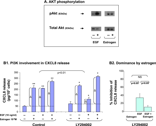
Figure 4.
The additive up-regulation of CXCL8 induced by EGF + estrogen: Involvement of PI3K. (A) AKT phosphorylation by EGF and estrogen. MCF-7 cells were stimulated by EGF (10 ng/ml, 4–7 minutes) or estrogen (10-8 M; the results are of 20 to 35 minutes of stimulation; no substantial phosphorylation was obtained at shorter or longer exposure times; time points were selected based on kinetics analyses, as described in Materials and Methods). AKT phosphorylation was determined by Western blot analysis. A representative experiment of n > 3 is presented. (B) The involvement of PI3K in EGF-, estrogen-, and EGF + estrogen-induced up-regulation of CXCL8 expression. MCF-7 cells were grown as detailed in Figure 1A. Two hours before addition of EGF, the cells were pretreated with conventional concentrations of the specific PI3K inhibitor LY294002 (20 εM) or DMSO (control = the drug's solubilizer). Then, the growth of the cells was continued in the absence or presence of the drugs (or DMSO as control). (B1) CXCL8 extracellular expression was determined in the supernatants of the cells by ELISA, and was analyzed in the linear range of absorbance. Δ, The net amount of CXCL8 added to cell supernatant on stimulation. These values were used for statistical evaluations of differences between the control group and the inhibitor-treated group. A representative experiment of n > 3 is presented. *P < .05, **P < .01 for the difference between cells stimulated by EGF/estrogen/EGF + estrogen and untreated cells. (B2) Dominance of estrogen over EGF in combined stimulation. The figure demonstrates a summary of the results of the experiments presented in B1, using LY294002. An average inhibition value (% ± SD) of n > 3 is presented.

