

Abstract
Protein thiol oxidation and modification by nitric oxide and glutathione are emerging as common mechanisms to regulate protein function and to modify protein structure. Also, thiol oxidation is a probable outcome of cellular oxidative stress and is linked to degenerative disease progression. We assessed the effect of the oxidants hypochlorous acid and chloramines on the cytoskeletal protein tubulin. Total cysteine oxidation by the oxidants was monitored by labeling tubulin with the thiol-selective reagent, 5-iodoacetamidofluorescein, by reaction with Ellman’s reagent, 5,5′dithiobis(2-nitrobenzoic acid), and by detecting interchain tubulin disulfides by Western blot under nonreducing conditions. Whereas HOCl induced both cysteine and methionine oxidation of tubulin, chloramines were predominantly cysteine oxidants. Cysteine oxidation of tubulin, rather than methionine oxidation, was associated with loss of microtubule polymerization activity and treatment of oxidized tubulin with disulfide reducing agents restored a considerable portion of the polymerization activity that was lost after oxidation. By comparing the reactivity of hypochlorous acid and chloramines with the previously characterized oxidants, peroxynitrite and the nitroxyl donor, Angeli’s salt, we have identified tubulin thiol oxidation, not methionine oxidation or tyrosine nitration, as a common outcome responsible for decreased polymerization activity.
Keywords: cysteine oxidation, hypochlorous acid, tubulin, methionine oxidation, chloramine, nitroxyl, peroxynitrite
Introduction
Extensive pathologic and cellular data has connected cumulative oxidative damage to proteins with the development of Alzheimer’s disease, Parkinson’s disease and amylotrophic lateral sclerosis.[1–4] For example, immunohistochemical studies detected high levels of nitrotyrosine and protein carbonyls, two markers of oxidative damage to proteins, in Alzheimer’s disease brain.[3, 5] The markers were further localized to neurons containing neurofibrillary tangles, a lesion characteristic of the disease. In addition to oxidative damage, cytoskeletal abnormalities have been detected in Alzheimer’s disease brain.[6] In neurons containing neurofibrillary tangles, the number of microtubules, composed of tubulin and microtubule-associated proteins, is reduced by 50%.[2] Cytoskeletal proteins like tubulin and its associated proteins are likely targets for modification by oxidants because they are among the most abundant proteins in a neuron.
Tubulin cysteine modifications including oxidation to disulfides, S-glutathionylation and S-nitrosation, have been identified in several proteomics studies using cell lines and tissue samples.[7–9] In both central nervous system and endothelial cells, tubulin S-glutathionylation, a modification in which glutathione is attached to proteins via a mixed disulfide, has been detected.[10, 11] Using brain homogenates, Jaffrey et al. identified both subunits of tubulin as targets for S-nitrosation.[12]
Our in vivo work with microtubule proteins shows that cysteine oxidation by peroxynitrite is associated with a marked decrease in microtubule polymerization activity. Tubulin, a heterodimer composed of similar 50 kDa α- and β-subunits, contains 20 reduced cysteines (12 in α-tubulin and 8 in β-tubulin).[13, 14] In neurons, tubulin constitutes 10–15% of total cellular protein and tubulin thiols could be present in sufficiently high concentrations to function as a redox buffer.[15, 16] Because some tubulin cysteine oxidation (~1–2 mol cys) by peroxynitrite is tolerated before microtubule polymerization is severely compromised, microtubule protein thiols may be capable of protecting other cellular targets from oxidation.[17, 18] This hypothesis is reinforced by our published data showing that the disulfides in tubulin and microtubule-associated proteins that are a result of peroxynitrite oxidation can be repaired by both the thioredoxin and glutaredoxin reductase systems.[17, 19]
Our current interest in the strong oxidant, hypochlorous acid (HOCl) stems from recent reports that myeloperoxidase, the enzyme that generates it from H2O2 and chloride ion, is aberrantly expressed in Alzheimer’s disease brain.[20, 21] A metabolite generated by myeloperoxidase, 3-chlorotyrosine, was elevated 3-fold in Alzheimer’s disease brain relative to control brain. Moreover, myeloperoxidase immunoreactivity co-localized with neurofibrillary tangles in neurons of Alzheimer’s disease brain.
HOCl, like peroxynitrite, oxidizes protein thiols and, if produced in neurons, will likely affect tubulin cysteines.[22, 23] Therefore, we investigated the effects of HOCl and two chloramines on the cysteines of purified porcine tubulin. Our continued focus on tubulin allows us to rank oxidants in terms of their potency and specificity for cysteine oxidation rather than other types of protein damage. Total cysteine oxidation and effects on microtubule polymerization by HOCl and chloramines as well as previously characterized oxidants, peroxynitrite and Angeli’s salt, an HNO donor, are presented.[24]
Materials and Methods
Materials
Porcine brains were obtained from Smithfield Packing Company in Smithfield, Virginia. Angeli’s salt was from Cayman Chemicals (Ann Arbor, MI). Bicinchoninic acid (BCA) protein assay reagent, West Pico chemiluminescence detection system, Tris(2-carboxyethyl)phosphine (TCEP) and 5-iodoacetomido-fluorescein (IAF) were from Thermo Pierce. The mouse anti-β-tubulin antibody (clone TUB 2.1) and the goat anti-mouse secondary antibody HRP conjugate were from Sigma. All other chemicals were from Fisher or Sigma. The concentration of HOCl was determined by measuring the absorbance at 292 nm (ε292 = 350 M−1 cm−1) in 0.1 M NaOH.[25] A solution of Angeli’s salt was prepared immediately prior to use in 0.01 M NaOH and stored on ice.
Preparation of thionitrobenzoic acid
Thionitrobenzoic acid (TNB) was prepared from 5,5′dithiobis(2-nitrobenzoic acid) (DTNB) as described with some modification.[26, 27] DTNB (0.5 g) in 25 ml 0.5M Tris–HCl, pH 8.8, was treated with 2.5 ml β-mercaptoethanol. The pH of the solution was adjusted to 1.5 with 6M HCl. Orange crystals of TNB formed after 6–8 h at 4 °C. The crystals were filtered and washed with ice cold 0.1 M HCl. The purity of TNB was 99.5% as determined by HPLC with detection at 320 nm.[27] Solid TNB was stable indefinitely at −20 °C.
Synthesis of chloramines
Glycine (GC) or taurine chloramines (TC) were prepared immediately prior to use as described [28]. HOCl in water was combined with the amines in 0.1 M phosphate buffer (PB) pH 7.4. The final molar ratio of HOCl to amine was 1:10. GC, TC and HOCl dilutions were assayed with TNB to determine concentration and stability. TNB (1 mM, 20 μl) in 0.1 M PB pH 7.4 was treated with 2 μl each oxidant to achieve final concentrations of 50, 100, 250 and 500 μM. Reaction mixtures were transferred to a 96 well plate and diluted to 200 μl with PB pH 7.4. TNB concentrations were determined from a TNB standard curve.
Synthesis of ONOO−
ONOO− was synthesized from acidified H2O2 and sodium nitrite as described and stored at −80 °C. The concentration of ONOO− was determined by measuring the absorbance at 302 nm (ε302 = 1670 M−1 cm−1) in 0.1 M NaOH.[29]
Purification of porcine brain tubulin
Tubulin was purified from porcine brain by two cycles of temperature-dependent polymerization and depolymerization and subsequent phosphocellulose chromatography as described.[30] Tubulin (typically 3.0 – 4.0 μg/μl) in PME buffer (0.1 M PIPES, pH 6.9, 1 mM MgSO4, 2 mM EGTA) containing 0.1 mM GTP was aliquoted and stored at −80 °C. Tubulin concentrations were determined by the BCA protein assay (Pierce).
Labeling of tubulin cysteines with IAF
Tubulin was diluted in either 0.1 M PB pH 7.4 or PME buffer pH 6.9 and then treated with each oxidant for 10 min at 25 °C in a total reaction volume of 20–30 μl. Either methionine (for HOCl and chloramines) or monochlorodimedone (for HOCl) was added to 1 mM to quench any remaining oxidant. IAF in DMF was added to final concentrations of 1.5–2.5 mM and samples were incubated at 37 °C for an additional 30 min. Protei ns were resolved by SDS-PAGE on 7.5% gels under reducing conditions and gel images were captured using a Kodak EDAS290 system and a UV transilluminator. The intensity of the fluorescein-labeled protein bands was measured using Kodak 1D Image Analysis software. Alternatively, IAF-labeled tubulin was precipitated with 80% ethanol, incubated on ice for 20 min and then the protein pellet was collected at 16,000 x g for 20 min. Pellets were washed twice with 80% ethanol and then resuspended in 3 M guanidine HCl in 0.1 M Tris pH 8.8. Fluorescein in each protein sample was quantitated relative to a fluorescein standard curve prepared in 3 M guanidine HCl in 0.1 M Tris pH 8.8. Absorbance at 490 nm for standards and samples was measured in a 96 well plate.
Detection of interchain disulfides by Western Blot
Following treatment with HOCl, GC or TC as described above, tubulin species (10–20 μg protein per lane) were separated by SDS-PAGE on 7.5% polyacrylamide gels under nonreducing conditions. Proteins were transferred to PVDF membranes, blocked with 3% milk for 30 min and probed with a mouse monoclonal anti-β-tubulin antibody (1:2000) for two hours. The β-tubulin/antibody complex was detected using a goat anti-mouse HRP conjugate (1 hr, 1:10,000) and the Pierce West Pico chemiluminescent substrate.
Microtubule polymerization assays
Purified tubulin, diluted with PME, was treated with 1.5–5.0 μl oxidant, for 10 min at 25 °C (50 μl, 25 μM final protein concentration). Any excess oxidant was scavenged with 1 mM methionine. For repair assays, DTT or TCEP was added for an additional 10 min at 25 °C. To induce polymerization, GTP (1 mM final) was added and the samples were incubated at 37 °C for 20–25 min. Microtubule polymer was collected by centrifugation at 16,000 × g for 20 min. The supernatants were removed and the protein concentration in each was determined by the BCA protein assay. Control polymerization activity was set at 100% for those samples containing GTP but no oxidant. Control samples without GTP were used to establish 0% activity. Samples of protein supernatants were also analyzed by SDS-PAGE with Coomassie Blue staining.
Detection of tubulin thiols with DTNB
DTNB, known as Ellman’s reagent, reacts with protein thiols to produce thionitrobenzoic acid (TNB), a yellow product with maximum absorbance at 412 nm.[31] Tubulin samples (20 μl total) were treated with oxidant for 10 min and any excess oxidant was scavenged with 1 mM methionine. Samples were transferred to a 96 well plate, diluted with 0.1 M PB pH 7.4 and treated with 500 μM DTNB (final well volume = 200 μl). In some cases, protein samples were first incubated with 6 M guanidine-HCl in 0.1 M PB pH 7.4 to unfold the protein completely. TNB was detected using a 405 nm filter within 10–15 min. Protein thiol concentrations were calculated from a GSH standard curve.
CNBr cleavage to detect methionine oxidation
Tubulin samples (12.5 μM, 250 μM cysteines, 325 μM methionines) were treated with each oxidant for up to 30 min at 25 °C. Samples were subsequently acidified to pH 2.5 with 70% formic acid and treated with 35–40 mM CNBr for 16 hours. The concentration of CNBr was at least 100-fold higher than the concentration of methionine target. Samples were neutralized to pH 7.4 – 7.6 with NH4OH and subjected to SDS-PAGE under reducing conditions on a 7.5% polyacrylamide gel. Proteins were transferred to PVDF, blocked with 3% milk and incubated with mouse anti-β-tubulin (1:2000) for 2 h. The tubulin-antibody complex was visualized using a goat anti-mouse HRP conjugate (1:10000, 1 hr) and a chemiluminescent substrate.
Detection of methionine and methionine sulfoxide by thin-layer chromatography
Methionine and methionine sulfoxide were detected by TLC on silica gel plates (5 × 10 cm). The solvent system used was acetonitrile, water, and glacial acetic acid (80:20:1). Methionine (1 mM) in 0.1 M PB pH 7.4 was treated with 5–50 equivalents of oxidant for 5–30 min. Amino acids were detected by spraying the plate with 1% ninhydrin in 90% methanol followed by heating on a hot plate. Rf values for methionine and methionine sulfoxide were 0.31 and 0.52, respectively.
Results
We routinely assess protein cysteine oxidation using 5-iodoacetamidofluorescein (IAF), a thiol-selective reagent. Figure 1A shows that IAF labeling of purified porcine tubulin decreases as the concentration of hypochlorous acid (HOCl) increases. The pKa of HOCl is 7.5; thus, both HOCl and the hypochlorite anion (OCl−) will be present for all experiments presented.[25] For consistency and simplicity, we use HOCl herein to represent both species. Likewise, because the pKa of peroxynitrous acid (ONOOH) is 6.8, a mixture of ONOOH and peroxynitrite anion (ONOO−) will be present at physiological pH. Herein, the term peroxynitrite represents a mixture of both. Though the final concentration of tubulin in Figure 1A is only 3 μM, the concentration of cysteine target in tubulin is 60 μM. The range of HOCl concentrations was 25 – 500 μM. In our experience, the exclusive products of tubulin cysteine oxidation are protein disulfides (both inter- and intra-chain); thus, for every molecule of HOCl, two cysteines are oxidized. Oxidation of tubulin cysteines by HOCl is not stoichiometric at this ratio of oxidant to protein; if so, complete oxidation and therefore, no IAF labeling would be observed at only 50 μM. In fact, 500 μM HOCl is required to completely oxidize 60 μM tubulin cysteines (Figure 1A – lane 6). Because the HOCl was present in excess, it was important to add an oxidant scavenger to achieve the reported reaction time (10 min in this case). Either methionine or monochlorodimedone was added in excess over HOCl.
Figure 1. Modification of tubulin cysteines by HOCl and chloramines.
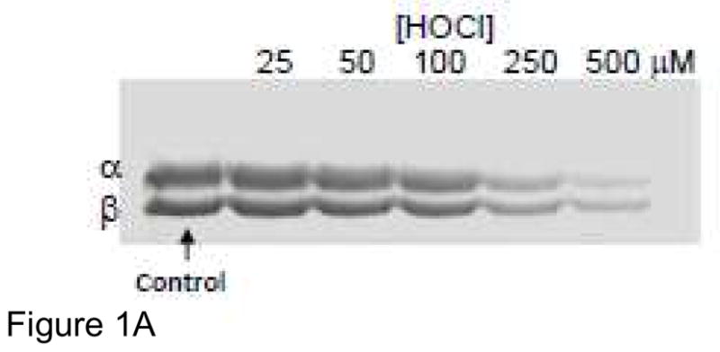

A) Tubulin samples (3 μM, 60 μM cys) were treated with 25, 50, 100, 250 or 500 μM HOCl for 10 min. at RT. Samples were incubated with 1.5 mM IAF for 30 min at 37 °C and then subjected to SDS-PAGE under reducing conditions on 7.5% polyacrylamide gels. Gel images were captured using a UV transilluminator and a Kodak DC290 imaging system. B) Tubulin samples (25 μM, 500 μM cys) were treated with 50, 100, 250 and 500 μM HOCl, GC or TC for 10 min at RT. Excess oxidant was scavenged with 1 mM met or monochlorodimedone (for HOCl). Samples were incubated with 2.5 mM IAF for 30 min at 37 °C and then protein was precipitated with 80% ethanol. Fluorescein-labeled protein pellets were collected, washed with 80% ethanol and resuspended in 3 M guanidine HCl in 0.1 M Tris pH 8.8. Fluorescein absorbance at 490 nm for standards and samples was measured in a 96 well plate. These data represent the mean +/− standard error of three independent experiments.
IAF labeling of oxidant-treated tubulin followed by SDS-PAGE is important because it allows us to assess any subunit selectivity by a particular oxidant. As shown in Figure 1A, the intensities of both the α- and β-tubulin bands decreased in a dose-dependent manner. Thus, HOCl does not show any selectivity for cysteines on a particular subunit. This is consistent with our published work with peroxynitrite, HNO and NO donors.[24]
Under physiological conditions, it is likely that HOCl will react with cellular amines to yield chloramines, RNHCl. Unlike HOCl, chloramines are reported to preferentially react with cysteines rather than methionines.[23] Figure 1B summarizes the effects of HOCl and two frequently studied chloramines, glycine chloramine (GC) and taurine chloramine (TC) on tubulin cysteines as assessed by IAF labeling, protein precipitation and quantitation of fluorescein incorporation. In this case, a much higher tubulin concentration was used (25 μM, 500 μM cysteine). One observes a dose-dependent decrease in tubulin cysteines for all three oxidants; however, consistent with Figure 1A, oxidation of tubulin cysteines is not stoichiometric.
Of note, the porcine tubulin preparation contains 100 μM GTP, an essential component of the purification buffer that ensures that the GTP binding sites on each tubulin subunit are occupied. Without GTP, the protein is much less stable resulting in a loss of function. When stock tubulin was diluted for oxidation assays presented herein, the final concentration of guanine nucleotides was ~50–60 μM. Several reports confirm that HOCl and chloramines react with guanine; thus even if GTP were hydrolyzed to GDP, it would be a target for oxidation.[32, 33] All oxidation assays with identical amounts of protein will contain the same concentration of guanine nucleotides.
Chloramines were prepared immediately prior to use by combining HOCl with a 10 molar excess of glycine or taurine. The excess amine limits the formation of dichloro compounds RNCl2.[23, 28] Therefore, it was essential to assess any effects of the high amine concentration on IAF labeling. Controls were prepared in which tubulin was treated with the amines only and subsequently with IAF. Quantitation of tubulin IAF labeling showed that the amines alone consistently decreased protein labeling and at the highest amine concentration tested (5 mM), labeling decreased by 4–7%. The amine control values were subtracted from the GC and TC data to obtain the results shown in Figure 1B. Although IAF is described as a thiol-selective reagent, it is not unexpected that the amines of glycine and taurine, present in high concentration relative to protein thiols, would react with it to some extent.[34] For this reason, controls with the amines are essential.
Using TNB to detect unreacted oxidant, we determined that none remained after 10 min for tubulin samples (25 μM, 500 μM cysteine) treated with 50, 100 or 250 μM HOCl, GC or TC. However, there was a decrease in TNB absorbance for those tubulin samples treated with 500 μM oxidant for 10 min. From the decrease in TNB absorbance, we calculated a residual oxidant concentration of approximately 100 μM. For this reason, it was essential to add an oxidant scavenger that would not interfere with subsequent analyses. Addition of 1 mM methionine to tubulin samples treated with 500 μM oxidant consumed the excess and no change in TNB absorbance was observed.
If HOCl reacted with tubulin lysines to yield stable protein-bound chloramines, then those lysine chloramines would have reacted with TNB resulting in an absorbance decrease. Because no decrease in TNB absorbance was observed at 50, 100 or 250 μM HOCl, we conclude that no stable protein-bound chloramines form when tubulin is treated with HOCl.
We also quantitated tubulin cysteine oxidation by HOCl, GC and TC using the DTNB assay. We and others consistently detect all 20 tubulin cysteines using multiple cysteine-specfic reagents.[35, 36] For these experiments, the tubulin concentration was 25 μM, a protein concentration that yields a theoretical protein thiol concentration of 500 μM and a methionine concentration of 650 μM (tubulin contains 26 methionines). Controls were performed using glycine and taurine alone to determine if the excess amines present in the chloramine solutions reacted with DTNB or altered the TNB absorbance to any extent. Though the oxidants, GC and TC, reacted to a limited extent with DTNB, the amines alone did not. Given that all GC and TC would have been consumed in their reactions with tubulin or quenched with methionine, their residual reaction with DTNB was inconsequential.
Figure 2 shows that HOCl, GC and TC oxidize tubulin cysteines in a dose-dependent manner. We detected all 20 tubulin cysteines by the DTNB assay; therefore, controls contained 500 μM cys. In some cases, assays were performed in the presence of 6 M guanidine HCl to ensure that tubulin was denatured. At all concentrations tested, the three oxidants were very similar in their reactivity with tubulin cysteines. TC was the most effective cysteine oxidant at 500 μM. There was no difference in the extent of cysteine oxidation at the other oxidant concentrations tested. Samples treated with 250 μM oxidant all showed about 60% of control cysteines which corresponds to detection of 12 cysteines and therefore oxidation of 8 mol cys/mol tubulin.
Figure 2. Detection of protein thiols using DTNB.
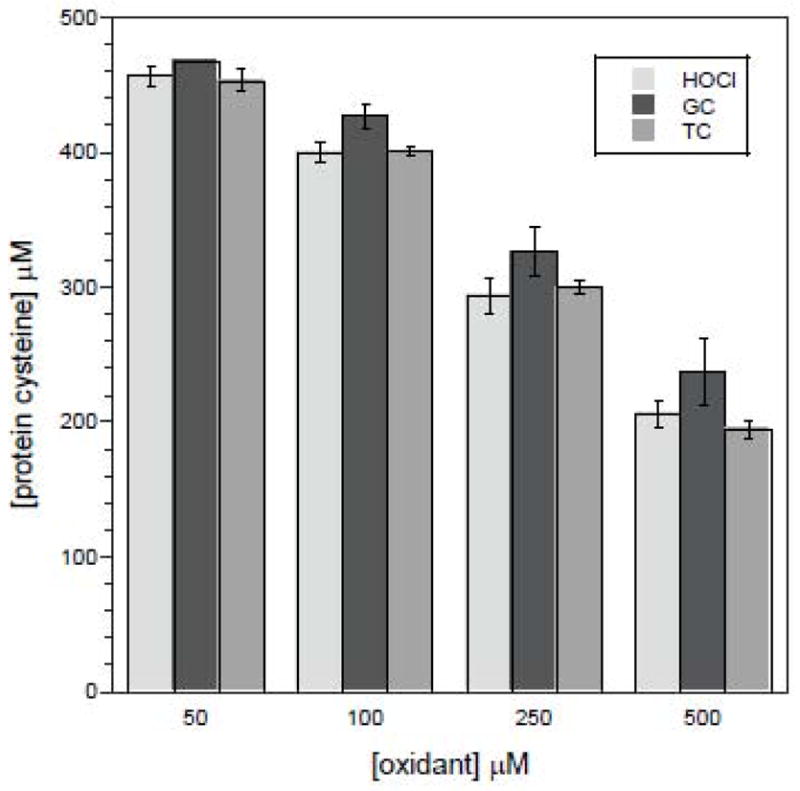
Tubulin samples (25 μM, 500 μM cys) were oxidized with 50, 100, 250 or 500 μM HOCl, GC or TC for 10 min at RT. Excess oxidant was scavenged with 1 mM met. Oxidized protein samples (20 μL) were transferred to a 96 well plate, diluted with 0.1 M PB pH 7.4 and treated with 0.5 mM DTNB in 200 μl. Maximum absorbance at 405 nm was recorded after 10–15 min. The results represent the mean +/− standard error of at least three independent experiments performed in duplicate.
Comparison of HOCl, GC and TC with peroxynitrite, an oxidant that we have previously studied in detail, shows that all three are superior in their ability to oxidize tubulin cysteines.[17, 18] For example, we previously reported that 500 μM peroxynitrite oxidized 7 mol cys/mol tubulin.[18] However, a direct comparison is challenging because when peroxynitrite, stored in dilute NaOH, is added to an aqueous buffer near physiological pH, it either reacts with its target or decomposes within seconds to form nitrate.[29, 37] Samples containing tubulin and oxidants were typically incubated for 10 min and it is certain that no peroxynitrite remained.
Given their greater ability to oxidize tubulin cysteines, it was not surprising to observe that HOCl, GC and TC were more effective inhibitors of microtubule polymerization than peroxynitrite. The data in Figure 3 shows that HOCl and GC are equivalent at all concentrations in their ability to inhibit polymerization. TC shows enhanced inhibition relative to HOCl and GC at all concentrations tested though the difference is most noticeable at 250 and 500 μM TC with essentially no microtubule polymer forming. To compare oxidant effectiveness, IC50 values were estimated from Figure 3. Comparisons of oxidant potency must take into consideration the ratio of oxidant to cysteine target in the protein preparation. In Figure 3, the concentration of cysteine target is 500 μM. For HOCl and GC, the IC50 is about 160 μM; whereas the IC50 for TC is 110 μM. For peroxynitrite, the IC50 determined from Figure 3 is 500 μM, a value identical to what we reported previously.[17, 24]. In those previous experiments with peroxynitrite, the cysteine target concentration was also 500 μM. The IC50 value for Angeli’s salt (AS), an HNO donor was 200 μM.[24]
Figure 3. Effect of oxidants on microtubule polymerization.
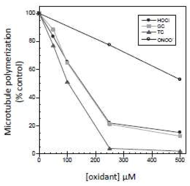
Tubulin samples (25 μM, 500 μM cys, final reaction volume = 50 μl) were treated with each oxidant for 10 min at RT and excess oxidant was scavenged with 1 mM met. GTP (1 mM final) was added and the samples were incubated at 37 °C for 22 min. Microtubule polymer was collected by centrifugation at 16000 x g for 20 min. The protein concentration in each supernatant was measured using the BCA reagent. The results represent the mean of at least three independent experiments performed in duplicate.
As with other oxidants we have tested in the past, there was no oxidant-induced protein aggregate formation. If there had been, protein pellets would have increased in size with increasing oxidant but they did not. Controls were performed to assess any effects of the excess amines on polymerization. Though a very slight inhibition (1–2%) was observed with both glycine and taurine, it was not sufficient to warrant subtracting out the effect.
Another method we employ to assess tubulin cysteine oxidation involves detection of interchain disulfides between tubulin subunits. Oxidized samples were subjected to SDS-PAGE under nonreducing conditions and higher molecular weight species were detected by Western blot using an anti-β-tubulin antibody. Figure 4A shows that HOCl oxidizes tubulin cysteines to form the characteristic dimers and tetramers that we observe with peroxynitrite and NO/HNO donors.[24] The concentrations of HOCl used ranged from 50 μM to 1 mM (lanes 2–6) whereas the tubulin concentration was only 8 μM (160 μM cys). Even control tubulin (lane 1) contains some tubulin dimers indicating that tubulin cysteines can air oxidize if stored in the absence of a reducing agent. At HOCl concentrations greater than 100 μM, there is a significant decrease in the monomer β-tubulin band indicating that tubulin is oxidized to form large disulfide-linked species that do not enter the separating gel, but that do not pellet upon centrifugation. The higher oxidant: protein ratio was used to illustrate that all tubulin can be oxidized to form disulfide-linked species if the oxidant concentration is sufficiently high.
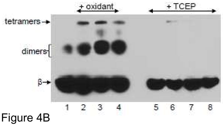
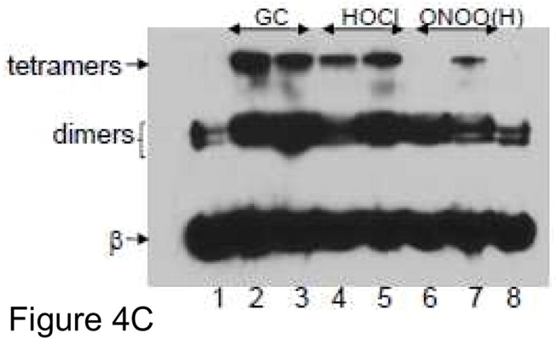
Figure 4. Detection of interchain tubulin disulfides.
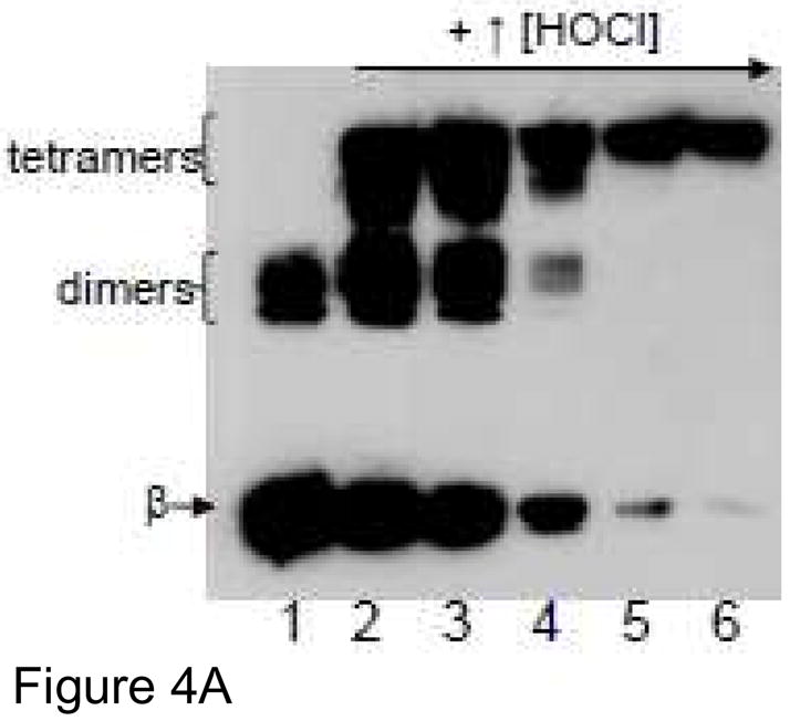
Oxidized tubulin species were separated by SDS-PAGE under nonreducing conditions on a 7.5% polyacrylamide gel and transferred to PVDF and probed with anti-β-tubulin. Monomeric β-tubulin (50 kDa) is labeled as well as dimers and tetramers. A) Tubulin samples (8 μM protein, 160 μM cysteines) were treated with 50, 100, 250, 500 and 1 mM HOCl for 10 min at 37 °C (Lanes 2–6). B) Tubulin (25 μM protein, 500 μM cys) was treated with 250 μM HOCl, GC or TC (lanes 2–4) for 10 min at RT. Identical oxidized tubulin samples were subsequently treated with 2.5 mM TCEP (lanes 6–8) for 5 min prior to electrophoresis. Control tubulin is in lanes 1 and 5 (+/− TCEP). C) Tubulin (25 μM protein, 500 μM cys) was treated with 100 or 250 μM GC (lanes 2–3), HOCl (lanes 4–5) and peroxynitrite (lanes 6–7) for 10 min at RT. Control tubulin is in lanes 1 and 8.
In Figure 4B, 25 μM tubulin was treated with 250 μM HOCl, GC or TC in lanes 2–4. These concentrations are the same as those used in Figures 1B, 2 and 3. Addition of the disulfide reducing agent, TCEP, eliminated all higher molecular-weight species (lanes 5–8). In additional Western blot assays, tubulin was treated with up to 1 mM HOCl followed by TCEP or DTT and no non-disulfide higher molecular weight species were detected (data not shown). This control was important because HOCl has been reported to induce the formation of noncovalent protein oligomers that are detected even in the presence of reducing agents.[38]
To further compare oxidants, tubulin was treated with 100 and 250 μM GC, HOCl and peroxynitrite (Figure 4C). GC (lanes 2 and 3) and HOCl (lanes 4 and 5) show more disulfides, tetramers especially, than peroxynitrite (lanes 6 & 7). As expected, controls in lanes 1 and 8 show some tubulin disulfides. These results are consistent with our previous work confirming that peroxynitrite is a less effective cysteine oxidant.[24]
In addition to cysteine oxidation, HOCl oxidizes methionine, another abundant target present in porcine tubulin.[23] The reaction of an oxidant with a protein depends on the relative abundance and accessibility of a protein’s side chains; thus, we must consider the relative concentrations of cysteines and methionines, the most readily oxidized amino acids in proteins. The most common tubulin isoform contains 26 methionines and 20 cysteines.[13, 39]
To assess methionine oxidation of tubulin by HOCl and chloramines, we used a cyanogen bromide (CNBr) cleavage assay. If methionines are oxidized, tubulin cannot be digested with CNBr, a reagent that reacts with the methionine thioether under acidic conditions resulting in peptide bond cleavage. As shown in Figure 5, oxidized methionines in tubulin were detected using 250 μM HOCl (lane 6) but not with 250 μM GC or TC (lanes 4&5). The darker β-tubulin band in the HOCl-treated sample indicates greater methionine oxidation resulting in decreased protein cleavage by CNBr. Thus, for tubulin treated with HOCl, both cysteine and methionine oxidation are detected. This observation is consistent with published reports showing that HOCl oxidizes both cysteine and methionine whereas chloramines are more specific cysteine oxidants.[23] Additional experiments showed some methionine oxidation by 500 μM GC and TC, but it was consistently lower than that observed for HOCl (data not shown). No methionine oxidation was detected when tubulin was treated with the same concentrations of H2O2 or peroxynitrite (data not shown).
Figure 5. Detection of methionine oxidation.
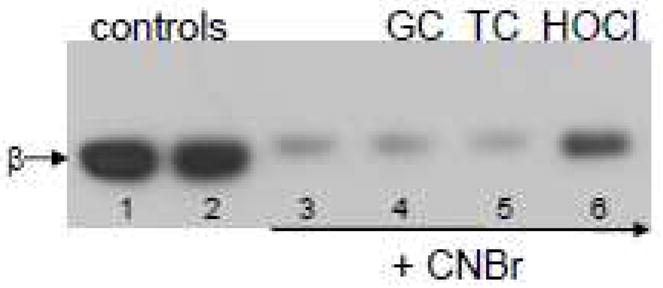
Tubulin samples (12.5 μM, 250 μM cysteines, 325 μM methionines) were treated with 250 μM of each oxidant for 30 min at RT. Samples were subsequently acidified with formic acid, and treated with CNBr for 16 hours. Samples were neutralized with NH4OH to pH 7.4–7.6 and subjected to SDS-PAGE under reducing conditions on a 7.5% polyacrylamide gel. Proteins were transferred to PVDF and probed with anti-β-tubulin. Lane 1 contains control tubulin, lane 2, control tubulin treated with formic acid and NH4OH, lane 3, tubulin treated with CNBr in the same manner as the oxidized tubulin samples in lanes 4–6.
Prior to this work, the most effective tubulin cysteine oxidant we had identified was Angeli’s salt (AS), Na2N2O3, an HNO donor.[24] To rank HOCl and TC, we compared their reactivity to AS in terms of the extent of tubulin cysteine oxidation, its effect on microtubule polymerization and the ability of TCEP to restore polymerization following treatment by the oxidants. The results of this comparison are shown in Figure 6A. We chose to use lower oxidant concentrations, 75 and 150 μM, because we wanted to more closely mimic physiological concentrations and with less oxidation, we expected that it would be easier to restore activity with a disulfide reductant.
Figure 6. Comparison of HOCl, AS and TC.

A) Tubulin samples (25 μM, 500 μM cys, final volume = 50 μl) were treated with 150 μM HOCl or AS for 10 min at RT. For polymerization, GTP (1 mM final) was added and the samples were incubated at 37 °C for 22 min. Microtubule polymer was collected by centrifugation at 16000 × g for 20 min. The protein concentration in each supernatant was measured using the BCA reagent. For the DTNB assay, protein samples (20 μL) were transferred to a 96 well plate, diluted with 0.1 M PB pH 7.4 and treated with 0.2 mM DTNB in 200 μl. Absorbance at 405 nm was recorded after 10 min. The results represent the mean +/− standard error of at least three independent experiments. For + TCEP samples, tubulin was oxidized, reduced with TCEP and polymerization was initiated with 1 mM GTP. Supernatants were analyzed by SDS-PAGE with Coomassie Blue staining. Band intensities from Figure 6B were used to quantitate repair.
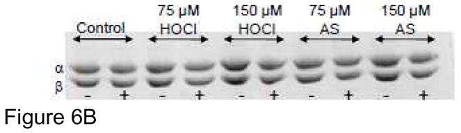
B) Effect of TCEP on microtubule polymerization following oxidant treatment Tubulin samples (25 μM, 500 μM cys, final volume = 50 μl) were treated with 75 and 150 μM HOCl or 75 and 150 μM AS for 10 min at RT. TCEP (2.5 mM final) was then added for an additional 10 min. GTP (1 mM final) was added and the samples were incubated at 37 °C for 22 min. Microtubule polymer was collected by centrifugation at 16000 × g for 20 min. Supernatant protein (5 μl of each) for samples treated with (+TCEP) or without (-TCEP) reducing agent was analyzed by SDS-PAGE under reducing conditions and detected by Coomassie Blue staining.
Figure 6A shows that 150 μM HOCl and AS are nearly identical with respect to their effects on tubulin cysteines. AS treatment oxidized slightly more cysteines than HOCl by the DTNB assay, but inhibition of polymerization was essentially the same. For both AS and HOCl, polymerization activity was restored following treatment with TCEP but not to control levels. Figure 6A shows polymerization activity decreases to 40% of control activity following tubulin oxidation by 150 μM AS or HOCl. Reduction with TCEP or DTT restores activity to 83% of control for HOCl and 81% of control for AS (Figure 6A). In comparison, a dose of 250 μM HOCl decreases activity to 22 % of control (Figure 2) and activity is only restored to 65% of control (data not shown). This is consistent with our previous findings using peroxynitrite as the oxidant.[17, 18, 40]
TC was the best inhibitor of polymerization but did not oxidize appreciably more cysteines than AS or HOCl at 150 μM. Polymerization was not restored by TCEP to the same extent for the TC treated samples (only 62% of control - Figure 6A). Both 1) the enhanced inhibition of polymerization by TC and 2) the decreased reversal of inhibition by reducing agents support a mechanism of inhibition not solely based on cysteine or methionine oxidation. Given that TC and GC are both chloramines with similar reactivities toward cysteine, we were surprised to observe that TC was both a better tubulin cysteine oxidant at high concentrations (Figure 2) and a better inhibitor of tubulin polymerization (Figure 3). Taurine alone, at the concentrations present in the prepared TC, did not inhibit tubulin polymerization. Further, we treated tubulin with other oxidants including AS, HOCl and GC for 5 min and then added taurine for an addition 5 min to determine if oxidized protein was susceptible to increased inhibition. We did not observe any change in inhibition by AS, HOCl or GC when taurine was added either before or after oxidant treatment. In the case of HOCl as oxidant, the combination of HOCl plus taurine showed the same level of oxidation as HOCl alone, rather than the enhanced inhibition observed for TC which would have formed when both HOCl and taurine were present.
In Figure 6B, we show that TCEP restores polymerization activity that was lost following tubulin oxidation by 75 and 150 μM HOCl and AS. Oxidized tubulin does not yield as much microtubule polymer (Figure 3) and therefore more protein is present in the supernatant after polymerization and centrifugation. In all cases, oxidized samples contain more protein than those treated with TCEP as indicated by the darker protein bands in the (−) lanes following Coomassie Blue staining. Quantitation of protein concentration for the 150 μM HOCl and AS samples yielded the +TCEP data in Figure 6A. Identical results were obtained when DTT was used as the disulfide reductant. The TCEP and DTT concentrations used, 2.5 mM for 10 minutes, were sufficient to reduce all interchain tubulin disulfides that were observed on Western blots following oxidant treatment (Figure 4B and data not shown). However, treatment with TCEP or DTT under these conditions would not reverse methionine oxidation. In an independent experiment, we observed that incubation of the free amino acid methionine sulfoxide with 250 equivalents of DTT for two days at 37 °C yields methionine.
Herein, we have observed both cysteine and methionine oxidation of tubulin by HOCl; however HNO, generated from AS, does not oxidize methionine. We confirmed this by treating the amino acid methionine with up to 50 equivalents of AS for 30 min. No methionine oxidation products were detected by thin-layer chromatography. Given that the half life of AS in neutral solution is roughly 4 min and because we routinely incubated tubulin with oxidants for 10 min, we did not attempt longer incubation times than 30 min for AS and methionine.
To lend support to the hypothesis that cysteine oxidation is largely responsible for the observed inhibition of microtubule polymerization, we analyzed some supernatant fractions following polymerization for remaining reduced cysteines. DTNB assays showed that all supernatants, regardless of the oxidant used, contained more oxidized cysteines on a mol cys/mol protein basis relative to the reactions in Figure 2 (which were not separated into microtubule pellet and supernatant fractions). For example, tubulin treated with 250 μM HOCl contained 12.3 mol cys/mol tubulin by the DTNB assay as shown in Figure 2. But only 8.5 mol cys/mol tubulin were detected in the supernatant fraction. In our previous work with peroxynitrite, we also observed greater cysteine oxidation in the supernatant fractions.[18] If the supernatant protein is more oxidized, then the microtubule polymer fractions must contain less oxidized protein.
Discussion
Cysteine oxidation of tubulin by HOCl and chloramines is associated with inhibition of tubulin polymerization. Three different methods were used to detect cysteine oxidation: 1) IAF labeling of remaining cysteine thiols after oxidant treatment (Figures 1A and 1B); 2) the DTNB assay in which remaining cysteines react with DTNB to yield the yellow TNB product (Figure 2) and 3) a Western blot method in which higher molecular weight tubulin species were detected under nonreducing conditions (Figures 4A–C). All three methods have been employed in our laboratory to characterize other cysteine oxidants including peroxynitrite, GSNO, NO donors and Angeli’s salt, a source of HNO.[41]
The IAF data in Figure 1B comparing HOCl and the chloramines, GC and TC shows a dose-dependent decrease in tubulin cysteines; however this assay was complicated by the observation that the amines, glycine and taurine, present in the chloramine preparations decreased IAF labeling. We chose to assess IAF labeling both by SDS-PAGE and by a more quantitative assay involving precipitation of IAF-labeled protein and quantitation of fluorescein incorporation. SDS-PAGE analysis was essential to show that cysteines of both tubulin subunits were oxidized by HOCl, GC and TC.
The IAF data in Figure 1B should be identical to the DTNB results in Figure 2 but there are some differences with respect to the extent of tubulin thiol oxidation by HOCl, GC and TC. The IAF assay (Figure 1B) is complicated by the presence of amines, both protein and nonprotein, that can react with iodoacetamides.[34] GC and TC preparations contain excess amines and a number of controls with the amines only were performed and subtracted from the IAF labeling data in Figure 1B. However, upon oxidation, changes in tubulin conformation may alter the accessibility and reactivity of protein amines. Figure 4B clearly shows that disulfide-linked tubulin dimers and tetramers form at the oxidant concentrations used in Figures 1B and 2. Greater IAF labeling of oxidized tubulin, thereby yielding less observed cysteine oxidation, may be the result of increased protein amine reactivity with IAF especially in the case of HOCl because no competing amines are present.
DTNB is specific for reaction with protein thiols and has been used extensively in our laboratory to quantitate thiol oxidation. For these reasons, we consider the DTNB assay data in Figure 2 to more accurately reflect the number of cysteines oxidized.
Treatment of oxidized tubulin with DTT and TCEP reduced all cysteines to the thiol form; thus, disulfides or sulfenic acids are the only possible cysteine oxidation products. The Western blot method shown in Figure 4 only detects disulfides between tubulin subunits but it is very likely that disulfides form within subunits. Our previous work showed that the thioredoxin reductase system restored all peroxynitrite-oxidized cysteines providing strong evidence that only disulfides and no stable sulfenic acids, are present in tubulin following oxidation.[17] Sulfenic acids (RSOH) are the initial product of cysteine oxidation by oxidants like peroxynitrite and the subsequent formation of a disulfide requires a reaction with a nearby cysteine to yield the disulfide.[37] For HOCl and chloramines, the initial cysteine oxidation product is the sulfenyl chloride (RSCl) which, like RSOH, reacts with a second cysteine to yield a disulfide.[23] Peskin and Winterbourn reported that the reaction of chloramines with thiols is dependent on the pKa of the thiol. The rates of reaction increase with decreasing thiol pKa implicating the thiolate anion as the reactive species.[23] Their observation suggests that chloramines may target specific cysteines within a protein in a sequence/charge-dependent fashion.
Although DTT and TCEP reduce all cysteines that are damaged by HOCl and chloramines (Figure 4B), not all microtubule polymerization activity is restored. This observation is consistent with previously reported studies from our laboratory in which peroxynitrite was used as the oxidant.[17, 18] As the concentration of oxidant increases, the fraction of polymerization activity that is restored decreases. At the higher concentrations of oxidant, there are multiple consequences that could affect reversibility. First, other types of protein damage in addition to cysteine oxidation can occur. Treatment of tubulin with 250 μM HOCl clearly caused methionine oxidation (Figure 5) that would not be reversed by DTT or TCEP treatment under the time and concentration conditions used herein.
In our previous work, we suspected that tyrosine nitration of tubulin by peroxynitrite contributed to the inhibition of polymerization and it would not be reversible.[18] Given that our results with other cysteine oxidants like AS, HOCl and chloramines are strikingly similar to what we observed with peroxynitrite and because these oxidants are not capable of nitrating tyrosines, our hypothesis must be revised. Likewise, in comparing AS and HOCl, we note that AS cannot oxidize methionines but HOCl can. The common connection between all oxidants studied in our laboratory is their ability to oxidize tubulin cysteines.
In comparison, HOCl-mediated oxidation of actin methionines alters both protein structure and function.[42] However, actin contains 16 methionines and only 5 cysteines (only 1 of which is solvent exposed). The predominant tubulin gene products contain 26 methionines and 20 cysteines. While all tubulin cysteines are detectable and able to be modified by many reagents, from examination of the tubulin structure, it appears that very few of the methionines would be solvent accessible.[39]
Therefore, it is likely that cysteine oxidation and subsequent reduction would cause changes in protein conformation that are not reversible. The result would be a subset of tubulin proteins that contain fully reduced cysteines but are no longer able to form microtubules. For example, oxidation and reduction could alter the GTP binding site on the tubulin subunits. Under physiologic conditions, proteins like tubulin would likely be exposed to lower oxidant concentrations generated over time rather than the relatively high concentrations used in this work. Thus, it is probable that a disulfide could form in monomeric tubulin and then be rapidly reduced by reductases before microtubule formation was compromised.
In our experiments, 1 mM GTP, the energy source to drive polymerization, is not added until after oxidation and reduction; therefore, changes in its concentration are not to blame for the inability to restore all activity. However, our tubulin assays are also complicated by the presence of ~50–60 μM guanine nucleotides during oxidation because it is an essential component of the purification buffer. Tubulin manipulations such as desalting compromise protein function; thus, to study functional consequences of protein oxidation, we must consider guanine oxidation as a competing reaction. The presence of low concentrations of guanine nucleotides has been consistent in all our tubulin oxidation studies to date including all with peroxynitrite and NO donors.
To compare oxidant potency, it is important to acknowledge that peroxynitrite degrades rapidly in solution near physiological pH and therefore the effective dose is not necessarily equal to the reported concentration. A comparison of rate constants is not possible because the rate constant for the reaction of tubulin with peroxynitrite is not known. To compare oxidant potency, it is essential to consider the ratio of oxidant to protein cysteine target. In this manuscript and others we have published, the concentration of tubulin is 25 μM yielding a cysteine concentration of 500 μM.[17, 24] The observed rate, and thereby the extent of reaction of any oxidant with tubulin cysteines, is dependent on the absolute concentrations of both.
AS, a source of HNO, is similar to peroxynitrite in that it decays in solution at neutral pH. The measured half-life for AS decomposition to HNO at neutral pH is about 4 minutes.[43] A direct concentration comparison shows AS to be equivalent to HOCl as a tubulin cysteine oxidant and inhibitor of polymerization (Figure 6A). HOCl is added as a bolus and does not degrade in solution like peroxynitrite or AS.
Although taurine chloramine was the best cysteine oxidant and polymerization inhibitor at 500 μM, we suspect that this effect is not strictly a result of oxidation of tubulin amino acids. We performed a number of control experiments to rule out taurine effects on polymerization. Taurine alone, at the concentrations present in the TC preparation did not inhibit polymerization. Taurine also did not enhance the inhibition observed with AS, HOCl or GC. Taurine contains a sulfonic acid group with a very low pKa of ~ 0 whereas glycine has a carboxylic acid. We suspect that the combination of the chloramine functional group and the sulfonic acid on the same molecule enhances the ability of TC to both oxidize cysteines and inhibit polymerization at high concentrations.
With 20 reduced cysteines and 26 methionines, the tubulin heterodimer is a likely target for oxidation in vivo. Because our brain tubulin preparation is composed of multiple α- and β-tubulin gene products and multiple post-translational modifications, sequence analysis is very challenging.[44, 45] Although we have not indentified individual oxidized amino acids following oxidant treatment, we have determined that tubulin cysteines are very similar in reactivity toward oxidants. When we compared HPLC traces of tryptic digests of IAF-labeled control and oxidant-treated tubulin, we observed that labeling of all peaks, and therefore all cysteines, decreased by a comparable amount. Thus, no one cysteine is selectively oxidized and quantitation by the DTNB assay detects partial oxidation of multiple cysteines. Luduena and Roach proposed that one or more of the 20 are “assembly-critical” and once those are modified, polymerization will not occur.[14] The most readily oxidized tubulin cysteines may not be the “assembly-critical” ones.
Our microtubule polymerization assays require much higher concentrations of protein than typical studies that examine enzyme inhibition by oxidants. For this reason, the concentrations of oxidants used are also high but not excessive. A 25 μM protein solution contains 500 μM cysteine and 650 μM methionine (Figures 1B, 2, 3, 4B, 4C, 6A). The highest oxidant concentration used was only 500 μM, a concentration of TC that completely inhibited polymerization (Figure 3).
We previously showed that peroxynitrite, GSNO and AS oxidized tubulin cysteines resulting in considerable loss of polymerization activity. The present study using HOCl and two chloramines shows that cysteines and methionines are oxidized, but that cysteine oxidation is associated with a loss of polymerization activity. The fact that disulfide reducing agents restore polymerization activity following HOCl and chloramine oxidation supports our hypothesis that cysteine oxidation is primarily responsible for the observed inhibition. Through our studies of oxidants that yield potentially different oxidation products and with differing reaction rates, we have identified tubulin cysteine oxidation as a common outcome. This observation supports our hypothesis that tubulin cysteines may play vital roles under conditions of oxidative stress in vivo because tubulin disulfides are readily repaired by reductase enzymes. Likewise, it is reasonable to suggest that microtubule polymerization could be regulated by fluctuations in the redox state of cells.
Acknowledgments
The authors acknowledge support from the National Institute of Neurological Disorders and Stroke (R15-NS38885 to LML) and the Petroleum Research Fund (Grant # 44091-B4 to LML).
ABBREVIATIONS
- AS
Angeli’s salt
- BCA
bicinchoninic acid
- CNBr
cyanogen bromide
- DTNB
5,5′dithiobis(2-nitrobenzoic acid)
- DTT
dithiothreitol
- GC
glycine chloramine
- GSNO
S-nitrosoglutathione
- HNO
nitroxyl
- HOCl
hypochlorous acid
- IAF
5-iodoacetomido-fluorescein
- PB
phosphate buffer
- PME
0.1 M PIPES pH 6.9, 1 mM MgSO4, 2 mM EGTA
- TC
taurine chloramine
- TCEP
Tris(2-carboxyethyl)phosphine
- TLC
thin-layer chromatography
- TNB
thionitrobenzoic acid
Footnotes
Publisher's Disclaimer: This is a PDF file of an unedited manuscript that has been accepted for publication. As a service to our customers we are providing this early version of the manuscript. The manuscript will undergo copyediting, typesetting, and review of the resulting proof before it is published in its final citable form. Please note that during the production process errors may be discovered which could affect the content, and all legal disclaimers that apply to the journal pertain.
References
- 1.Sponne I, Fifre A, Drouet B, Klein C, Koziel V, Pincon-Raymond M, Olivier JL, Chambaz J, Pillot T. Apoptotic neuronal cell death induced by the non-fibrillar amyloid beta peptide proceeds through an early reactive oxygen species-dependent cytoskeletal perturbation. Journal of Biological Chemistry. 2003;278:3437–3445. doi: 10.1074/jbc.M206745200. [DOI] [PubMed] [Google Scholar]
- 2.Paula-Barbosa M, Tavares MA, Cadete-Leite A. A quantitative study of frontal cortex dendritic microtubules in patients with Alzheimer’s disease. Brain Research. 1987;417:139–142. doi: 10.1016/0006-8993(87)90188-0. [DOI] [PubMed] [Google Scholar]
- 3.Good PF, Werner P, Hsu A, Olanow CW, Perl DP. Evidence for neuronal oxidative damage in Alzheimer’s disease. American Journal of Pathology. 1996;149:21–28. [PMC free article] [PubMed] [Google Scholar]
- 4.Lyras L, Cairns NJ, Jenner A, Jenner P, Halliwell B. As assessment of oxidative damage to proteins, lipids and DNA in brain from patients with Alzheimer’s disease. Journal of Neurochemistry. 1997;68:2061–2069. doi: 10.1046/j.1471-4159.1997.68052061.x. [DOI] [PubMed] [Google Scholar]
- 5.Butterfield DA, Perluigi M, Sultana R. Oxidative stress in Alzheimer’s disease brain: New insights from redox proteomics. European Journal of Pharmacology. 2006;545:39–50. doi: 10.1016/j.ejphar.2006.06.026. [DOI] [PubMed] [Google Scholar]
- 6.Cash AD, Aliev G, Seidlak SL, Nunomura A, Fujioka H, Zhu X, Tabaton M, Johnson AB, Paula-Barbosa M, Avila J, Jones PK, Castellani RJ, Smith MA, Perry G. Microtubule reduction in Alzheimer’s disease and aging is independent of tau filament formation. American Journal of Pathology. 2003;162:1623–1627. doi: 10.1016/s0002-9440(10)64296-4. [DOI] [PMC free article] [PubMed] [Google Scholar]
- 7.Cumming RC, Andon NL, Haynes PA, Park M, Fischer WH, Schubert D. Protein disulfide bond formation in the cytoplasm during oxidative stress. Journal of Biological Chemistry. 2004;279:21749–21758. doi: 10.1074/jbc.M312267200. [DOI] [PubMed] [Google Scholar]
- 8.Pamplona R, Dalfo E, Ayala V, Bellmunt MJ, Prat J, Ferrer I, Portero-Otin M. Proteins in human brain cortex are modified by oxidation, glycoxidation, and lipoxidation. Journal of Biological Chemistry. 2005;280:21522–21530. doi: 10.1074/jbc.M502255200. [DOI] [PubMed] [Google Scholar]
- 9.Brennan JP, Wait R, Begum S, Bell JR, Dunn MJ, Eaton P. Detection and mapping of widespread intermolecular protein disulfide formation during cardiac oxidative stress using proteomics with diagonal electrophoresis. Journal of Biological Chemistry. 2004;279:41352–41360. doi: 10.1074/jbc.M403827200. [DOI] [PubMed] [Google Scholar]
- 10.Sparaco M, Gaeta LM, Tozzi G, Bertini E, Pastore A, Simonati A, Santorelli FM, Piemonte F. Protein glutathionylation in human central nervous system: potential role in redox regulation of neuronal defense against free radicals. Journal of Neuroscience Research. 2006;83:256–263. doi: 10.1002/jnr.20729. [DOI] [PubMed] [Google Scholar]
- 11.Lind C, Gerdes R, Hamnell Y, Shuppe-Koistinen, Brockenhuus von Lowenhielm H, Holmgren A, Cotgreave IA. Identification of S-glutathionylated cellular proteins during oxidative stress and constitutive metabolism by affinity purification and proteomic analysis. Archives of Biochemistry and Biophysics. 2002;406:229–240. doi: 10.1016/s0003-9861(02)00468-x. [DOI] [PubMed] [Google Scholar]
- 12.Jaffrey SR, Erdjument-Bromage H, Ferris CD, Tempst P, Snyder SH. Protein S-nitrosylation: a physiological signal for neuronal nitric oxide. Nature Cell Biology. 2001;3:193–197. doi: 10.1038/35055104. [DOI] [PubMed] [Google Scholar]
- 13.Nogales E, Wolf SG, Downing KH. Structure of the ab- tubulin dimer by electron crystallography. Nature. 1998;391:199–203. doi: 10.1038/34465. [DOI] [PubMed] [Google Scholar]
- 14.Luduena RF, Roach MC. Tubulin sulfhydryl groups as probes and targets for antimitotic and antimicrotubule agents. Pharmacological Therapeutics. 1991;49:133–152. doi: 10.1016/0163-7258(91)90027-j. [DOI] [PubMed] [Google Scholar]
- 15.Olmsted JB. Tubulin pools in differentiating neuroblastoma cells. Journal of Cell Biology. 1981;89:418–423. doi: 10.1083/jcb.89.3.418. [DOI] [PMC free article] [PubMed] [Google Scholar]
- 16.Anderson PJ. The structure and amount of tubulin in cells and tissues. Journal of Biological Chemistry. 1979;254:2168–2171. [PubMed] [Google Scholar]
- 17.Landino LM, Iwig JS, Kennett KL, Moynihan KL. Repair of peroxynitrite damage to tubulin by the thioredoxin reductase system. Free Radical Biology and Medicine. 2004;36:497–506. doi: 10.1016/j.freeradbiomed.2003.11.026. [DOI] [PubMed] [Google Scholar]
- 18.Landino LM, Hasan R, McGaw A, Cooley S, Smith AW, Masselam K, Kim G. Peroxynitrite oxidation of tubulin sulfhydryls inhibits microtubule polymerization. Archives of Biochemistry and Biophysics. 2002;398:213–220. doi: 10.1006/abbi.2001.2729. [DOI] [PubMed] [Google Scholar]
- 19.Landino LM, Moynihan KL, Todd JV, Kennett KL. Modulation of the redox state of tubulin by the glutathione/glutaredoxin reductase system. Biochemical and Biophysical Research Communications. 2004;314:555–560. doi: 10.1016/j.bbrc.2003.12.126. [DOI] [PubMed] [Google Scholar]
- 20.Green PS, Mendez AJ, Jacob JS, Crowley JR, Growdon W, Hyman BT, Heinecke JW. Neuronal expression of myeloperoxidase is increased in Alzheimer’s disease. Journal of Neurochemistry. 2004;90:724–733. doi: 10.1111/j.1471-4159.2004.02527.x. [DOI] [PubMed] [Google Scholar]
- 21.Maki RA, Tyurin VA, Lyon RC, Hamilton RL, DeKosky ST, Kagan VE, Reynolds WF. Aberrant expression of myeloperoxidase in astrocytes promotes phospholipid oxidation and memory deficits in a mouse model of Alzheimer Disease. Journal of Biological Chemistry. 2009;284:3158–3169. doi: 10.1074/jbc.M807731200. [DOI] [PMC free article] [PubMed] [Google Scholar]
- 22.Peskin AV, Winterbourn CC. Taurine chloramine is more selective than hypochlorous acid at targeting critical cysteines and inactivating creatine kinase and glyceraldehyde-3-phosphate dehydrogenase. Free Radical Biology and Medicine. 2006;40:45–53. doi: 10.1016/j.freeradbiomed.2005.08.019. [DOI] [PubMed] [Google Scholar]
- 23.Peskin AV, Winterbourn CC. Kinetics of the reactions of hypochlorous acid and amino acid chloramines with thiols, methionine and ascorbate. Free Radical Biology and Medicine. 2001;30:572–579. doi: 10.1016/s0891-5849(00)00506-2. [DOI] [PubMed] [Google Scholar]
- 24.Landino LM, Koumas MT, Mason CE, Alston JA. Modification of tubulin cysteines by nitric oxide and nitroxyl donors alters tubulin polymerization activity. Chemical Research in Toxicology. 2007;20:1693–1700. doi: 10.1021/tx7001492. [DOI] [PubMed] [Google Scholar]
- 25.Winterbourn CC, Brennan SO. Characterization of the oxidation products of the reaction between reduced glutathione and hypochlorous acid. Biochemical Journal. 1997;326:87–92. doi: 10.1042/bj3260087. [DOI] [PMC free article] [PubMed] [Google Scholar]
- 26.Miron T, Rabinkov A, Mirelman D, Weiner L, Wilchek M. A spectrophotometric assay for allicin and alliinase (alliin lyase) activity: Reaction of 2-nitro-thiobenzoate with thiosulfinates. Analytical Biochemistry. 1998;265:317–325. doi: 10.1006/abio.1998.2924. [DOI] [PubMed] [Google Scholar]
- 27.Landino LM, Mall CB, Nicklay JJ, Dutcher SK, Moynihan KL. Oxidation of 5-thio-2-nitrobenzoic acid by the biologically relevant oxidants peroxynitrite anion, hydrogen peroxide and hypochlorous acid. Nitric Oxide. 2008;18:11–18. doi: 10.1016/j.niox.2007.09.087. [DOI] [PMC free article] [PubMed] [Google Scholar]
- 28.Thomas EL, Grisham MB, Jefferson MM. Preparation and characterization of chloramines. Methods in Enzymology. 1986;132:569–585. doi: 10.1016/s0076-6879(86)32042-1. [DOI] [PubMed] [Google Scholar]
- 29.Beckman JS, Chen J, Ischiropoulos H, Crow JP. Oxidative chemistry of peroxynitrite. Methods in Enzymology. 1994;233:229–240. doi: 10.1016/s0076-6879(94)33026-3. [DOI] [PubMed] [Google Scholar]
- 30.Williams RC, Lee JC. Preparation of tubulin from brain. Methods in Enzymology. 1982;85:376–385. doi: 10.1016/0076-6879(82)85038-6. [DOI] [PubMed] [Google Scholar]
- 31.Riddles PW, Blakeley RL, Zerner B. Reassessment of Ellman’s reagent. Methods in Enzymology. 1983;91:49–60. doi: 10.1016/s0076-6879(83)91010-8. [DOI] [PubMed] [Google Scholar]
- 32.Stanley NR, Pattison DI, Hawkins CL. Ability of hypochlorous acid and N-chloramines to chlorinate DNA and its constituents. Chemical Research in Toxicology. 2010;23:1293–1302. doi: 10.1021/tx100188b. [DOI] [PubMed] [Google Scholar]
- 33.Hawkins CL, Davies MJ. Hypochlorite-induced damage to nucleosides: formation of chloramines and nitrogen-centered radicals. Chemical Research in Toxicology. 2001;14:1071–1081. doi: 10.1021/tx010071r. [DOI] [PubMed] [Google Scholar]
- 34.Gurd FRN. Carboxymethylation. Methods in Enzymology. 1967;11:532–541. [Google Scholar]
- 35.Britto PJ, Knipling L, Wolff J. The local electrostatic environment determines cysteine reactivity of tubulin. Journal of Biological Chemistry. 2002;277:29018–29027. doi: 10.1074/jbc.M204263200. [DOI] [PubMed] [Google Scholar]
- 36.Britto PJ, Knipling L, McPhie P, Wolff J. Thiol-disulphide interchange in tubulin: kinetics and the effect on polymerization. Biochemical Journal. 2005;389:549–558. doi: 10.1042/BJ20042118. [DOI] [PMC free article] [PubMed] [Google Scholar]
- 37.Radi R, Beckman JS, Bush KM, Freeman BA. Peroxynitrite oxidation of sulfhydryls. Journal of Biological Chemistry. 1991;266:4244–4250. [PubMed] [Google Scholar]
- 38.Chapman ALP, Winterbourn CC, Brennan SO, Jordan TW, Kettle AJ. Characterization of non-covalent oligomers of proteins treated with hypochlorous acid. Biochemical Journal. 2003;375:33–40. doi: 10.1042/BJ20030685. [DOI] [PMC free article] [PubMed] [Google Scholar]
- 39.Lowe J, Li H, Downing KH, Nogales E. Refined structure of ab-tubulin at 3.5 A resolution. Journal of Molecular Biology. 2001;313:1045–1057. doi: 10.1006/jmbi.2001.5077. [DOI] [PubMed] [Google Scholar]
- 40.Landino LM, Skreslet TE, Alston JA. Cysteine oxidation of tau and microtubule-associated protein-2 by peroxynitrite: modulation of microtubule assembly kinetics by the thioredoxin reductase system. Journal of Biological Chemistry. 2004;279:35101–35105. doi: 10.1074/jbc.M405471200. [DOI] [PubMed] [Google Scholar]
- 41.Landino LM. Protein thiol modification by peroxynitrite anion and nitric oxide donors. Methods in Enzymology. 2008;440:95–109. doi: 10.1016/S0076-6879(07)00805-1. [DOI] [PubMed] [Google Scholar]
- 42.Dalle-Donne I, Rossi R, Giustarini D, Gagliano N, Di Simplicio P, Colombo R, Milzani A. Methionine oxidation as the major cause of the functional impairment of oxidized actin. Free Radical Biology and Medicine. 2002;32:927–937. doi: 10.1016/s0891-5849(02)00799-2. [DOI] [PubMed] [Google Scholar]
- 43.Miranda KM, Paolocci N, Katori T, Thomas DD, Ford E, Bartberger MD, Espey MG, Kass DA, Fukuto JM, Wink DA. A biochemical rationale for the discrete behavior of nitroxyl and nitric oxide in the cardiovascular system. Proceedings of the National Academy of Science USA. 2003;100:9197–9201. doi: 10.1073/pnas.1430507100. [DOI] [PMC free article] [PubMed] [Google Scholar]
- 44.Luduena RF. Multiple forms of tubulin: different gene products and covalent modifications. Internation review of cytology. 1998;178:207–275. doi: 10.1016/s0074-7696(08)62138-5. [DOI] [PubMed] [Google Scholar]
- 45.McKean PG, Vaughan S, Gull K. The extended tubulin superfamily. Journal of Cell Science. 2001;114:2723–2733. doi: 10.1242/jcs.114.15.2723. [DOI] [PubMed] [Google Scholar]